Abstract
Gastrointestinal stromal tumors (GISTs), the most common mesenchymal tumors of the gastrointestinal tract, arise from interstitial cells of Cajal. Most of them are composed of spindle cells and mimic leiomyoma morphologically. Nearly all GISTs demonstrate positive staining for CD-117 at immunohistochemistry examination, which helps to distinguish them from true leiomyomas. We describe the CT findings of a GIST of small bowel. At plain CT scan, the tumor was of homogeneous iso-attenuation. After intravenous administration of contrast materials, a well-defined exophytic growth mass with avid homogeneous enhancement was demonstrated. The mucosal layer over the mass was intact and fat plane between the mass and adjacent GI wall was preserved. A diagnosis of GIST was suggested and was then confirmed by positive staining for CD-117.
Keywords: Gastrointestinal stromal tumors, CD-117, CT findings
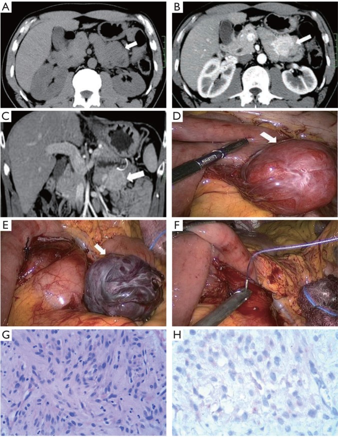
A 43-year-old man presented with upper abdominal discomfort and complained for a palpable mass in the left upper abdomen. His medical history and laboratory examination were unremarkable. Contrast enhanced CT examination was performed to further explore the abdominal mass. Plain CT revealed a suspected mass in the left lower quadrant (Figure 1A), but it was impossible to distinguish it from crimpy small bowel loops. Contrast-enhanced CT demonstrated an avid homogeneously enhanced mass (Figure 1B). It was well-defined and the mucosal layer covering the mass was intact (Figure 1C). Fat plane surrounding the mass was preserved. All these characteristics suggested that the mass was exophytic and arisen from the submucosal layer of GI. A diagnosis of GIST was proposed. The patient underwent laparoscopy surgery to resect the mass. Laparoscopy revealed a well-defined mass with intact capsule arising from jejunum (Figure 1D). The mass was completely excised (Figure 1E) and the wound was sewed under laparoscopy (Figure 1F). At pathological examinations, the tumor was consisted of spindle cells stained highly positive for CD-117 (Figure 1G, H). The diagnosis of GIST was confirmed.
Figure 1. A. Transversal plain CT scan depicts a homogeneous iso-attenuation mass (arrow), but it could not be differentiated from crimpy bowel loops; B. Transversal contrast-enhanced CT demonstrates a homogeneous avidly enhanced mass (arrow); C. Coronal multi-planar reformation of contrast-enhanced CT shows the mass is well-defined (arrow). The musical layer covering the mass (curve arrow) is intact and the fat plane between the mass and surrounding tissue is preserved; D. Laparoscopic photograph shows an exophytic mass with intact capsule; E. Laparoscopic photograph show the mass was completely excised; F. Laparoscopic photograph shows the wound was sewed; G. Photomicrograph (original magnification, ×100; hematoxylin-eosin stain) of the tumor shows spindle cells with pale eosinophilic cytoplasm; H. Immunohistochemistry demonstrates positive staining for CD-117 (original magnification, ×200).
Footnotes
No potential conflict of interest.


