

Abstract
Copper is one of the most interesting elements for various biomedical applications. Copper compounds show vast array of biological actions, including anti-inflammatory, anti-proliferative, biocidal and other. It also offers a selection of radioisotopes, suitable for nuclear imaging and radiotherapy. Quick progress in nanotechnology opened new possibilities for design of copper based drugs and medical materials. To date, copper has not found many uses in medicine, but number of ongoing research, as well as preclinical and clinical studies, will most likely lead to many novel applications of copper in the near future.
Keywords: Copper, Nuclear medicine, Nanotechnology, Drug development
Introduction
Copper (Cu) is a transition metal with atomic number 29, known since ancient times. It is an important trace element for most organisms in all kingdoms. In humans, copper plays role as a cofactor for numerous enzymes, such as Cu/Zn-superoxide dismutase, cytochrome c oxidase, tyrosinase, ceruloplasmin and other proteins, crucial for respiration, iron transport and metabolism, cell growth, hemostasis (Puig and Thiele 2002; Bertini et al. 2010). With the progress in medical sciences, copper has gained a lot of attention. The number of publications concerning copper and its compounds for potential medical applications, have reached tens of thousands. There are several reasons that render this element so attractive for drug development. Generally, simple inorganic salts of copper are toxic, but as a transition metal, with unsaturated d shell, it forms a large number of complexes. Coordination chemistry of copper is well-studied and “straightforward” in comparison to many other elements. From three known oxidation states, +1 and +3 are mostly unstable in biological systems, but on +2 state, Cu forms stable complexes with coordination number of 4, 5 or 6. Administration of copper in a form of organometallic complexes can be done in order to selectively deliver copper ions or radionuclides to diseased tissues, or to modify pharmacokinetics and/or pharmacodynamics of ligands. Moderate amounts of metal ions that could be liberated from biological degradation or transchelation of Cu complexes can be managed by organism, as copper is an important microelement, in contrary to many other transition metals, whose leakage from their compounds can lead to accumulation and toxic effects. Copper has several radioisotopes, five of them are particularly interesting for radiotherapy and imaging applications. Continuous progress of nanotechnology made it possible to exploit novel physicochemical properties of copper-containing nanoparticles and molecules. This article reviews current trends in various fields of medicine, in development of copper based pharmaceuticals and medical materials.
Biological activity of complexes of stable copper isotopes
Inflammation
In folklore it is believed that wearing copper bracelets and jewellery can ease the pain in rheumatoid arthritis. This belief had drawn attention to possible anti-inflammatory properties of copper ions and complexes. This issue was extensively researched in past century by Sorenson (1976, 1982, 1987, 1989). Hostýnek et al. (2006) found that metallic copper can indeed penetrate skin, after being oxidized on air. Anti-inflammatory effect of Cu can be linked with modulation of prostaglandin synthesis (Sakuma et al. 1996; Franco and Doria 1997; Sakuma et al. 1999), interleukin IL-2 expression (Hopkins and Failla 1999), neutralization of reactive oxygen radicals by Cu/Zn-superoxide dismutase and other. Though copper deficiency is known to impair immunity, the exact mechanism is unclear (Huang and Failla 2000).
In the past decade, several authors reported copper(II) complexes with potential anti-inflammatory properties. For treatment of rheumatoid arthritis, chelating agents that can facilitate transport of Cu(II) ions to sites of inflammation were researched (1–13).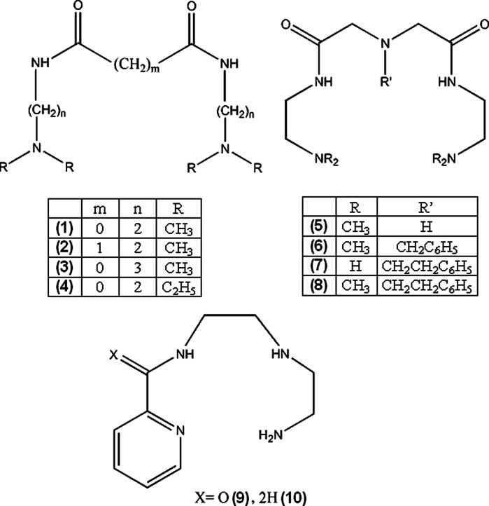
Jackson et al. (2000) attempted to design linear polyamine ligands that can mobilize copper in organism. The complexes cannot be too stable, because they would be quickly excreted with urine in unchanged form. Ligands 1–4 formed neutral complexes only above pH 7.0 and were too labile for systemic administration, but still could be used to facilitate dermal absorption of copper. Complexes of 5–8, due to additional nitrogen atom were significantly more stable (~2 log units), 6–8 were also more lipophilic, but the stability was still suboptimal (Jackson et al. 2000). More promising results for dermally absorbed Cu complexes were achieved for ligands 9 and 10. The compounds show selectivity towards copper ions, good stability at physiological pH (formation constants at 25 °C in 0.15 M NaCl, for unprotonated ligands: log β = 11.51 for 9 and 18.62 for 10), low renal clearance and water/octanol partitioning indicating possible dermal absorption. An important feature of 9 and 10 is that they form more labile complexes with Ca2+ and Zn2+ ions (for 9 and 10 respectively: with zinc log β = 5.55 and 11.51, with calcium log β = 3.24 and 3.92), which are main competitors of copper in blood plasma. Simulations showed that Cu complexes of the ligands are stable in blood plasma, and effectively mobilize copper ions without affecting significantly other metal ions levels (Zvimba and Jackson 2007). Odisitse et al. (2007, 2009) also reported dermally absorbed complexes of copper with 11–13 ligands. The compounds showed approximately 24 h biological half-life which is desired for potential anti-inflammatory drugs. Simulation of behavior of 13 in blood plasma indicated that Ca2+ and Zn2+ ions concentration is sufficient to compete with Cu2+, even though 13 is more selective towards cupric ions. Therefore, the ligand can facilitate copper transport through skin, then release Cu2+ ions in bloodstream.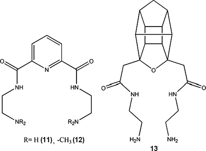
Copper-zinc-superoxide dismutase (SOD) is an important enzyme protecting cells against oxidative injury, scavenging and neutralizing reactive oxygen species. It has been shown that SOD can significantly reduce inflammation induced in laboratory animals (Emerit et al. 1991; Zhang et al. 2002; Garcia-González et al. 2009). Many complexes of copper(II) have similar to SOD ability to neutralize superoxides (14–27). These SOD-mimicking complexes of copper were proposed as non-analgesic anti-inflammatory drugs by various authors: Cu complexes of aromatic acids (14–16) (Suksrichavalit et al. 2008), saccharinate and pyridine derivates (17–18) (Ferrer et al. 2010), tolfenamic acid (19) (Kovala-Demertzi et al. 2004), 2-amino-2-thiazoline and polyamines (20–25) (Pontiki et al. 2006), o-vanillin (26) (González-Baró et al. 2010), oxaprozinate (27) (Dutta et al. 2004).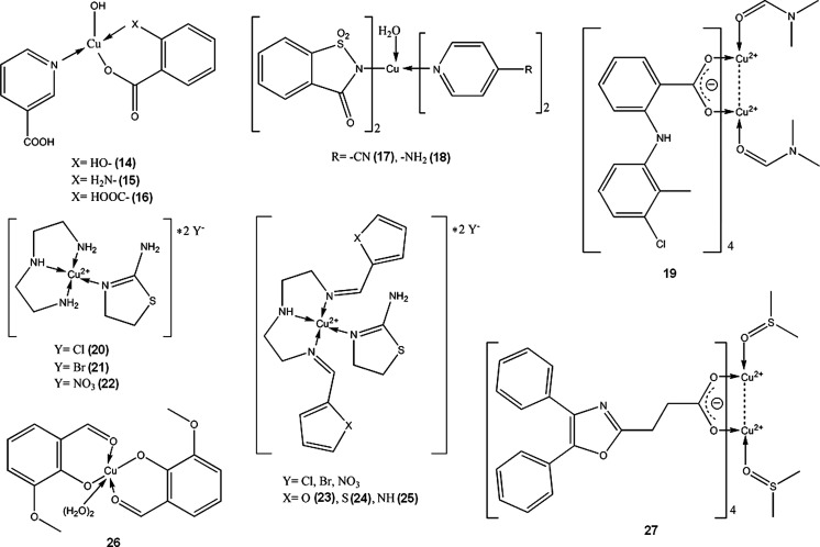
However, there is one important hindrance, as both SOD and SOD-like Cu-complexes can cleave cellular DNA by reacting with hydrogen peroxide and generating hydroxyl radicals by Fenton-type reaction (Sang and Yang 2005; Han et al. 2007; Seng et al. 2009; Ghosh et al. 2010; Ibrahim et al. 2011). This aspect requires thorough dose–response studies, before this group of potential anti-inflammatory drugs can emerge.
In the last 20–30 years of 20th century, there was a lot of interest in copper(II) complexes with NSAIDs (Non-Steroidal Anti-Inflammatory Drugs), such as acetylsalicylic acid, indomethacin, piroxicam, ibuprofen, diclofenac, naproxen and others. These complexes were reported to possess increased activity and lower ulcerogenic effect than respective NSAID and copper administered separately. However, none of the substances has been approved for internal therapy of humans. Exhaustive review of the subject was written by Weder et al. (2002) Currently, there are still a number of publications each year on Cu-NSAIDs complexes, but mainly concerning the structural and physicochemical aspects of the compounds; structure–activity and biological studies are sparse, therefore it can be assumed that this group of potential drugs will not make any impact on medicine in the nearest future.
It should be noted that copper-indomethacine complex (28, Fig. 1) underwent some biological evaluation, and is currently used in veterinary in Australia, New Zealand and some other countries. Similarly to other NSAID-Cu complexes, copper indomethacinate retains parent drug anti-inflammatory activity but have lower ulcerogenic effect, probably due to free radical scavenging ability (Bertrand et al. 1999). Cu-indomethacin-dimethylformamide complex shows good solution stability at pH 7.4 (<8 % decomposition after 3 days), which can be further increased by micellar solubilisation of the complex with Span 80 and tetraglycol (Weder et al. 1999). In Australia, Cu-salicylate was available until recently for external use in humans, in a form of topical anti-inflammatory gel.
Fig. 1.

Copper-indomethacine-N,N-dimethylformamide complex (Weder et al. 1999). Data from Cambridge Crystallographic Data Centre
Cancer
Since cisplatin was introduced for chemotherapy of cancer, a search for other transition metal complexes with anti-proliferative activity has started. Various copper(II) complexes were found to be cytotoxic, with the most common ligands being NSAIDs or Schiff bases (29–38). As mentioned in above section, many Cu(II) complexes possess catalytic activity towards reactive oxygen species and can induce breakage of DNA strand. This can explain cytotoxicity of some of the compounds. 29 (Fig. 2) in aqueous solution without presence of any external reducing factors, forms bis-(1,10-phenantroline)copper(I) which oxidatively degrades nucleic acids (Barceló-Oliver et al. 2007). However, in many cases, probably more sophisticated mechanisms are involved.
Fig. 2.

Crystal structure of Cu-o-iodohippurate-1,10-phenantroline complex(29) (Barceló-Oliver et al. 2007) Data from Cambridge Crystallographic Data Centre
Meloxicam (30) and piroxicam (31, Fig. 3) form stable in physiological pH Cu complexes (K = 3.2 × 10⁹ and 9.8 × 10⁹ M−2 respectively) that are able to strongly bind with DNA, disrupting its structure and stopping transcription as a result (Roy et al. 2006; Cini et al. 2007). Copper N-(2-hydroxyacetophenone) glycinate (32) is an interesting immunomodulatory agent, capable of inducing apoptosis in multidrug-resistant cancer cells by stimulating production of cytokines, such as interferon γ or TNF-α (Tumor Necrosis Factor α) (Mookerjee et al. 2006). Guo et al. (2010) suggested that salicylaldehyde-amino acid Schiff base copper chelates (33, 34) trigger cancer cell’s apoptosis by downregulation of overexpressed mutant type P53 protein.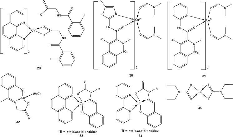
Fig. 3.

Copper piroxicam-DMF complex crystal structure (Cini et al. 1990) Data from Cambridge Crystallographic Data Centre
Disulfiram, a drug used in alcoholism treatment, forms in vivo a copper complex (35) which acts as a proteasome inhibitor, and selectively induces apoptosis in breast tumors (Chen et al. 2006). Disulfiram and copper gluconate are currently under phase I trials for treatment of solid tumors with metastases in liver (ClinicalTrials.gov 2012). Compound 36 shows high in vitro and in vivo activity towards MCF-7, PC3 and HEK293 cell lines. Its proposed mode of action is multidirectional and includes apoptosis induction via caspase pathway, DNA fragmentation and antioxidant enzymes inhibition. 36 is more effective than cisplatin in breast tumor models (about 20-fold lower IC50) and shows minimal toxicity (Chakraborty et al. 2010). Other type Cu(II) complexes with both antimicrobial and antitumor properties were reported by Singh et al. (2009)(37,38).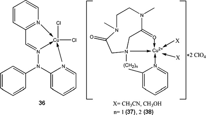
Antimicrobial
Copper, both in metallic form and in many chemical compounds, possess antimicrobial activity, which was already used by ancients. Cupric ions exhibit non-specific biocidal activity, although weaker than silver. Copper-silver electrolytic ionization systems are used in many hospitals to decrease number of Legionella residing in hot water pipes. Metals and alloys used in orthopedic implants can be doped with copper ions, in order to reduce risk of infection after prosthetic surgery. The tradeoff is reduced to some extent corrosion resistance of the resulting materials, but still on a reasonable level (Wan et al. 2007). Due to non-specific toxicity, for the use of copper as an antibacterial therapeutic, the metal should be administered in a form of complex compounds, rather than simple inorganic salts. Nature of chelating agent, however, plays very important role, as there can be no simple correlation between antibacterial activity and complex stability (Azenha et al. 1995). Many various Cu(II) complexes with different ligands were reported to possess antibacterial and antifungal activity (39–48) (Gölcü et al. 2005; Shakir et al. 2006; Singh et al. 2008; Sreedaran et al. 2008; Kumar and Arunachalam 2009; Suksrichavalit et al. 2009). Singh et al. (2008) utilized an approach to use ligands which already have antimicrobial activity and enhance it by complexation with copper (39–41). Antihypertensive drug pindolol, when complexed with Cu (41) (complex stability constant log β = 11.28 in water-dioxan 40:60 at 25 °C), exhibits notable antimicrobial activity towards some bacterial and fungal strains (Gölcü et al. 2005). Water soluble, polymeric complex 47 shows good antimicrobial activity and is also capable of binding DNA (Kumar and Arunachalam 2009). The complexes (39–48) were only screened for antibiotic properties, and to the best of our knowledge no further evaluations for medical applicability were performed.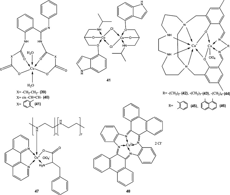
Other uses
Copper (I)-Cl-(nicotinic acid)2 (polymeric) is able to notably reduce gastrointestinal mucosa lesion caused by NSAIDs such as acetylsalicylic acid. The complex shows antioxidative, antiapoptotic, secretolytic and antihemorrhagic activity and can be a good alternative for currently used anti-ulcer drugs, proton pump inhibitors, which increase gastrin level (Tuorkey and Abdul-Aziz 2009). It is also a rare example of Cu(I) compound proposed for medical use. Toyota et al. (2005) described a series of copper and iron complexes acting as thrombin inhibitors. One of these compounds, Cu(II) complex with 4-formyl-3-hydroxybenzamidine and d-tryptophane (49), had the highest inhibitory activity (Ki value 2.7 × 10⁻⁸ M), comparable to registered anticoagulant drug, argatroban (Ki 1.9 × 10⁻⁸ M) (Toyota et al. 2005). Tian et al. (2009) suggested copper-taurine as possible compound able to facilitate wounds healing by stimulating process of tissue regeneration and by preventing infections.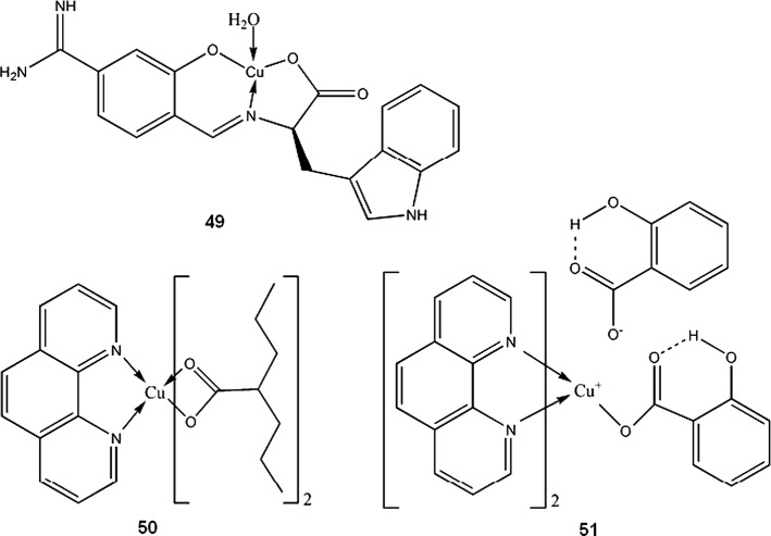
Work by Sylla-Iyarreta Veitía et al. (2009) is a good example, how complexation with copper of clinically used drug, valproic acid in that case, can lead to novel, more potent compound. Bis-valproinato(1,10-phenanthroline)copper(II) (50) was found to be very effective in preventing Minimal Clonic seizures (ED50 8 μmol/kg). 1,10-phenantroline and salicylate Cu complex (51, Fig. 4) and bis(1,10-phenanthroline)-μ-bis(salicylato)dicopper(II) with anticonvulsant activity effective against MES (maximal electroshock) induced seizures, were reported earlier by Lemoine et al. (2002). Despite different structure in solid state, both complexes showed similar anticonvulsant activity, probably due to formation of the same species in the dilute solutions. The compounds lose salicylate and one phenantroline ligand in dilute N,N-dimethylformamide (DMF) solution to form [Cu(1,10-phenantroline)DMF4]2+. The results can only be interpolated to biological systems, since both complexes are insoluble in water.
Fig. 4.

Crystal structure of 51 (Lemoine et al. 2002) Data from Cambridge Crystallographic Data Centre
Copper palmitate may be useful in preventing skin photosensitivity induced by porphyrins in patients who underwent photodynamic therapy. Liposomal topical cream with Cu-palmitate effectively prevented skin inflammation in photosensitized rats exposed to light (Bilgin et al. 2005). Taggar et al. (2006) successfully prepared liposomal form of anticancer drugs, topotecan and irinotecan. Copper(II) sulphate loaded liposomes accumulated and retained drug molecules due to formation of copper complex inside the liposome. The authors also reported improved therapeutic activity of this drug formulation (Taggar et al. 2006). Sreedhara et al. (2000) reported Cu-aminoglycosides complexes (of neamine and kanamycin A) as efficient deoxyribonucleases with reaction kinetics similar to enzymes. Notably, the DNA cleavage was achieved by hydrolytic pathway, without generation of free radicals (Sreedhara et al. 2000). Copper-l-histidine complex (52) is in phase II clinical trials for treatment of Menkes disease, a genetic disorder in Cu transport, leading to copper deficiency (ClinicalTrials.gov 2012).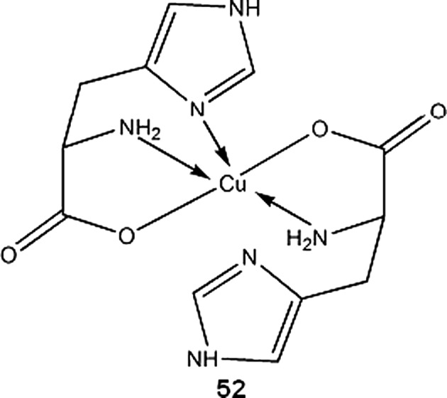
Copper radioisotopes in nuclear medicine
Natural copper comprises two stable isotopes: 63Cu (69.17 %) and 65Cu (30.83 %). Of 27 known copper radioisotopes, five are particularly interesting for nuclear medicine: 60Cu, 61Cu, 62Cu, 67Cu, and especially 64Cu. Their nuclear characteristics are given in Table 1.
Table 1.
Decay properties of medically important Cu radioisotopes
| Isotope | T1/2 | β− (MeV) | β+ (MeV) | EC (%) | γ (MeV) |
|---|---|---|---|---|---|
| 60Cu | 23.7 min | – |
1.91 (11.6 %) 1.98 (49 %) 2.95 (15 %) 3.77 (5 %) |
7.2 |
0.511 (185 %) 0.826 (21.7 %) 1.33 (88 %) 1.79 (45.4 %) 3.12 (4.8 %) |
| 61Cu | 3.33 h | – |
0.932 (5.5 %) 1.22 (51 %) |
36 |
0.283 (12.2 %) 0.373 (2.1 %) 0.511 (123 %) 0.656 (10.8 %) 1.19 (3.7 %) |
| 62Cu | 9.67 min | – | 2.93 (97.2 %) | 2 | 0.511 (195 %) |
| 64Cu | 12.7 h | 0.579 (38.5 %) | 0.653 (17.6 %) | 40 |
0.511 (35.2 %) 1.35 (0.5 %) |
| 67Cu | 61.83 h |
0.377 (57 %) 0.468 (22 %) 0.562 (20 %) |
– | – |
0.093 (16.1 %) 0.185 (48.7 %) 0.3 (0.8 %) |
Values taken from National Nuclear Data Center (Brookhaven National Laboratory 2012)
β−, β+, γ—electron, positron and gamma emission respectively, EC-electron capture
Decay characteristics of copper radionuclides make them suitable for numerous medical applications, such as Positron Emission Tomography(PET) imaging, radioimmunological tracing and radiotherapy of cancer. For widespread use in medicine of any radioisotope, two factors are essential: availability of the isotope and effective modes of binding with an appropriate chemical carrier. Efficient production of copper isotopes was extensively researched over past 20–30 years, and also many potential chelators were developed during that time. Methods of production, applications in nuclear medicine and chelating agents for copper radioisotopes were reviewed by Blower et al. (1996), Williams et al. (2005), Rowshanfarzad et al. (2006), Hao et al. (2009), Wadas et al. (2010), and Ding et al. (2011).
60Cu
PET is a three dimensional imaging technique which utilizes simultaneous detection of two oppositely moving photons, resulting from annihilation of positron with electron. Positron comes from decay of a radioisotope incorporated into targeting molecule which can selectively accumulate in desired tissues, organs or tumors. The most popular PET tracer is 18F in a form of 2-deoxy-2-(18F)fluoro-d-glucose. Metallic radioisotopes have advantage over fluorine-18, as they can be easily introduced into a targeting molecule by forming a coordination compound with it. 60Cu is a β+ emitter with decay properties making it possible candidate for PET tracer. 60Cu can be produced using small cyclotrons at relatively low costs from 60Ni target (McCarthy et al. 1999). Relatively high energy positron and gamma emissions, compared to 62Cu, are the most important disadvantages of 60Cu isotope as PET imaging agent.
Copper bis-thiosemicarbazones complexes, mainly 60/61/62/64Cu-diacetyl-bis(N4-methylthiosemicarbazone) (60/61/62/64Cu-ATSM 53), 60/61/62/64Cu-pyruvaldehyde-bis(N4-methylthiosemicarbazone) (60/61/62/64Cu-PTSM 54) and 60/61/62/64Cu-ethylglyoxal bis(thiosemicarbazone) (60/61/62/64Cu-ETS 55), are the most widely studied copper radioisotopes compounds for use in PET. 60/61/62/64Cu-ATSM and 60/61/62/64Cu-ETS, due to their specific redox properties, can be useful for detection and imaging of hypoxic tumor cells. Mechanism of action of copper-thiosemicarbazones in broad outline is as follows: the complex enters cell where it is spontaneously reduced from Cu(II) to Cu(I) state, then it can either be reoxidized by molecular oxygen and diffuse from the cell, or in hypoxic conditions, it irreversibly decomposes and stays trapped within cell (Dearling and Packard 2010). It should be noted that nonradioactive Cu-ATSM has been recently found to be neuroprotective agent, and can be used for Parkinson’s disease treatment (Hung et al. 2012).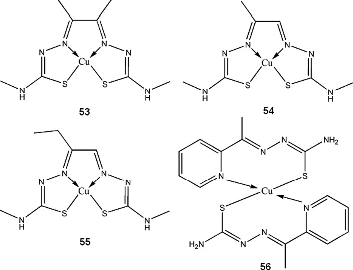
60Cu-ATSM was clinically studied for monitoring tumor hypoxia in lung and cervical cancer, and found to be feasible for prediction of tumor response to therapy (Dehdashti et al. 2003; 2008). Analogous pilot clinical study for rectal cancer was carried by Dietz et al. (2008), and also confirmed possible applicability of 60Cu-ATSM. Chao et al. (2001) suggested that PET images obtained with 60Cu-ATSM can be used for intensity-modulated radiation therapy of head and neck cancer. Since hypoxia of the tumor makes it resistant to radiotherapy, localization with 60Cu-ATSM can be used to accurately deliver higher radiation doses needed for destroying cancer cells (Chao et al. 2001).
61Cu
61Cu isotope can be produced from zinc, nickel or cobalt targets. Necessity of highly enriched Ni and Zn targets or high energy particle beams limited accessibility of 61Cu for biomedical use, until more economic production methods from natural Zn or Co were developed (Rowshanfarzad et al. 2006; Hao et al. 2009; Das et al. 2012). Longer half-life than that of 60Cu and 62Cu makes 61Cu better choice for prolonged imaging of processes with slower kinetics. This isotope, however, is much less popular in today’s biomedical studies than the other copper radioisotopes.
61Cu-APTS (2-acetylpyridine thiosemicarbazone) complex (56), for PET imaging of cancer, was proposed by Jalilian et al. (2006) Using pyridine thiosemicarbazone as a ligand, can give additional antiproliferative activity to the compound, which was previously observed by other authors (Belicchi-Ferrari et al. 2005). Hao et al. (2009) found 61Cu-1,4,7,10-tetraazacyclododecane-1,4,7,10-tetraacetic acid (DOTA)–human serum albumin to be good blood pool imaging agent and suggested its application in antiangiogenic therapy monitoring. Novel approach for therapy of multinodular goitre, using human chorionic gonadotropin (hCG) directly labeled with ionic 61Cu (or some other β+ emitters) was proposed by Maiti et al. (2011) Initial studies indicates that copper-hCG complex half-life is shorter than that of a hCG –TSH (thyroid-stimulating hormone) receptor complex, thus hyperactive thyroid cells can be destroyed before internalization of the receptor occurs. More in vitro and in vivo studies are required to assess usefulness of this therapy.
62Cu
62Cu has unique properties being almost pure β+ emitter(97.2 %) with short half-life of 9,67 min. It is easily obtainable from 62Zn/62Cu generators (Fukumura et al. 2006; El-Azony 2011), however relatively short half-life of parent 62Zn makes these generators operable for not more than three days. This isotope is currently the most intensively studied copper radioisotope besides 64Cu. 62Cu-PTSM is extensively researched 62Cu radiopharmaceutical that can be used for monitoring renal, myocardial and cerebral perfusion. Mathias et al. (1995) observed high species dependent variability in binding 62Cu-PTSM and 62Cu-ATSM by serum albumin. This can render problems when predicting behavior of copper thiosemicarbazones in human system, basing on animal data. 62Cu-ETS (55) complex is proposed as an alternative to 60/61/62/64Cu-PTSM for PET perfusion imaging (Mathias et al. 1995; Green et al. 2007; Basken et al. 2008). 62Cu-PTSM can be used together with 62Cu-ATSM to obtain complementary data on tumor hypoxia and blood circulation in a single PET session (Black et al. 2008; Wong et al. 2008). 62Cu-ATSM complex is widely researched for PET imaging of tumor hypoxia (Laforest et al. 2005; Wong et al. 2008; Minagawa et al. 2011), myocardial (Takahashi et al. 2001) and cerebral ischaemia (Isozaki et al. 2011).
Other than imaging clinical application of 62Cu was proposed by Chan et al. (2000) Balloons filled with 62Cu solution have been found effective for intravascular treatment preventing coronary restenosis in porcine model.
64Cu
The most versatile isotope, 64Cu has found its application in: in vivo studies of copper metabolism, radiotracing biodistribution of potential therapeutics, PET imaging, cancer diagnosing and radiotherapy(preclinical and clinical trials). Although there are many methods of 64Cu production, the most important are those which do not require high energy beams, unattainable for typical small medical cyclotrons (Obata et al. 2003; Szajek et al. 2005; Le et al. 2009). However, such methods need enriched targets, which increase overall costs. Using natural zinc as a starting material, 64Cu can be produced with reasonable purity, but many highly radioactive byproducts of the reaction need to be removed and handled properly (Bonardi et al. 2003). 64Cu half-life allows it to be transported to locations remote of the production site, and currently this isotope is commercially available from several producers around the world.
Similarly to 60Cu, 61Cu and 62Cu isotopes, 64Cu-ATSM is subject to many ongoing research as selective tumor hypoxia imaging agent. It is in phase II clinical trials for PET/CT monitoring of therapeutic progress in patients with cervical cancer (ClinicalTrials.gov 2012). Similar compound, 64Cu-ATSE (Cu-diacetyl-bis(N4-ethylthiosemicarbazone)) (57), has wider tissue-oxygenation level specificity than 64Cu-ATSM. Increased uptake of 64Cu-ATSM by cell cultures occurs between oxygen concentration 0.1–0.5 %, while for the 64Cu-ATSE it happens between 0.1 and 5 %, which can make it more suitable imaging agent for less extreme hypoxias in myocardial and nervous tissues (McQuade et al. 2005). Because 64Cu is also β− emitter, 64Cu-bis-thiosemicarbazones can be used for radiotherapy. Yoshii et al. (2011) showed that 64Cu-ATSM administration reduces volume and metastatic abilities of Colon-26 tumor in mice. Advantage of this treatment over other cancer therapies comes from the fact that 64Cu-ATSM reduced number of CD133+ (prominin-1 positive) cells within tumor. CD133+ cells contributes to ineffectiveness of cancer therapies, being chemo- and radioresistant, and also highly tumorigenic. 64Cu-ATSM decreases number of CD133+ cells not by specific interactions, but rather by accumulating within regions of tumor with high abundance of CD133+ cells, which results in higher doses of radiation in that areas (Yoshii et al. 2011). To increase cytotoxic effectiveness of 64Cu-ATSM, Aft et al. 2003 administered it together with 2-deoxy-d-glucose to mice bearing EMT-6 mammary carcinoma cell line. 2-deoxyglucose accumulates in tumor cells and potentiate effects of radiation therapy. In the study, pretreatment with 2-deoxyglucose increased tumor uptake of 64Cu-ATSM. Continuing daily administration (2 mg/g) of 2-deoxyglucose after single dose of 64Cu-ATSM increased survival time of the animals (Aft et al. 2003). Other than thiosemicarbazone ligands for 64Cu, based on 2-nitroimidazole (another hypoxia-selective compound), were evaluated in vivo by Engelhardt et al. (2002) and were found suitable for imaging of tumor hypoxia. More recently, Bonnitcha et al. (2010) explored an idea to conjugate thiosemicarbazones with nitroimidazoles, since these compounds have the same biological targets. Copper complexes of the ligands (58) synthesized by the authors showed excellent selectivity for hypoxic EMT-6 cells.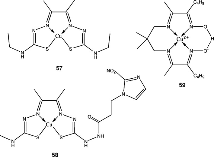
PET and SPECT (Single-Photon Emission Computed Tomography) techniques are used in mapping brain activity in behavioral studies on animals and humans. Many social behaviors cannot be monitored in immobilized test subjects. Compounds containing long living β+ emitters, such as 64Cu-PTSM, are suitable for monitoring cerebral perfusion in freely moving subjects (Holschneider and Maarek 2004). 5,13-dioximino-6,9,9,12-tetramethyl-7,11-diazaheptadeca-6,11-diene complex of copper-64 (59) synthesized by Packard et al. (2002) can be potentially used as myocardial perfusion imaging agent, and also for multidrug resistance screening. The complex shows tumor uptake similar to 99mTc-MIBI (hexakis(2-methoxy-2-methylpropylisonitrile) technetium (99mTc)), a compound used for predicting drug resistance of tumors associated with P-glycoprotein expression (Packard et al. 2002).
64Cu labeled peptides for targeted cancer therapy/imaging are one of the largest group of copper radiopharmaceuticals currently researched. They are built of a targeting peptide such as bombesin or octreotide analogue, a linker, and a bifunctional chelator (BFC), commonly tetraazamacrocycle derivate, like TETA or DOTA (Fig. 5). The peptide binds to a specific receptor expressed by cancer cells while copper isotope-BFC moiety allows localization of the tumor by positron emission detection. β− radiation of 64Cu can also be exploited for selective irradiation of malignant cells. Attractiveness of peptides for targeted radiotherapy, in comparison to monoclonal antibodies, comes from their good tissue distribution, fast clearance, low immunogenicity, and inexpensive, automated production. By modifying amino acid composition of a peptide, one can adjust hydrophobicity, pKa, resistance to proteolysis, and other parameters of the peptide to form a suitable diagnostic agent. Table 2 lists the most popular peptides which were modified to be used with 64Cu for cancer imaging and therapy.
Fig. 5.

DOTA and TETA, the two most common bifunctional chelators used for labeling biomolecules
Table 2.
Targeting peptides for 64Cu PET tracers
| Peptide | Properties | Cancer type | Reference |
|---|---|---|---|
| Bombesin | Amphibian homologue of mammalian gastrin-releasing peptide (GRP) |
Prostate (PC-3) Lung Breast (T-47D) |
Yang et al. (2006), Hoffman and Smith (2009), Prasanphanich et al. (2009), and Lane et al. (2010) |
| Tyr3-octreotide | Somatostatin analog | Neuroendocrine tumors | Sprague et al. (2004) and Eiblmaier et al. (2007) |
| Arg-Gly-Asp (RGD) peptides | Ligands for αvβ3 integrin, expressed during angiogenesis | Metastatic cancers | Chen et al. (2004), Wei et al. (2009), Galibert et al. (2010), and Jin et al. (2011) |
| VIP | Vasoactive intestinal peptide |
Breast Colorectal Prostate |
Thakur et al. (2004) and Zhang et al. (2007a) |
| PACAP | Pituitary adenylate cyclase activating peptide | Breast cancer | Zhang et al. (2007a) |
| α-MSH | Melanocyte stimulating hormone | Melanoma | Cheng et al. (2007) and Wei et al. (2007) |
| Ac-Cys-ZEGFR:1907 | Affibody for epidermal growth factor receptor | Various types | (Miao et al. (2010) |
Zhang et al. approached other than oncological use of such type of compounds. They designed a 64Cu-labeled peptide targeting neutrophils that can be used for non-invasive detection of acute, neutrophilic inflammation (Zhang et al. 2007c).
Monoclonal antibodies (mAbs) are vast group of biotechnologically produced proteins, with constantly rising number of applications in immunotherapy, targeted drug delivery, and in vivo/in vitro diagnostics. In this compounds group, we can distinguish intact immunoglobulins (murine, chimeric, humanized and human) and fragments of heavy chain antibodies (nanobodies, domain-deleted mAbs, hypervariable domain region peptides, minibodies, affibodies and other). Transforming mAbs into radiopharmaceuticals is relatively simple. When using radioisotopes such as iodine-131 or fluorine-18, small molecule labeled with atom/atoms of the isotope, coupled with a linker is attached to amino acids residues (mostly randomly) of the antibody. Similarly, if using metallic radioisotopes, a bifunctional chelator with linker is coupled to the antibody, then solution of the radioisotope salt is added to form a complex. In most cases, mAbs for radiotherapy can be formulated in a convenient form of kits, for preparation of the radiopharmaceutical right before administration to a patient (Reilly 2010). Examples of 64Cu-labeled antibodies for PET imaging are trastuzumab (breast cancers expressing human epidermal growth factor receptor 2 or HER2, in clinical trials) (Sampath et al. 2010; ClinicalTrials.gov 2012), 12A8 (c-kit expressing tumors) (Yoshida et al. 2011), etaracizumab (antibody against human αvβ3 integrin) (Cai et al. 2006), cetuximab (targeting EGFR-epidermal growth-factor receptor expressing tumors) (Li et al. 2008).
67Cu
67Cu is the longest living copper radioisotope and also one of the most difficult to produce, since it requires fast neutron flux reactor or high-energy proton beams and costly 68Zn target (Katabuchi et al. 2008). This isotope of copper, owning to interesting decay properties, is widely acknowledged as potentially useful for radioimmunotherapy, but due to limited availability, the number of research that actually use this isotope is low, compared to other Cu isotopes. Dynamic growth of radioimmunotherapy, can increase demand for this isotope. Medvedev et al. (2012) reported an attempt to produce 67Cu in a larger scale, which gives perspectives for wider commercial availability of the isotope in the near future.
The possibility to change imaging agents into therapeutics is very attractive in copper radiopharmaceuticals. This can be achieved by replacing positron emitting nuclides of 60/61/62/64Cu with electron emitting 67Cu, without changing pharmacokinetics of the compounds. Since biodistribution of 60/61/62/64Cu-labeled substances can be monitored using PET, the data can be directly translated to 67Cu compounds (Cai et al. 2006). 67Cu is one of the best suited isotopes for radioimmunotherapy, because of its half-life long enough to allow good biodistribution within tumor (similar to biological half-life of many mAbs), relatively low gamma radiation abundance (lower whole body dose for patient and safer for medical personnel), higher tumor uptake (compared to iodine-131) and simple radiolabeling procedure (Carrel et al. 1997; Delaloye et al. 1997; DeNardo et al. 2000; Novak-Hofer and Schubiger 2002). Examples of 67Cu-labelled mAbs are chCE7, an anti-L1-cell adhesion molecule antibody for neuroblastoma, ovarian, and some renal carcinoma therapy (Zimmermann et al. 1999; Zimmermann et al. 2003; Knogler et al. 2007), Lym-1 for non-Hodgkin’s lymphoma (DeNardo et al. 1999; Mirick et al. 1999; DeNardo et al. 2000), C595 an anti-MUC1 mucin antibody for bladder cancer treatment (Hughes et al. 2000). Possible applications of 67Cu are not limited to radiotherapy; gamma radiation of the isotope can be used for Single-Photon Emission Computed Tomography (SPECT) (Engelhardt et al. 2002).
To date, no radiopharmaceutical containing copper isotope is approved for use in humans. Although many promising results were obtained during studies on Cu radiopharmaceuticals, several problems also emerged. Therefore current research have to focus on overcoming these obstacles.
Of five discussed isotopes, only 62Cu can be obtained from generator. 64Cu and 67Cu have half-life long enough to be transported from remote locations, but 60Cu and 61Cu require cyclotron access. Therefore, availability of copper radioisotopes is still the main limitation for their wider application.
High energy of emitted positrons of 60/61/62Cu in relative to standard PET imaging radionuclide 18F, is cause of the loss of spatial resolution of the resulting image. Recovery of three-dimensional data, when imaging with high-resolution PET camera, requires development of dedicated analysis algorithms (Ruangma et al. 2006; Liu et al. 2009).
Burgman et al. (2005) found that 64Cu-ATSM shows cell line dependent pharmacokinetics, therefore obtained imaging data in some cases can be irrelevant to tumor hypoxia.
Theoretical calculations made by O’Donoghue et al. (1995) indicate that tumors 2–3 mm in diameter are optimal for effective treatment with 67Cu. Thus, usefulness of this isotope in cancer therapy is limited only to small tumors.
In vivo studies of first generation of Cu-radioisotope labeled peptides showed poor stability of these compounds and liberation of copper from the complexes (Mirick et al. 1999; Bass et al. 2000; Boswell et al. 2004; Sprague et al. 2006). Monocycylic tetraazamacrocycle based BFCs, such as TETA or DOTA, are not sufficiently inert in blood serum and should not be considered in designing new copper radiopharmaceuticals. Cross-bridged macrocycles are currently replacing other type chelating agents (Ma et al. 2002; Boswell et al. 2004; Anderson et al. 2008).
Radiolabeled peptides and antibodies show high retention in kidneys which receive larger dose of radiation than other organs. To prevent kidneys damage, either dose of the radiopharmaceuticals has to be reduced, which can lead to ineffectiveness of the therapy, or additional substances reducing renal uptake need to be administered simultaneously (Vegt et al. 2010).
Main problem of radioimmunotherapy with intact mAbs is their heterogenous biodistribution within solid tumors, resulting in insufficient dose delivered to some of the malignant cells. Therefore, it is necessary to develop other strategies for use of monoclonal antibodies in cancer radiotherapy, such as pretargeting techniques, reduction of the size of the antibody or increasing capillary permeability (Tempero et al. 2000; Goldenberg and Sharkey 2006; Reilly 2006; Thurber et al. 2008).
Copper in nanomedicine
Past ten to twenty years are the time of rapid progress in nanotechnology and nanomedicine. Term nanotechnology generally refers to chemistry and physics of 1–100 nm sized particles, however, the term has become overused for synthesis and rational design of large molecule compounds, polymeric and colloidal materials. Reduction of size has opened new possibilities for use of metallic elements and their compounds in medicine. Metal nanosized particles or quantum dots (colloidal metal chalcogenides, consisting of core and external shell), exhibit novel physicochemical properties that cannot be observed in macroscale. Cations of metal can be complexed with multi-part macromolecular ligands, so the resulting chemical constructs can overcome limitations in distribution, bioavailability and binding specificity of simple compounds (Balogh et al. 2007; Studer et al. 2010; Gunawan et al. 2011; Webster 2011).
Biocidal properties of copper and its compounds have been known since ancient times and include antibacterial, antifungal, molluscicidal, nematocidal, antiviral and other (Borkow and Gabbay 2005). Mechanism of antimicrobial action of copper is complex and not fully understood; Cu2+ ions disrupt permeability of cell’s membrane, cause lipid peroxidation and proteins inactivation (Ohsumi et al. 1988; Nan et al. 2008; Raffi et al. 2010; Wu et al. 2011). Antibacterial properties of nanometer sized copper particles come mainly from ions liberation, however, the size plays important role in adsorption on bacterial cell surface (Raffi et al. 2010). It is possible to construct polymers doped with metallic or ionic copper. Such polymers can be used to make dressings, sutures, bandages and other medical materials with anti-infection, anti-inflammatory and healing-accelerating properties (Zhang et al. 2007b; Borkow et al. 2009; Grace et al. 2009; Sheikh et al. 2011). Similarly to copper nanoparticles, copper oxide nanoparticles are known to be nonspecifically cytotoxic. The activity comes from intracellular, amino acids mediated liberation of copper ions, which form complexes inducing formation of reactive oxygen species (Studer et al. 2010; Gunawan et al. 2011). Socks impregnated with copper oxide are effective in treatment of tinea pedis (fungal infection caused by Trichophyton genus) (Zatcoff et al. 2008). The socks can also be used for preventing so called hand and foot syndrome in capecitabine treated patients; relevant clinical studies have started (ClinicalTrials.gov 2012). Respiratory face masks with CuO offer very good protection against human influenza virus H1N1 (Borkow et al. 2010). A number of copper containing textiles and materials are already commercially available.
Most of today’s contraceptive intrauterine devices (IUDs) contain metallic copper in a form of sheet or wire. Rapid release of cupric ions in the first few days after implantation of IUD can cause adverse effects such as pelvic inflammatory disease, bleeding and expulsion (Timonen 1976; Farley et al. 1992; Mora et al. 2002). Low density polyethylene-copper nanoparticles composites show sustained, zero-order kinetic of copper ions release, therefore can be used to replace conventional IUDs (Cai et al. 2005).
Bhattacharya et al. (2006) suggested that metal nanoparticles can be used for selective precipitation and conformational alterations in proteins. They found that copper nanoparticles clusters precipitate with human hemoglobin mutant HbE, and can serve as a screening agent for hemoglobinopathies such as β-thalassemia (Bhattacharya et al. 2006). Photothermal ablation is one of the newest methods of cancer treatment. Microscopic spheres, built of dielectric core and metal shell, accumulate passively or actively (after functionalization with antibodies) in tumors, and destroy them with heat which the particles emit when excited by near infrared light. Modified gold nanoparticles are commonly researched for this purpose (Cai et al. 2008; Chen et al. 2010; Choi et al. 2011). As an alternative to costly gold, Li et al. (2010) proposed copper sulfide nanoparticles which have very good optical properties, minimal cytotoxicity and low production costs.
Quantum dots (QDs) are nanoparticles that have received much attention in medicine as tumor detection and imaging agents (Zhang et al. 2008). Coating QDs with amphiphilic polymers and functionalizing their surface with antibodies, peptides, oligonucleotides or small-molecule drugs can be done in order to facilitate targeted delivery and to reduce non-specific binding of these nanoparticles (Gao et al. 2005). To achieve quantitative imaging of tumor vasculature in deep tissues, Chen et al. (2008) successfully developed dual optical/PET tracer by functionalizing QDs with 64Cu-DOTA. There is little known, however, about QDs toxicity, which is an important matter, since most of QDs contain hazardous elements such as cadmium, selenium, tellurium and arsenic (Rzigalinski and Strobl 2009). Oxidation of CdSe cores and liberation of Cd2+ ions take place even in coated QDs (Derfus et al. 2004). Development of cadmium-free QDs could be a solution to this problem. Using copper-indium sulfide based QDs, Yong et al. (2010) achieved very promising results for novel, non-toxic, highly sensitive cancer imaging agent.
Superparamagnetic iron oxide nanoparticles are another type of nanostructures that can be functionalized in a similar manner to quantum dots. Their magnetic properties can be used for magnetic resonance imaging (MRI) of cancer. Several authors have exploited the idea of dual MRI/PET tracing to obtain complementary data on tumor localization, using 64Cu-DOTA labeled iron oxide particles (Jarrett et al. 2008; Lee et al. 2008).
Carbon nanotubes (CNTs) have been successfully applied in various areas of science, technology and in medicine. CNTs are very promising as multifunctional platforms for targeted therapy and imaging. A good example of such CNT construct was synthesized and tested in vivo by Liu et al. (2007). The authors used single walled CNTs coated by phospholipids-polyethyleneglycol for water solubility, functionalized with RGD peptide for targeted delivery and labeled with 64Cu-DOTA for PET imaging. The resulting construct showed good biodistribution and selectivity towards αvβ3-positive cancer (Liu et al. 2007). CNTs do not cause acute toxicity, but there is no sufficient knowledge yet about long term exposure and distant effects on human health (Liu et al. 2008; Firme and Bandaru 2010).
Medical sensing devices are very helpful for diagnosing and for monitoring patient’s pharmacotherapy. Various authors have prepared copper nanoparticles-based electrodes for determination of glucose and other carbohydrates (Male et al. 2004; Xu et al. 2006; Jiang and Zhang 2010), drugs such as sotalol or acetaminophen (Boopathi et al. 2004; Heli et al. 2009) and amino acids (Zen et al. 2004; Dong et al. 2010). Cai et al. (2003) demonstrated that gold covered copper nanoparticles, functionalized with oligonucleotides can be used for electrochemical detection of characteristic DNA sequences present in pathogenic microorganisms or mutated genes. Nanocrystals of CuS conjoined with immunoglobulin, was a part of multiple protein detection system, developed by Liu et al. (2004) The system allows sensitive, simultaneous, electrochemical detection of proteins, and can be used to construct novel diagnosing devices.
Advances in modern polymer sciences have opened new horizons for targeted drug delivery systems and diagnostic tools development. One of the most promising and extensively studied groups of compounds are dendrimers. Dendrimers are globular shaped, branched polymers with fixed molecular weight that can be modified with various functional groups on their surface. Moreover, there are empty spaces between polymer branches that can be fitted with small molecules. There has been a lot of interest in dendrimers as potential drug carriers and artificial enzymes (Kofoed and Reymond 2005). By utilizing the ‘click chemistry’, dendrimers can be easily synthesized and functionalized. Dijkgraaf et al. (2007) used this approach to synthesize dendrimers conjoined with RGD peptides and DOTA, for use in tumor imaging after complexation with radionuclides such as 111In or 64Cu. Some dendritic copper complexes were tested for antimicrobial activity by Refat et al. (2009) and showed moderate strength of action on selected microorganisms. Poly(amidoamine)-Schiff base dendrimers synthesized by Zhao et al. (2010) form multinuclear complexes with CuCl2, which show good antiproliferative activity against MOLT-4 leukemia and cisplatin resistant MCF-7 breast cancer cells.
There are known several dendritic copper complexes that exhibit catalytic properties ranging from Lewis acid catalyzed addition reactions to free radical induced hydrolysis (Yang et al. 2003; Fujita et al. 2006; Kao et al. 2011). One particular example is nuclease activity of copper(II) complexes of a pyridine-modified poly(amidoamine) dendrimers. These compounds have ability to induce formation of oxygen radicals leading to cleavage of nucleic acid strand (Kao et al. 2011). Artificial nucleases can be used as anticancer drugs or for sequencing DNA and RNA. On the other hand, natural recombined nucleases, such as dornase alpha, are used in lung diseases (cystic fibrosis, chronic bronchitis), reducing viscosity of mucus in respiratory tract. An unexplored to date possibility is the use of dendritic deoxyribonucleases as potential therapeutics in aforementioned diseases.
Dendrimers can be also used as protective colloids, acting as templates, in the synthesis of copper nanoparticles with regular shape and size (Jin et al. 2008).
Another type of polymeric nanoparticles are aggregates formed by controlled self assembly of diblock amphiphilic copolymers. Many shapes can be achieved, such as spheres, rods, discs, helices, tubes, but the spheres are generally the easiest to attain and are also the most versatile. These structures can be stabilized by cross-linking and variously functionalized (O’Reilly et al. 2006). Rossin et al. (2005) synthesized shell cross-linked nanoparticles with folic acid and 64Cu-TETA moieties attached on the surface which can be used for early diagnosing and therapy of tumors overexpressing folate receptor.
Binding copper(II) with small peptides in some cases can induce formation of nanoaggregates of resulting complexes (Yang et al. 2008; Ren et al. 2008; Li et al. 2011). Complexing with metal ions can reinforce the biological activity of various peptides because such complexes have more rigid structure, and therefore less possible conformations (Tian and Bartlett 1996; Taraszka et al. 2000; Salvati et al. 2008). Li et al. (2011) synthesized four Cu(II)-RGD-octapeptides and found that these compounds have significantly higher anti-thrombotic activity in vivo than free RGD-octapeptides. Similar results are reported in the paper by Ren et al.(2008), in which several tripeptide-Cu(II) complexes were found to have increased thrombolytic activity both in vivo and in vitro, along with additional vasodilatation effect.
Conclusion
Versatility of copper and its compounds has given it a strong position in development of new pharmaceuticals. Although currently there are only a few applications of Cu in medicine, numerous ongoing studies will most likely result in novel uses in the future. Copper radiopharmaceuticals will be probably the first to be approved for clinical use. Cu-containing materials and nanomaterials also hold a great promise and should soon find many applications in various fields of medicine.
Acknowledgments
This work was supported by Medical University in Lodz, No. 503/3-015-01/503-01.
Open Access
This article is distributed under the terms of the Creative Commons Attribution License which permits any use, distribution, and reproduction in any medium, provided the original author(s) and the source are credited.
References
- Aft RL, Lewis JS, Zhang F, Kim J, Welch MJ. Enhancing targeted radiotherapy by copper(II)diacetyl-bis(N4-methylthiosemicarbazone) using 2-deoxy-d-glucose. Cancer Res. 2003;63:5496–5504. [PubMed] [Google Scholar]
- Anderson CJ, Wadas TJ, Wong EH, Weisman GR. Cross-bridged macrocyclic chelators for stable complexation of copper radionuclides for PET imaging. Q J Nucl Med Mol Imaging. 2008;52:185–192. [PMC free article] [PubMed] [Google Scholar]
- Azenha M, Vasconcelos MT, Cabral JPS. Organic ligands reduce copper toxicity in Pseudomonas syringae. Environ Toxicol Chem. 1995;14:369–373. [Google Scholar]
- Balogh L, Nigavekar SS, Nair BM, Lesniak W, Zhang C, Sung LY, Kariapper MST, El-Jawahri A, Llanes M, Bolton B, Mamou F, Tan W, Hutson A, Minc L, Khan MK. Significant effect of size on the in vivo biodistribution of gold composite nanodevices in mouse tumor models. Nanomedicine. 2007;4:281–296. doi: 10.1016/j.nano.2007.09.001. [DOI] [PubMed] [Google Scholar]
- Barceló-Oliver M, García-Raso Á, Terrón Á, Molins E, Prieto MJ, Moreno V, Martínez J, Lladó V, López I, Gutiérrez A, Escribá PV. Synthesis and mass spectroscopy kinetics of a novel ternary copper(II) complex with cytotoxic activity against cancer cells. J Inorg Biochem. 2007;101:649–659. doi: 10.1016/j.jinorgbio.2006.12.008. [DOI] [PubMed] [Google Scholar]
- Basken NE, Mathias CJ, Lipka AE, Green MA. Species dependence of [64Cu]Cu-Bis(thiosemicarbazone) radiopharmaceutical binding to serum albumins. Nucl Med Biol. 2008;35:281–286. doi: 10.1016/j.nucmedbio.2007.11.009. [DOI] [PMC free article] [PubMed] [Google Scholar]
- Bass LA, Wang M, Welch MJ, Anderson CJ. In vivo transchelation of copper-64 from TETA-octreotide to superoxide dismutase in rat liver. Bioconjug Chem. 2000;11:527–532. doi: 10.1021/bc990167l. [DOI] [PubMed] [Google Scholar]
- Belicchi-Ferrari M, Bisceglie F, Casoli C, Durot S, Morgenstern-Badarau I, Pelosi G, Pilotti E, Pinelli S, Tarasconi P. Copper(II) and cobalt(III) pyridoxal thiosemicarbazone complexes with nitroprusside as counterion: syntheses, electronic properties, and antileukemic activity. J Med Chem. 2005;48:1671–1675. doi: 10.1021/jm049529n. [DOI] [PubMed] [Google Scholar]
- Bertini I, Cavallaro G, McGreevy KS. Cellular copper management-a draft user’s guide. Coord Chem Rev. 2010;254:506–524. doi: 10.1016/j.ccr.2009.07.024. [DOI] [Google Scholar]
- Bertrand V, Guessous F, Le Roy AL, Viossat B, Fessi H, El Abbouyi A, Giroud JP, Roch-Arveiller M. Copper-indomethacinate associated with zwitterionic phospholipids prevents enteropathy in rats: effect on inducible NO synthase. Dig Dis Sci. 1999;44:991–999. doi: 10.1023/A:1026668816189. [DOI] [PubMed] [Google Scholar]
- Bhattacharya J, Choudhuri U, Siwach O, Sen P, Dasgupta AK. Interaction of hemoglobin and copper nanoparticles: implications in hemoglobinopathy. Nanomedicine. 2006;2:191–199. doi: 10.1016/j.nano.2006.07.001. [DOI] [PubMed] [Google Scholar]
- Bilgin MD, Elçin AE, Elçin YM. Topical use of liposomal copper palmitate formulation blocks porphyrin-induced photosensitivity in rats. J Photochem Photobiol B. 2005;80:107–114. doi: 10.1016/j.jphotobiol.2005.03.009. [DOI] [PubMed] [Google Scholar]
- Black NF, McJames S, Rust TC, Kadrmas DJ. Evaluation of rapid dual-tracer 62Cu-PTSM + 62Cu-ATSM PET in dogs with spontaneously occurring tumors. Phys Med Biol. 2008;53:217–232. doi: 10.1088/0031-9155/53/1/015. [DOI] [PMC free article] [PubMed] [Google Scholar]
- Blower PJ, Lewis JS, Zweit J. Copper Radionuclides and Radiopharmaceuticals in Nuclear Medicine. Nucl Med Biol. 1996;23:957–980. doi: 10.1016/S0969-8051(96)00130-8. [DOI] [PubMed] [Google Scholar]
- Bonardi ML, Groppi F, Birattari C, Gini L, Mainardi C, Ghioni A, Menapace E, Abbas K, Holzwarth U, Stroosnijder MF. Thin-target excitation functions and optimization of simultaneous production of NCA copper-64 and gallium-66,67 by deuteron induced nuclear reactions on a natural zinc target. J Radioanal Nucl Chem. 2003;257:229–241. doi: 10.1023/A:1024703022762. [DOI] [Google Scholar]
- Bonnitcha PD, Bayly SR, Theobald MBM, Betts HM, Lewis JS, Dilworth JR. Nitroimidazole conjugates of bis(thiosemicarbazonato)64Cu(II) – Potential combination agents for the PET imaging of hypoxia. J Inorg Biochem. 2010;104:126–135. doi: 10.1016/j.jinorgbio.2009.10.009. [DOI] [PMC free article] [PubMed] [Google Scholar]
- Boopathi M, Won M-S, Shim Y-B. A sensor for acetaminophen in a blood medium using a Cu(II)-conducting polymer complex modified electrode. Anal Chim Acta. 2004;512:191–197. doi: 10.1016/j.aca.2004.03.005. [DOI] [Google Scholar]
- Borkow G, Gabbay J. Copper as a biocidal tool. Curr Med Chem. 2005;12:2163–2175. doi: 10.2174/0929867054637617. [DOI] [PubMed] [Google Scholar]
- Borkow G, Zatcoff RC, Gabbay J. Reducing the risk of skin pathologies in diabetics by using copper impregnated socks. Med Hypotheses. 2009;73:883–886. doi: 10.1016/j.mehy.2009.02.050. [DOI] [PubMed] [Google Scholar]
- Borkow G, Zhou SS, Page T, Gabbay J. A novel anti-influenza copper oxide containing respiratory face mask. PLoS ONE. 2010;5:e11295. doi: 10.1371/journal.pone.0011295. [DOI] [PMC free article] [PubMed] [Google Scholar]
- Boswell CA, Sun X, Niu W, Weisman GR, Wong EH, Rheingold AL, Anderson CJ. Comparative in Vivo Stability of Copper-64-Labeled Cross-Bridged and Conventional Tetraazamacrocyclic Complexes. J Med Chem. 2004;47:1465–1474. doi: 10.1021/jm030383m. [DOI] [PubMed] [Google Scholar]
- Brookhaven National Laboratory (2012) National Nuclear Data Center. http://www.nndc.bnl.gov. Accessed 5 March 2012
- Burgman P, O’Donoghue JA, Lewis JS, Welch MJ, Humm JL, Ling CC. Cell line-dependent differences in uptake and retention of the hypoxia-selective nuclear imaging agent Cu-ATSM. Nucl Med Biol. 2005;32:623–630. doi: 10.1016/j.nucmedbio.2005.05.003. [DOI] [PubMed] [Google Scholar]
- Cai H, Zhu N, Jiang Y, He P, Fang Y. Cu@Au alloy nanoparticle as oligonucleotides labels for electrochemical stripping detection of DNA hybridization. Biosens Bioelectron. 2003;18:1311–1319. doi: 10.1016/S0956-5663(03)00084-8. [DOI] [PubMed] [Google Scholar]
- Cai S, Xia X, Xie C. Corrosion behavior of copper/LDPE nanocomposites in simulated uterine solution. Biomaterials. 2005;26:2671–2676. doi: 10.1016/j.biomaterials.2004.08.003. [DOI] [PubMed] [Google Scholar]
- Cai W, Wu Y, Chen K, Cao Q, Tice DA, Chen X. In vitro and In vivo Characterization of 64Cu-Labeled Abegrin™, a Humanized Monoclonal Antibody against Integrin αvβ3. Cancer Res. 2006;66:9673–9681. doi: 10.1158/0008-5472.CAN-06-1480. [DOI] [PubMed] [Google Scholar]
- Cai W, Gao T, Hong H, Sun J. Applications of gold nanoparticles in cancer nanotechnology. Nanotechnol Sci Appl. 2008;1:17–32. doi: 10.2147/NSA.S3788. [DOI] [PMC free article] [PubMed] [Google Scholar]
- Carrel F, Amstutz H, Novak-Hofer I, Schubiger PA. Evaluation of radioiodinated and radiocopper labeled monovalent fragments of monoclonal antibody chCE7 for targeting of neuroblastoma. Nucl Med Biol. 1997;24:539–546. doi: 10.1016/S0969-8051(97)00034-6. [DOI] [PubMed] [Google Scholar]
- Chakraborty A, Kumar P, Ghosh K, Roy P. Evaluation of a Schiff base copper complex compound as potent anticancer molecule with multiple targets of action. Eur J Pharmacol. 2010;647:1–12. doi: 10.1016/j.ejphar.2010.08.003. [DOI] [PubMed] [Google Scholar]
- Chan RC, Lacy JL, Bhargava B, Collins SD, Cates P, Cottin Y, Kollum M, Yang N, Haynes NG, Martin CS, Nayak N, Vodovotz Y, Kim HS, Waksman R. Anti-restenotic effect of copper-62 liquid-filled balloon in porcine coronary arteries: novel use of a short half-life positron emitter. Int J Radiat Oncol Phys. 2000;48:583–592. doi: 10.1016/S0360-3016(00)00649-0. [DOI] [PubMed] [Google Scholar]
- Chao KSC, Bosch WR, Mutic S, Lewis JS, Dehdashti F, Mintun MA, Dempsey JF, Perez CA, Purdy JA, Welch MJ. A novel approach to overcome hypoxic tumor resistance: Cu-ATSM-guided Intensity-Modulated Radiation Therapy. Int J Radiat Oncol Biol Phys. 2001;49:1171–1182. doi: 10.1016/S0360-3016(00)01433-4. [DOI] [PubMed] [Google Scholar]
- Chen X, Liu S, Hou Y, Tohme M, Park R, Bading JR, Conti PS. MicroPET Imaging of Breast Cancer αv-Integrin Expression with 64Cu-Labeled Dimeric RGD Peptides. Mol Imag Biol. 2004;6:350–359. doi: 10.1016/j.mibio.2004.06.004. [DOI] [PubMed] [Google Scholar]
- Chen D, Cui QC, Yang H, Dou QP. Disulfiram, a clinically used anti-alcoholism drug and copper-binding agent, induces apoptotic cell death in breast cancer cultures and xenografts via inhibition of the proteasome activity. Cancer Res. 2006;66:10425–10433. doi: 10.1158/0008-5472.CAN-06-2126. [DOI] [PubMed] [Google Scholar]
- Chen K, Li Z-B, Wang H, Cai W, Chen X. Dual-modality optical and positron emission tomography imaging of vascular endothelial growth factor receptor on tumor vasculature using quantum dots. Eur J Nucl Med Mol Imaging. 2008;35:2235–2244. doi: 10.1007/s00259-008-0860-8. [DOI] [PubMed] [Google Scholar]
- Chen J, Glaus C, Laforest R, Zhang Q, Yang M, Gidding M, Welch MJ, Xia Y. Gold Nanocages as Photothermal Transducers for Cancer Treatment. Small. 2010;6:811–817. doi: 10.1002/smll.200902216. [DOI] [PMC free article] [PubMed] [Google Scholar]
- Cheng Z, Xiong Z, Subbarayan M, Chen X, Gambhir SS. 64Cu-Labeled Alpha-Melanocyte-Stimulating Hormone Analog for MicroPET Imaging of Melanocortin 1 Receptor Expression. Bioconjug Chem. 2007;18:765–772. doi: 10.1021/bc060306g. [DOI] [PMC free article] [PubMed] [Google Scholar]
- Choi WI, Kim JY, Kang C, Byeon CC, Kim YH, Tae G. Tumor Regression In Vivo by Photothermal Therapy Based on Gold-Nanorod-Loaded, Functional Nanocarriers. ACS Nano. 2011;5:1995–2003. doi: 10.1021/nn103047r. [DOI] [PubMed] [Google Scholar]
- Cini R, Giorgi G, Cinquantini A, Rossi C, Sabat M. Metal Complexes of the Antiinflammatory Drug Piroxicam. Inorg Chem. 1990;29:5197–5200. doi: 10.1021/ic00351a012. [DOI] [Google Scholar]
- Cini R, Tamasi G, Defazio S, Hursthouse MB. Unusual coordinating behavior by three non-steroidal anti-inflammatory drugs from the oxicam family towards copper(II). Synthesis, X-ray structure for copper(II)–isoxicam, –meloxicam and –cinnoxicam-derivative complexes, and cytotoxic activity for a copper(II)–piroxicam complex. J Inorg Biochem. 2007;101:1140–1152. doi: 10.1016/j.jinorgbio.2007.04.015. [DOI] [PubMed] [Google Scholar]
- ClinicalTrials.gov (2012) http://clinicaltrials.gov. Accessed 4 March 2012
- Das SS, Chattopadhyay S, Barua L, Das MK. Production of 61Cu using natural cobalt target and its separation using ascorbic acid and common anion exchange resin. Appl Radiat Isot. 2012;70:365–368. doi: 10.1016/j.apradiso.2011.10.011. [DOI] [PubMed] [Google Scholar]
- Dearling JLJ, Packard AB. Some thoughts on the mechanism of cellular trapping of Cu(II)-ATSM. Nucl Med Biol. 2010;37:237–243. doi: 10.1016/j.nucmedbio.2009.11.004. [DOI] [PubMed] [Google Scholar]
- Dehdashti F, Mintun MA, Lewis JS, Bradley J, Govindan R, Laforest R, Welch MJ, Siegel BA. In vivo assessment of tumor hypoxia in lung cancer with 60Cu-ATSM. Eur J Nucl Med Mol Imaging. 2003;30:844–850. doi: 10.1007/s00259-003-1130-4. [DOI] [PubMed] [Google Scholar]
- Dehdashti F, Grigsby PW, Lewis JS, Laforest R, Siegel BA, Welch MJ. Assessing Tumor Hypoxia in Cervical Cancer by PET with 60Cu-Labeled Diacetyl-Bis(N4-Methylthiosemicarbazone) J Nucl Med. 2008;49:201–205. doi: 10.2967/jnumed.107.048520. [DOI] [PubMed] [Google Scholar]
- Delaloye AB, Delaloye B, Buchegger F, Vogel CA, Gillet M, Mach JP, Smith A, Schubiger PA. Comparison of copper-67- and iodine-125-labeled anti-CEA monoclonal antibody biodistribution in patients with colorectal tumors. J Nucl Med. 1997;38:847–853. [PubMed] [Google Scholar]
- DeNardo SJ, DeNardo GL, Kukis DL, Shen S, Kroger LA, DeNardo DA, Goldstein DS, Mirick GR, Salako Q, Mausner LF, Srivastava SC, Meares CF. 67Cu-21T-BAT-Lym1-Pharmacokinetics, Radiation Dosimetry, Toxicity and Tumor Regression in Patients with Lymphoma. J Nucl Med. 1999;40:302–310. [PubMed] [Google Scholar]
- DeNardo GL, DeNardo SJ, O’Donnell RT, Kroger LA, Kukis DL, Meares CF, Goldstein DS, Shen S. Are Radiometal-Labeled Antibodies Better Than Iodine-131–Labeled Antibodies: comparative Pharmacokinetics and Dosimetry of Copper-67–, Iodine-131–, and Yttrium-90–Labeled Lym-1 Antibody in Patients with Non-Hodgkin’s Lymphoma. Clin Lymphoma. 2000;1:118–126. doi: 10.3816/CLM.2000.n.010. [DOI] [PubMed] [Google Scholar]
- Derfus AM, Chan WCW, Bhatia SN. Probing the cytotoxicity of semiconductor quantum dots. Nano Lett. 2004;4:11–18. doi: 10.1021/nl0347334. [DOI] [PMC free article] [PubMed] [Google Scholar]
- Dietz DW, Dehdashti F, Grigsby PW, Malyapa RS, Myerson RJ, Picus J, Ritter J, Lewis JS, Welch MJ, Siegel BA. Tumor hypoxia detected by positron emission tomography with 60Cu-ATSM as a predictor of response and survival in patients undergoing Neoadjuvant chemoradiotherapy for rectal carcinoma: a pilot study. Dis Colon Rectum. 2008;51:1641–1648. doi: 10.1007/s10350-008-9420-3. [DOI] [PMC free article] [PubMed] [Google Scholar]
- Dijkgraaf I, Rijnders AY, Soede A, Dechesne AC, van Esse GW, Brouwer AJ, Corstens FH, Boerman OC, Rijkers DT, Liskamp RM. Synthesis of DOTA-conjugated multivalent cyclic-RGD peptide dendrimers via 1,3-dipolar cycloaddition and their biological evaluation: implications for tumor targeting and tumor imaging purposes. Org Biomol Chem. 2007;5:935–944. doi: 10.1039/b615940k. [DOI] [PubMed] [Google Scholar]
- Ding X, Xie H, Kang YJ. The significance of copper chelators in clinical and experimental application. J Nutr Biochem. 2011;22:301–310. doi: 10.1016/j.jnutbio.2010.06.010. [DOI] [PubMed] [Google Scholar]
- Dong YP, Pei LZ, Chu XF, Zhang WB, Zhang QF. Electrochemical behavior of cysteine at a CuGeO3 nanowires modified glassy carbon electrode. Electrochim Acta. 2010;55:5135–5141. doi: 10.1016/j.electacta.2010.04.020. [DOI] [Google Scholar]
- Dutta S, Padhye S, Mckee V. Structural characterization and SOD activity of copper–oxaprozinate. Inorg Chem Commun. 2004;7:1071–1074. doi: 10.1016/j.inoche.2004.07.022. [DOI] [Google Scholar]
- Eiblmaier M, Andrews R, Laforest R, Rogers BE, Anderson CJ. Nuclear Uptake and Dosimetry of 64Cu-Labeled Chelator–Somatostatin Conjugates in an SSTr2-Transfected Human Tumor Cell Line. J Nucl Med. 2007;48:1390–1396. doi: 10.2967/jnumed.107.039990. [DOI] [PubMed] [Google Scholar]
- El-Azony KM. Improving the separation of Cu(II) from Zn(II) based on an anion exchanger for the preparation a 62Zn/62Cu generator. Appl Radiat Isot. 2011;69:1176–1180. doi: 10.1016/j.apradiso.2011.04.001. [DOI] [PubMed] [Google Scholar]
- Emerit J, Pelletier S, Likforman J, Pasquier C, Thuillier A. Phase II trial of copper zinc superoxide dismutase (CuZn SOD) in the treatment of Crohn’s disease. Free Radic Res Commun. 1991;13:563–569. doi: 10.3109/10715769109145831. [DOI] [PubMed] [Google Scholar]
- Engelhardt EL, Schneider RF, Seeholzer SH, Stobbe CC, Chapman JD. The Synthesis and Radiolabeling of 2-Nitroimidazole Derivatives of Cyclam and Their Preclinical Evaluation as Positive Markers of Tumor Hypoxia. J Nucl Med. 2002;43:837–850. [PubMed] [Google Scholar]
- Farley TM, Rosenberg MJ, Rowe PJ, Chen JH, Meirik O. Intrauterine devices and pelvic inflammatory disease: an international perspective. Lancet. 1992;339:785–788. doi: 10.1016/0140-6736(92)91904-M. [DOI] [PubMed] [Google Scholar]
- Ferrer EG, Baeza N, Naso LG, Castellano EE, Piro OE, Williams PAM. Superoxidedismutase-mimetic copper(II) complexes containing saccharinate and 4-aminopyridine/4-cyanopyridine. J Trace Elem Med Biol. 2010;24:20–26. doi: 10.1016/j.jtemb.2009.09.002. [DOI] [PubMed] [Google Scholar]
- Firme CP, III, Bandaru PR. Toxicity issues in the application of carbon nanotubes to biological systems. Nanomedicine. 2010;6:245–256. doi: 10.1016/j.nano.2009.07.003. [DOI] [PubMed] [Google Scholar]
- Franco L, Doria D. Prostaglandins and Nitric Oxide in Copper-Complex Mediated Protection Against Ethanol-Induced Gastric Damage. Pharmacol Res. 1997;36:395–399. doi: 10.1006/phrs.1997.0247. [DOI] [PubMed] [Google Scholar]
- Fujita K, Muraki T, Hattori H, Sakakura T. Diels–Alder reaction using a dendritic copper(II) triflate-catalyst: a positive dendritic effect on the chemical yield. Tetrahedron Lett. 2006;47:4831–4834. doi: 10.1016/j.tetlet.2006.05.040. [DOI] [Google Scholar]
- Fukumura T, Okada K, Suzuki H, Nakao R, Mukai K, Szelecsényi F, Kovács Z, Suzuki K. An improved 62Zn/62Cu generator based on a cation exchanger and its fully remote-controlled preparation for clinical use. Nucl Med Biol. 2006;33:821–827. doi: 10.1016/j.nucmedbio.2006.05.003. [DOI] [PubMed] [Google Scholar]
- Galibert M, Jin ZH, Furukawa T, Fukumura T, Saga T, Fujibayashi Y, Dumy P, Boturyn D. RGD–cyclam conjugate: synthesis and potential application for positron emission tomography. Bioorg Med Chem Lett. 2010;20:5422–5425. doi: 10.1016/j.bmcl.2010.07.114. [DOI] [PubMed] [Google Scholar]
- Gao X, Yang L, Petros JA, Marshall FF, Simons JW, Nie S. In vivo molecular and cellular imaging with quantum dots. Curr Opin Biotechnol. 2005;16:63–72. doi: 10.1016/j.copbio.2004.11.003. [DOI] [PubMed] [Google Scholar]
- Garcia-González A, Lotz M, Ochoa JL. Anti-inflammatory activity of superoxide dismutase obtained from Debaryomyces hansenii on type II collagen induced arthritis in rats. Rev Invest Clin. 2009;61:212–220. [PubMed] [Google Scholar]
- Ghosh K, Kumar P, Tyagi N, Singh UP, Aggarwal V, Baratto MC. Synthesis and reactivity studies on new copper(II) complexes: DNA binding, generation of phenoxyl radical, SOD and nuclease activities. Eur J Med Chem. 2010;45:3770–3779. doi: 10.1016/j.ejmech.2010.05.026. [DOI] [PubMed] [Google Scholar]
- Gölcü A, Dolaz M, Dağcı EK. Synthesis of Binuclear Copper (II) Complex of the Antihypertensive Drug Pindolol. KSU J Sci Eng. 2005;8:4–9. [Google Scholar]
- Goldenberg DM, Sharkey RM. Advances in cancer therapy with radiolabeled monoclonal antibodies. Q J Nucl Med Mol Imaging. 2006;50:248–264. [PubMed] [Google Scholar]
- González-Baró AC, Pis-Diez R, Franca CA, Torre MH, Parajón-Costa BS. Physicochemical characterization of Cu(II) complexes with SOD-like activity, theoretical studies and biological assays. Polyhedron. 2010;29:959–968. doi: 10.1016/j.poly.2009.11.015. [DOI] [Google Scholar]
- Grace M, Chand N, Bajpai SK. Copper Alginate-Cotton Cellulose (CACC) Fibers with Excellent Antibacterial Properties. J Eng Fiber Fabr. 2009;4:24–35. [Google Scholar]
- Green MA, Mathias CJ, Willis LR, Handa RK, Lacy JL, Miller MA, Hutchins GD. Assessment of Cu-ETS as a PET radiopharmaceutical for evaluation of regional renal perfusion. Nucl Med Biol. 2007;34:247–255. doi: 10.1016/j.nucmedbio.2007.01.002. [DOI] [PubMed] [Google Scholar]
- Gunawan C, Teoh WY, Marquis CP, Amal R. Cytotoxic Origin of Copper(II) Oxide Nanoparticles: comparative Studies with Micron-Sized Particles, Leachate, and Metal Salts. ACS Nano. 2011;5:7214–7225. doi: 10.1021/nn2020248. [DOI] [PubMed] [Google Scholar]
- Guo AJ, Xu XS, Hu YH, Wang MZ, Tan X. Effects of ternary complexes of copper with salicylaldehyde-amino acid Schiff base coordination compounds on the proliferation of BGC823 cells. Chin J Cancer. 2010;29:277–282. doi: 10.5732/cjc.009.10155. [DOI] [PubMed] [Google Scholar]
- Han Y, Shen T, Jiang W, Xia Q, Liu C. DNA cleavage mediated by copper superoxide dismutase via two pathways. J Inorg Biochem. 2007;101:214–224. doi: 10.1016/j.jinorgbio.2006.09.014. [DOI] [PubMed] [Google Scholar]
- Hao G, Fukumura T, Nakao R, Suzuki H, Szelecsényi F, Kovács Z, Suzuki K. Cation exchange separation of 61Cu2+ from natCo targets and preparation of 61Cu–DOTA–HAS as a blood pool agent. Appl Radiat Isot. 2009;67:511–515. doi: 10.1016/j.apradiso.2008.12.004. [DOI] [PubMed] [Google Scholar]
- Heli H, Jabbari A, Zarghan M, Moosavi-Movahedi AA. Copper nanoparticles–carbon microparticles nanocomposite for electrooxidation and sensitive detection of sotalol. Sens Actuators, B. 2009;140:245–251. doi: 10.1016/j.snb.2009.04.021. [DOI] [Google Scholar]
- Hoffman TJ, Smith CJ. True radiotracers: Cu-64 targeting vectors based upon bombesin peptide. Nucl Med Biol. 2009;36:579–585. doi: 10.1016/j.nucmedbio.2009.03.007. [DOI] [PubMed] [Google Scholar]
- Holschneider DP, Maarek JMI. Mapping brain function in freely moving subjects. Neurosci Biobeh Rev. 2004;28:449–461. doi: 10.1016/j.neubiorev.2004.06.007. [DOI] [PMC free article] [PubMed] [Google Scholar]
- Hopkins RG, Failla ML. Transcriptional regulation of interleukin-2 gene expression is impaired by copper deficiency in Jurkat human T lymphocytes. J Nutr. 1999;129:596–601. doi: 10.1093/jn/129.3.596. [DOI] [PubMed] [Google Scholar]
- Hostýnek JJ, Dreher F, Maibach HI. Human stratum corneum penetration by copper: in vivo study after occlusive and semi-occlusive application of the metal as powder. Food Chem Toxicol. 2006;44:1539–1543. doi: 10.1016/j.fct.2006.04.003. [DOI] [PubMed] [Google Scholar]
- Huang ZL, Failla ML. Copper deficiency suppresses effector activities of differentiated U937 cells. J Nutr. 2000;130:1536–1542. doi: 10.1093/jn/130.6.1536. [DOI] [PubMed] [Google Scholar]
- Hughes ODM, Bishop MC, Perkins AC, Wastie ML, Denton G, Price MR, Frier M, Denley H, Rutherford R, Schubiger PA. Targeting Superficial Bladder Cancer by the Intravesical Administration of Copper-67–Labeled Anti-MUC1 Mucin Monoclonal Antibody C595. J Clin Oncol. 2000;18:363–370. doi: 10.1200/JCO.2000.18.2.363. [DOI] [PubMed] [Google Scholar]
- Hung LW, Villemagne VL, Cheng L, Sherratt NA, Ayton S, White AR, Crouch PJ, Lim SC, Leong SL, Wilkins S, George J, Roberts BR, Pham CLL, Liu X, Chiu FCK, Shackleford DM, Powell AK, Masters CL, Bush AI, O’Keefe G, Culvenor JG, Cappai R, Cherny RA, Donnelly PS, Hill AF, Finkelstein DI, Barnham KJ. The hypoxia imaging agent CuII(atsm) is neuroprotective and improves motor and cognitive functions in multiple animal models of Parkinson’s disease. JEM. 2012;209:837–854. doi: 10.1084/jem.20112285. [DOI] [PMC free article] [PubMed] [Google Scholar]
- Ibrahim MM, Ramadan AMM, Mersal GAM, El-Shazly SA. Synthesis, superoxide dismutase, nuclease, and anticancer activities of copper(II) complexes incorporating bis(2-picolyl)amine with different counter anions. J Mol Struct. 2011;998:1–10. doi: 10.1016/j.molstruc.2011.04.009. [DOI] [Google Scholar]
- Isozaki M, Kiyono Y, Arai Y, Kudo T, Mori T, Maruyama R, Kikuta K, Okazawa H. Feasibility of 62Cu-ATSM PET for evaluation of brain ischaemia and misery perfusion in patients with cerebrovascular disease. Eur J Nucl Med Mol Imaging. 2011;38:1075–1082. doi: 10.1007/s00259-011-1734-z. [DOI] [PubMed] [Google Scholar]
- Jackson GE, Mkhonta-Gama L, Voye A, Kelly M. Design of copper-based anti-inflammatory drugs. J Inorg Biochem. 2000;79:147–152. doi: 10.1016/S0162-0134(99)00171-3. [DOI] [PubMed] [Google Scholar]
- Jalilian AR, Rowshanfarzad P, Sabet M, Shafiee A. Preparation of [61Cu]-2-acetylpyridine thiosemicarbazone complex as a possible PET tracer for malignancies. Appl Radiat Isot. 2006;64:337–341. doi: 10.1016/j.apradiso.2005.08.002. [DOI] [PubMed] [Google Scholar]
- Jarrett BR, Gustafsson B, Kukis DL, Louie AY. Synthesis of 64Cu-Labeled Magnetic Nanoparticles for Multimodal Imaging. Bioconjug Chem. 2008;19:1496–1504. doi: 10.1021/bc800108v. [DOI] [PMC free article] [PubMed] [Google Scholar]
- Jiang LC, Zhang WD. A highly sensitive nonenzymatic glucose sensor based on CuO nanoparticles-modified carbon nanotube electrode. Biosens Bioelectron. 2010;25:1402–1407. doi: 10.1016/j.bios.2009.10.038. [DOI] [PubMed] [Google Scholar]
- Jin L, Yang S-P, Tian Q-W, Wu H-X, Cai Y-J. Preparation and characterization of copper metal nanoparticles using dendrimers as protectively colloids. Mater Chem Phys. 2008;112:977–983. doi: 10.1016/j.matchemphys.2008.06.073. [DOI] [Google Scholar]
- Jin ZH, Furukawa T, Galibert M, Boturyn D, Coll JL, Fukumura T, Saga T, Dumy P, Fujibayashi Y. Noninvasive visualization and quantification of tumor αVβ3 integrin expression using a novel positron emission tomography probe, 64Cu-cyclam-RAFT-c(-RGDfK-)4. Nucl Med Biol. 2011;38:529–540. doi: 10.1016/j.nucmedbio.2010.11.008. [DOI] [PubMed] [Google Scholar]
- Kao CL, Tang YH, Lin YC, Chiu LT, Chen HT, Hsu SC, Hsieh KC, Lu CY, Chen YL. Copper complex of a pyridine-modified poly(amidoamine) dendrimer as a chemical nuclease: synthetic and catalytic study. Nanomedicine. 2011;7:273–276. doi: 10.1016/j.nano.2010.10.009. [DOI] [PubMed] [Google Scholar]
- Katabuchi T, Watanabe S, Ishioka NS, Iida Y, Hanaoka H, Endo K, Matsuhashi S. Production of 67Cu via the 68Zn(p,2p)67Cu reaction and recovery of 68Zn target. J Radioanal Nucl Chem. 2008;277:467–470. doi: 10.1007/s10967-007-7144-9. [DOI] [Google Scholar]
- Knogler K, Grünberg J, Zimmermann K, Cohrs S, Honer M, Ametamey S, Altevogt P, Fogel M, Schubiger PA, Novak-Hofer I. Copper-67 Radioimmunotherapy and Growth Inhibition by Anti-L1-Cell Adhesion Molecule Monoclonal Antibodies in a Therapy Model of Ovarian Cancer Metastasis. Clin Cancer Res. 2007;13:603–611. doi: 10.1158/1078-0432.CCR-06-1486. [DOI] [PubMed] [Google Scholar]
- Kofoed J, Reymond JL. Dendrimers as artificial enzymes. Curr Opin Chem Biol. 2005;9:656–664. doi: 10.1016/j.cbpa.2005.10.013. [DOI] [PubMed] [Google Scholar]
- Kovala-Demertzi D, Galani A, Demertzis MA, Skoulika S, Kotoglou C. Binuclear copper(II) complexes of tolfenamic: synthesis, crystal structure, spectroscopy and superoxide dismutase activity. J Inorg Biochem. 2004;98:358–364. doi: 10.1016/j.jinorgbio.2003.11.004. [DOI] [PubMed] [Google Scholar]
- Kumar RS, Arunachalam S. DNA binding and antimicrobial studies of polymer–copper(II) complexes containing 1,10-phenanthroline and l-phenylalanine ligands. Eur J Med Chem. 2009;44:1878–1883. doi: 10.1016/j.ejmech.2008.11.001. [DOI] [PubMed] [Google Scholar]
- Laforest R, Dehdashti F, Lewis JS, Schwarz SW. Dosimetry of 60/61/62/64Cu-ATSM: a hypoxia imaging agent for PET. Eur J Nucl Med Mol Imaging. 2005;32:764–770. doi: 10.1007/s00259-004-1756-x. [DOI] [PubMed] [Google Scholar]
- Lane SR, Nanda P, Rold TL, Sieckman GL, Figueroa SD, Hoffman TJ, Jurisson SS, Smith CJ. Optimization, biological evaluation and microPET imaging of copper-64-labeled bombesin agonists, [64Cu-NO2A-(X)-BBN(7–14)NH2], in a prostate tumor xenografted mouse model. Nucl Med Biol. 2010;37:751–761. doi: 10.1016/j.nucmedbio.2010.04.016. [DOI] [PubMed] [Google Scholar]
- Le VS, Howse J, Zaw M, Pellegrini P, Katsifis A, Greguric I, Weiner R. Alternative method for 64Cu radioisotope production. Appl Radiat Isot. 2009;67:1324–1331. doi: 10.1016/j.apradiso.2009.02.069. [DOI] [PubMed] [Google Scholar]
- Lee H-Y, Li Z, Chen K, Hsu AR, Xu C, Xie J, Sun S, Chen X. PET/MRI Dual-Modality Tumor Imaging Using Arginine-Glycine-Aspartic (RGD)–Conjugated Radiolabeled Iron Oxide Nanoparticles. J Nucl Med. 2008;49:1371–1379. doi: 10.2967/jnumed.108.051243. [DOI] [PubMed] [Google Scholar]
- Lemoine P, Viossat B, Morgant G, Greenaway FT, Tomas A, Dung NH, Sorenson JRJ. Synthesis, crystal structure, EPR properties, and anticonvulsant activities of binuclear and mononuclear 1,10-phenanthroline and salicylate ternary copper(II) complexes. J Inorg Biochem. 2002;89:18–28. doi: 10.1016/S0162-0134(01)00324-5. [DOI] [PubMed] [Google Scholar]
- Li WP, Meyer LA, Capretto DA, Sherman CD, Anderson CJ. Receptor-Binding, Biodistribution, and Metabolism Studies of 64Cu-DOTA-Cetuximab, a PET-Imaging Agent for Epidermal Growth-Factor Receptor-Positive Tumors. Cancer Biother Radiopharm. 2008;23:158–171. doi: 10.1089/cbr.2007.0444. [DOI] [PubMed] [Google Scholar]
- Li Y, Lu W, Huang Q, Huang M, Li C, Chen W. Copper sulfide nanoparticles for photothermal ablation of tumor cells. Nanomedicine. 2010;5:1161–1171. doi: 10.2217/nnm.10.85. [DOI] [PubMed] [Google Scholar]
- Li N, Kang G, Gui L, Zhao M, Wang W, Zhang J, Yue YF, Peng S. Novel Cu(II)-RGD-octapeptides: synthesis, coordination mode, in vitro anti-platelet aggregation/in vivo anti-thrombotic evaluation and correlation of sequence with nano-structure. Nanomedicine. 2011;7:403–409. doi: 10.1016/j.nano.2011.01.005. [DOI] [PubMed] [Google Scholar]
- Liu X, Laforest R. Quantitative small animal PET imaging with nonconventional nuclides. Nucl Med Biol. 2009;36:551–559. doi: 10.1016/j.nucmedbio.2009.01.019. [DOI] [PubMed] [Google Scholar]
- Liu G, Wang J, Kim J, Jan MR, Collins GE. Electrochemical coding for multiplexed immunoassays of proteins. Anal Chem. 2004;76:7126–7130. doi: 10.1021/ac049107l. [DOI] [PubMed] [Google Scholar]
- Liu Z, Cai W, He L, Nakayama N, Chen K, Sun X, Chen X, Dai H. In vivo biodistribution and highly efficient tumour targeting of carbon nanotubes in mice. Nat Nanotechnol. 2007;2:47–52. doi: 10.1038/nnano.2006.170. [DOI] [PubMed] [Google Scholar]
- Liu Z, Davis C, Cai W, He L, Chen X, Dai H. Circulation and long-term fate of functionalized, biocompatible single-walled carbon nanotubes in mice probed by Raman spectroscopy. Proc Natl Acad Sci USA. 2008;105:1410–1415. doi: 10.1073/pnas.0707654105. [DOI] [PMC free article] [PubMed] [Google Scholar]
- Ma D, Lu F, Overstreet T, Milenic DE, Brechbiel MW. Novel chelating agents for potential clinical applications of copper. Nucl Med Biol. 2002;29:91–105. doi: 10.1016/S0969-8051(01)00287-6. [DOI] [PubMed] [Google Scholar]
- Maiti M, Sen K, Sen S, Lahiri S. Studies on stabilities of some human chorionic gonadotropin complexes with β-emitting radionuclides. Appl Radiat Isot. 2011;69:316–319. doi: 10.1016/j.apradiso.2010.11.019. [DOI] [PubMed] [Google Scholar]
- Male KB, Hrapovic S, Liu Y, Wang D, Luong JHT. Electrochemical detection of carbohydrates using copper nanoparticles and carbon nanotubes. Anal Chim Acta. 2004;516:35–41. doi: 10.1016/j.aca.2004.03.075. [DOI] [Google Scholar]
- Mathias CJ, Bergmann SR, Green MA. Species-dependent binding of copper(II) bis(thiosemicarbazone) radiopharmaceuticals to serum albumin. J Nucl Med. 1995;36:1451–1455. [PubMed] [Google Scholar]
- McCarthy DW, Bass LA, Cutler PD, Shefer RE, Klinkowstein RE, Herrero P, Lewis JS, Cutler CS, Anderson CJ, Welch MJ. High purity production and potential applications of copper-60 and copper-61. Nucl Med Biol. 1999;26:351–358. doi: 10.1016/S0969-8051(98)00113-9. [DOI] [PubMed] [Google Scholar]
- McQuade P, Martin KE, Castle TC, Went MJ, Blower PJ, Welch MJ, Lewis JS. Investigation into 64Cu-labeled Bis(selenosemicarbazone) and Bis(thiosemicarbazone) complexes as hypoxia imaging agents. Nucl Med Biol. 2005;32:147–156. doi: 10.1016/j.nucmedbio.2004.10.004. [DOI] [PubMed] [Google Scholar]
- Medvedev DG, Mausner LF, Meinken GE, Kurczak SO, Schnakenberg H, Dodge CJ, Korach EM, Srivastava SC. Development of a large scale production of 67Cu from 68Zn at the high energy proton accelerator: closing the 68Zn cycle. Appl Radiat Isot. 2012;70:423–429. doi: 10.1016/j.apradiso.2011.10.007. [DOI] [PubMed] [Google Scholar]
- Miao Z, Ren G, Liu H, Jiang L, Cheng Z. Small-animal PET imaging of human epidermal growth factor receptor positive tumor with a 64Cu labeled affibody protein. Bioconjug Chem. 2010;21:947–954. doi: 10.1021/bc900515p. [DOI] [PubMed] [Google Scholar]
- Minagawa Y, Shizukuishi K, Koike I, Horiuchi C, Watanuki K, Hata M, Omura M, Odagiri K, Tohnai I, Inoue T, Tateishi U. Assessment of tumor hypoxia by 62Cu-ATSM PET/CT as a predictor of response in head and neck cancer: a pilot study. Ann Nucl Med. 2011;25:339–345. doi: 10.1007/s12149-011-0471-5. [DOI] [PubMed] [Google Scholar]
- Mirick GR, O’Donnell RT, DeNardo SJ, Shen S, Meares CF, DeNardo GL. Transfer of Copper from a Chelated 67Cu-Antibody Conjugate to Ceruloplasmin in Lymphoma Patients. Nucl Med Biol. 1999;26:841–845. doi: 10.1016/S0969-8051(99)00049-9. [DOI] [PubMed] [Google Scholar]
- Mookerjee A, Mookerjee Basu J, Dutta P, Majumder S, Bhattacharyya S, Biswas J, Pal S, Mukherjee P, Raha S, Baral RN, Das T, Efferth T, Sa G, Roy S, Choudhuri SK (2006) Overcoming drug-resistant cancer by a newly developed copper chelate through host-protective cytokine-mediated apoptosis. Clin Cancer Res 12:4339–4349 [DOI] [PubMed]
- Mora N, Cano E, Mora EM, Bastidas JM. Influence of pH and oxygen on copper corrosion in simulated uterine fluid. Biomaterials. 2002;23:667–671. doi: 10.1016/S0142-9612(01)00154-5. [DOI] [PubMed] [Google Scholar]
- Nan L, Yang W, Liu Y, Xu H, Li Y, Lü M, Yang K. Antibacterial Mechanism of Copper-bearing Antibacterial Stainless Steel against E.Coli. J Mater Sci Technol. 2008;24:197–201. doi: 10.1007/s10856-008-3444-z. [DOI] [PubMed] [Google Scholar]
- Novak-Hofer I, Schubiger PA. Copper-67 as a therapeutic nuclide for radioimmunotherapy. Eur J Nucl Med. 2002;29:821–830. doi: 10.1007/s00259-001-0724-y. [DOI] [PubMed] [Google Scholar]
- O’Donoghue JA, Bardiès M, Wheldon TE. Relationship between Tumor Size and Curability for Uniformly Targeted Therapy with Beta-Emitting Radionuclides. J Nucl Med. 1995;36:1902–1909. [PubMed] [Google Scholar]
- O’Reilly RK, Hawker CJ, Wooley KL. Cross-linked block copolymer micelles: functional nanostructures of great potential and versatility. Chem Soc Rev. 2006;35:1068–1083. doi: 10.1039/b514858h. [DOI] [PubMed] [Google Scholar]
- Obata A, Kasamatsu S, McCarthy DW, Welch MJ, Saji H, Yonekura Y, Fujibayashi Y. Production of therapeutic quantities of 64Cu using a 12 MeV cyclotron. Nucl Med Biol. 2003;30:535–539. doi: 10.1016/S0969-8051(03)00024-6. [DOI] [PubMed] [Google Scholar]
- Odisitse S, Jackson GE. In vitro and in vivo studies of the dermally absorbed Cu(II) complexes of N5O2 donor ligands – Potential anti-inflammatory drugs. Inorg Chim Acta. 2009;362:125–135. doi: 10.1016/j.ica.2008.03.092. [DOI] [Google Scholar]
- Odisitse S, Jackson GE, Govender T, Kruger HG, Singh A. Chemical speciation of copper(II) diaminediamide derivative of pentacycloundecane-a potential anti-inflammatory agent. Dalton Trans. 2007;11:1140–1149. doi: 10.1039/b614878f. [DOI] [PubMed] [Google Scholar]
- Ohsumi Y, Kitamoto K, Anraku Y. Changes induced in the permeability barrier of the yeast plasma membrane by cupric ion. J Bacteriol. 1988;170:2676–2682. doi: 10.1128/jb.170.6.2676-2682.1988. [DOI] [PMC free article] [PubMed] [Google Scholar]
- Packard AB, Kronauge JF, Barbarics E, Kiani S, Treves ST. Synthesis and biodistribution of a Lipophilic 64Cu-labeled monocationic Copper(II) complex. Nucl Med Biol. 2002;29:289–294. doi: 10.1016/S0969-8051(02)00285-8. [DOI] [PubMed] [Google Scholar]
- Pontiki E, Hadjipavlou-Litina D, Chaviara AT, Bolos CA. Evaluation of anti-inflammatory and antioxidant activities of mixed-ligand Cu(II) complexes of dien and its Schiff dibases with heterocyclic aldehydes and 2-amino-2-thiazoline. Bioorg Med Chem Lett. 2006;16:2234–2237. doi: 10.1016/j.bmcl.2006.01.045. [DOI] [PubMed] [Google Scholar]
- Prasanphanich AF, Retzloff L, Lane SR, Nanda PK, Sieckman GL, Rold TL, Ma L, Figueroa SD, Sublett SV, Hoffman TJ, Smith CJ. In vitro and in vivo analysis of [64Cu-NO2A-8-Aoc-BBN(7–14)NH2]: a site-directed radiopharmaceutical for positron-emission tomography imaging of T-47D human breast cancer tumors. Nucl Med Biol. 2009;36:171–181. doi: 10.1016/j.nucmedbio.2008.11.005. [DOI] [PMC free article] [PubMed] [Google Scholar]
- Puig S, Thiele DJ. Molecular mechanisms of copper uptake and distribution. Curr Opin Chem Biol. 2002;6:171–180. doi: 10.1016/S1367-5931(02)00298-3. [DOI] [PubMed] [Google Scholar]
- Raffi M, Mehrwan S, Bhatti TM, Akhter JI, Hameed A, Yawar W, ul Hasan MM (2010) Investigations into the antibacterial behavior of copper nanoparticles against Escherichia coli. Ann Microbiol 60:75–80
- Refat MS, El-Deen IM, Grabchev I, Anwer ZM, El-Ghol S. Spectroscopic characterizations and biological studies on newly synthesized Cu2+ and Zn2+ complexes of first and second generation dendrimers. Spectrochim Acta A. 2009;72:772–782. doi: 10.1016/j.saa.2008.11.024. [DOI] [PubMed] [Google Scholar]
- Reilly RM. Radioimmunotherapy of Solid Tumors: the Promise of Pretargeting Strategies Using Bispecific Antibodies and Radiolabeled Haptens. J Nucl Med. 2006;47:196–199. [PubMed] [Google Scholar]
- Reilly RM, editor. Monoclonal antibody and peptide-targeted radiotherapy of cancer. Hoboken: John Wiley and Sons; 2010. [Google Scholar]
- Ren X, Cui G, Zhao M, Wang C, Peng S. Coordination of thrombolytic Pro-Ala-Lys peptides with Cu (II): leading to nanoscale self-assembly, increase of thrombolytic activity and additional vasodilation. J Phys Chem B. 2008;112:8174–8180. doi: 10.1021/jp800645g. [DOI] [PubMed] [Google Scholar]
- Rossin R, Pan D, Qi K, Turner JL, Sun X, Wooley KL, Welch MJ. 64Cu-labeled folate-conjugated shell cross-linked nanoparticles for tumor imaging and radiotherapy: synthesis, radiolabeling, and biologic evaluation. J Nucl Med. 2005;46:1210–1218. [PubMed] [Google Scholar]
- Rowshanfarzad P, Sabet M, Jalilian AR, Kamalidehghan M. An overview of copper radionuclides and production of 61Cu by proton irradiation of natZn at a medical cyclotron. Appl Radiat Isot. 2006;64:1563–1573. doi: 10.1016/j.apradiso.2005.11.012. [DOI] [PubMed] [Google Scholar]
- Roy S, Banerjee R, Sarkar M. Direct binding of Cu(II)-complexes of oxicam NSAIDs with DNA backbone. J Inorg Biochem. 2006;100:1320–1331. doi: 10.1016/j.jinorgbio.2006.03.006. [DOI] [PubMed] [Google Scholar]
- Ruangma A, Bai B, Lewis JS, Sun X, Welch MJ, Leahy R, Laforest R. Three-dimensional maximum a posteriori (MAP) imaging with radiopharmaceuticals labeled with three Cu radionuclides. Nucl Med Biol. 2006;33:217–226. doi: 10.1016/j.nucmedbio.2005.11.001. [DOI] [PubMed] [Google Scholar]
- Rzigalinski BA, Strobl J. Cadmium-containing nanoparticles: perspectives on pharmacology and toxicology of quantum dots. Toxicol Appl Pharm. 2009;238:280–288. doi: 10.1016/j.taap.2009.04.010. [DOI] [PMC free article] [PubMed] [Google Scholar]
- Sakuma S, Fujimoto Y, Miyata Y, Ohno M, Nishida H, Fujita T. Effects of Fe2+, Zn2+, Cu2+ and Se4+ on the synthesis and catabolism of prostaglandins in rabbit gastric antral mucosa. Prostagland Leukot Essent Fatty Acids. 1996;54:193–197. doi: 10.1016/S0952-3278(96)90016-2. [DOI] [PubMed] [Google Scholar]
- Sakuma S, Fujimoto Y, Kitao A, Sakamoto H, Nishida H, Fujita T. Simultaneous measurement of prostaglandin and arachidonoyl CoA formed from arachidonic acid in rabbit kidney medulla microsomes: the roles of Zn2+ and Cu2+ as modulators of formation of the two products. Prostagland Leukot Essent Fatty Acids. 1999;61:105–112. doi: 10.1054/plef.1999.0078. [DOI] [PubMed] [Google Scholar]
- Salvati M, Cordero FM, Pisaneschi F, Melani F, Gratteri P, Cini N, Bottoncetti A, Brandi A. Synthesis, SAR and in vitro evaluation of new cyclic Arg-Gly-Asp pseudopentapeptides containing a s-cis peptide bond as integrin αvβ3 and αvβ5 ligands. Bioorg Med Chem. 2008;16:4262–4271. doi: 10.1016/j.bmc.2008.02.080. [DOI] [PubMed] [Google Scholar]
- Sampath L, Kwon S, Hall MA, Price RE, Sevick-Muraca EM. Detection of cancer metastases with a dual-labeled near-infrared/positron emission tomography imaging agent. Transl Oncol. 2010;3:307–317. doi: 10.1593/tlo.10139. [DOI] [PMC free article] [PubMed] [Google Scholar]
- Sang RL, Yang P. Efficient DNA cleavage by copper(II) complex derived from 1, 1′-dimethyl-2, 2′-biimidazole ligand. Chinese Chem Lett. 2005;16:1397–1400. [Google Scholar]
- Seng HL, Tan KW, Maah MJ, Tan WT, Hamada H, Chikira M, Ng CH. Copper(II) complexes of methylated glycine derivatives: effect of methyl substituent on their DNA binding and nucleolytic property. Polyhedron. 2009;28:2219–2227. doi: 10.1016/j.poly.2009.03.022. [DOI] [Google Scholar]
- Shakir M, Azim Y, Chishti HTN, Parveen S. Synthesis, characterization of complexes of Co(II), Ni(II), Cu(II) and Zn(II) with 12-membered Schiff base tetraazamacrocyclic ligand and the study of their antimicrobial and reducing power. Spectrochim Acta A. 2006;65:490–496. doi: 10.1016/j.saa.2005.11.029. [DOI] [PubMed] [Google Scholar]
- Sheikh FA, Kanjwal MA, Saran S, Chung WJ, Kim H. Polyurethane nanofibers containing copper nanoparticles as future materials. Appl Surf Sci. 2011;257:3020–3026. doi: 10.1016/j.apsusc.2010.10.110. [DOI] [Google Scholar]
- Singh BK, Bhojak N, Mishra P, Garg BS. Copper(II) complexes with bioactive carboxyamide: synthesis, characterization and biological activity. Spectrochim Acta A. 2008;70:758–765. doi: 10.1016/j.saa.2007.09.008. [DOI] [PubMed] [Google Scholar]
- Singh AP, Kaushik NK, Verma AK, Hundal G, Gupta R. Synthesis, structure and biological activity of copper(II) complexes of 4-(2-pyridylmethyl)-1,7-dimethyl-1,4,7-triazonane-2,6-dione and 4-(2-pyridylethyl)-1,7-dimethyl-1,4,7-triazonane-2,6-dione. Eur J Med Chem. 2009;44:1607–1614. doi: 10.1016/j.ejmech.2008.07.029. [DOI] [PubMed] [Google Scholar]
- Sorenson JRJ. Copper chelates as possible active forms of the antiarthritic agents. J Med Chem. 1976;19:135–148. doi: 10.1021/jm00223a024. [DOI] [PubMed] [Google Scholar]
- Sorenson JRJ (1982) In: Sigel H (ed) Inorganic drugs in deficiency and disease. Metal ions in biological systems, vol 14. Marcel Dekker, New York
- Sorenson JRJ, editor. Biology of copper complexes. Clifton: Humana Press; 1987. [Google Scholar]
- Sorenson JRJ. 6 copper complexes offer a physiological approach to treatment of chronic diseases. Prog Med Chem. 1989;26:437–568. doi: 10.1016/S0079-6468(08)70246-7. [DOI] [PubMed] [Google Scholar]
- Sprague JE, Peng Y, Sun X, Weisman GR, Wong EH, Achilefu S, Anderson CJ. Preparation and biological evaluation of copper-64-labeled Tyr3-octreotate using a cross-bridged macrocyclic chelator. Clin Cancer Res. 2004;10:8674–8682. doi: 10.1158/1078-0432.CCR-04-1084. [DOI] [PubMed] [Google Scholar]
- Sprague JE, Li WP, Liang K, Achilefu S, Anderson CJ. In vitro and in vivo investigation of matrix metalloproteinase expression in metastatic tumor models. Nucl Med Biol. 2006;33:227–237. doi: 10.1016/j.nucmedbio.2005.10.011. [DOI] [PubMed] [Google Scholar]
- Sreedaran S, Bharathi KS, Rahiman AK, Jagadish L, Kaviyarasan V, Narayanan V. Novel unsymmetrical macrocyclic dicompartmental binuclear copper(II) complexes bearing 4- and 6-coordination sites: electrochemical, magnetic, catalytic and antimicrobial studies. Polyhedron. 2008;27:2931–2938. doi: 10.1016/j.poly.2008.06.025. [DOI] [Google Scholar]
- Sreedhara A, Freed JD, Cowan JA. Efficient inorganic deoxyribonucleases greater than 50-million-fold rate enhancement in enzyme-like DNA cleavage. J Am Chem Soc. 2000;122:8814–8824. doi: 10.1021/ja994411v. [DOI] [Google Scholar]
- Studer AM, Limbach LK, Van Duc L, Krumeich F, Athanassiou EK, Gerber LC, Moch H, Stark WJ. Nanoparticle cytotoxicity depends on intracellular solubility: comparison of stabilized copper metal and degradable copper oxide nanoparticles. Toxicol Lett. 2010;197:169–174. doi: 10.1016/j.toxlet.2010.05.012. [DOI] [PubMed] [Google Scholar]
- Suksrichavalit T, Prachayasittikul S, Piacham T, Isarankura-Na-Ayudhya C, Nantasenamat C, Prachayasittikul V. Copper complexes of nicotinic-aromatic carboxylic acids as superoxide dismutase mimetics. Molecules. 2008;13:3040–3056. doi: 10.3390/molecules13123040. [DOI] [PMC free article] [PubMed] [Google Scholar]
- Suksrichavalit T, Prachayasittikul S, Nantasenamat C, Isarankura-Na-Ayudhya C, Prachayasittikul V. Copper complexes of pyridine derivatives with superoxide scavenging and antimicrobial activities. Eur J Med Chem. 2009;44:3259–3265. doi: 10.1016/j.ejmech.2009.03.033. [DOI] [PubMed] [Google Scholar]
- Sylla-Iyarreta Veitía M, Dumas F, Morgant G, Sorenson JRJ, Frapart Y, Tomas A. Synthesis, structural analysis and anticonvulsant activity of a ternary Cu(II) mononuclear complex containing 1,10-phenanthroline and the leading antiepileptic drug valproic acid. Biochimie. 2009;91:1286–1293. doi: 10.1016/j.biochi.2009.06.015. [DOI] [PubMed] [Google Scholar]
- Szajek LP, Meyer W, Plascjak P, Eckelman WC. Semi-remote production of [64Cu]CuCl2 and preparation of high specific activity [64Cu]Cu-ATSM for PET studies. Radiochim Acta. 2005;93:239–244. doi: 10.1524/ract.93.4.239.64070. [DOI] [Google Scholar]
- Taggar AS, Alnajim J, Anantha M, Thomas A, Webb M, Ramsay E, Bally MB. Copper–topotecan complexation mediates drug accumulation into liposomes. J Control Release. 2006;114:78–88. doi: 10.1016/j.jconrel.2006.05.019. [DOI] [PubMed] [Google Scholar]
- Takahashi N, Fujibayashi Y, Yonekura Y, Welch MJ, Waki A, Tsuchida T, Sadato N, Sugimoto K, Nakano A, Lee JD, Itoh H. Copper-62 ATSM as a hypoxic tissue tracer in myocardial ischemia. Ann Nucl Med. 2001;15:293–296. doi: 10.1007/BF02987849. [DOI] [PubMed] [Google Scholar]
- Taraszka JA, Li J, Clemmer DE. Metal-mediated peptide ion conformations in the gas phase. J Phys Chem B. 2000;104:4545–4551. doi: 10.1021/jp9939468. [DOI] [Google Scholar]
- Tempero M, Peter Leichner P, Baranowska-Kortylewicz J, Harrison K, Augustine S, Schlom J, Anderson J, Wisecarver J, Colcher D (2000) High-dose therapy with 90Yttrium-labeled monoclonal antibody CC49: A phase I trial. Clin Cancer Res 6:3095–3102 [PubMed]
- Thakur ML, Aruva MR, Gariepy J, Acton P, Rattan S, Prasad S, Wickstrom E, Alavi A. PET imaging of oncogene overexpression using 64Cu-vasoactive intestinal peptide (VIP) analog: comparison with 99mTc-VIP analog. J Nucl Med. 2004;45:1381–1389. [PMC free article] [PubMed] [Google Scholar]
- Thurber GM, Schmidt MM, Wittrup KD. Antibody tumor penetration: transport opposed by systemic and antigen-mediated clearance. Adv Drug Deliv Rev. 2008;60:1421–1434. doi: 10.1016/j.addr.2008.04.012. [DOI] [PMC free article] [PubMed] [Google Scholar]
- Tian ZQ, Bartlett PA. Metal coordination as a method for templating peptide conformation. J Am Chem Soc. 1996;118:943–949. doi: 10.1021/ja952475+. [DOI] [Google Scholar]
- Tian X, Zhang Z, Wang S, Diao Y, Zhao Z, Lv D. Copper–taurine (CT): a potential organic compound to facilitate infected wound healing. Med Hypotheses. 2009;73:1048–1050. doi: 10.1016/j.mehy.2009.06.051. [DOI] [PubMed] [Google Scholar]
- Timonen H. Copper release from copper-T intrauterine devices. Contraception. 1976;14:25–38. doi: 10.1016/S0010-7824(76)80005-4. [DOI] [PubMed] [Google Scholar]
- Toyota E, Sekizaki H, Takahashi Y, Itoh K, Tanizawa K. Amidino-containing Schiff base copper(II) and iron(III) chelates as a thrombin inhibitor. Chem Pharm Bull. 2005;53:22–26. doi: 10.1248/cpb.53.22. [DOI] [PubMed] [Google Scholar]
- Tuorkey MJF-A, Abdul-Aziz KK. A pioneer study on the anti-ulcer activities of copper nicotinate complex [CuCl (HNA)2] in experimental gastric ulcer induced by aspirin-pyloris ligation model (Shay model) Biomed Pharmacother. 2009;63:194–201. doi: 10.1016/j.biopha.2008.01.015. [DOI] [PubMed] [Google Scholar]
- Vegt E, de Jong M, Wetzels JFM, Masereeuw R, Melis M, Oyen WJG, Gotthardt M, Boerman OC. Renal toxicity of radiolabeled peptides and antibody fragments: mechanisms, impact on radionuclide therapy, and strategies for prevention. J Nucl Med. 2010;51:1049–1058. doi: 10.2967/jnumed.110.075101. [DOI] [PubMed] [Google Scholar]
- Wadas TJ, Wong EH, Weisman GR, Anderson CJ. Coordinating radiometals of copper, gallium, indium, yttrium, and zirconium for PET and SPECT imaging of disease. Chem Rev. 2010;110:2858–2902. doi: 10.1021/cr900325h. [DOI] [PMC free article] [PubMed] [Google Scholar]
- Wan YZ, Xiong GY, Liang H, Raman S, He F, Huang Y. Modification of medical metals by ion implantation of copper. Appl Surf Sci. 2007;253:9426–9429. doi: 10.1016/j.apsusc.2007.06.031. [DOI] [Google Scholar]
- Webster TJ (ed) (2011) Nanotechnology enabled in situ sensors for monitoring health. Springer, New York
- Weder JE, Hambley TW, Kennedy BJ, Lay PA, MacLachlan D, Bramley R, Delfs CD, Murray KS, Moubaraki B, Warwick B, Biffin JR, Regtop HL. Anti-inflammatory dinuclear copper(II) complexes with indomethacin. Synthesis, magnetism and epr spectroscopy. Crystal structure of the N,N-dimethylformamide adduct. Inorg Chem. 1999;38:1736–1744. doi: 10.1021/ic981100x. [DOI] [PubMed] [Google Scholar]
- Weder JE, Dillon CT, Hambley TW, Kennedy BJ, Lay PA, Biffin JR, Regtop HL, Davies NM. Copper complexes of non-steroidal anti-inflammatory drugs: an opportunity yet to be realized. Coord Chem Rev. 2002;232:95–126. doi: 10.1016/S0010-8545(02)00086-3. [DOI] [Google Scholar]
- Wei L, Butcher C, Miao Y, Gallazzi F, Quinn TP, Welch MJ, Lewis JS. Synthesis and biologic evaluation of 64Cu-labeled rhenium-cyclized α-MSH peptide analog using a cross-bridged cyclam chelator. J Nucl Med. 2007;48:64–72. [PubMed] [Google Scholar]
- Wei L, Ye Y, Wadas TJ, Lewis JS, Welch MJ, Achilefu S, Anderson CJ. 64Cu-Labeled CB-TE2A and diamsar-conjugated RGD peptide analogs for targeting angiogenesis: comparison of their biological activity. Nucl Med Biol. 2009;36:277–285. doi: 10.1016/j.nucmedbio.2008.12.008. [DOI] [PMC free article] [PubMed] [Google Scholar]
- Williams HA, Robinson S, Julyan P, Zweit J, Hastings D. A comparison of PET imaging characteristics of various copper radioisotopes. Eur J Nucl Med Mol Imaging. 2005;32:1473–1480. doi: 10.1007/s00259-005-1906-9. [DOI] [PubMed] [Google Scholar]
- Wong TZ, Lacy JL, Petry NA, Hawk TC, Sporn TA, Dewhirst MW, Vlahovic G. PET of hypoxia and perfusion with 62Cu-ATSM and 62Cu-PTSM using a 62Zn/62Cu generator. AJR. 2008;190:427–432. doi: 10.2214/AJR.07.2876. [DOI] [PMC free article] [PubMed] [Google Scholar]
- Wu Y, Han G, Gong Y, Zhang Y, Xia Y, Yue C, Wu D. Antibacterial property and mechanism of copper alginate fiber. Adv Mater Res. 2011;152–153:1351–1355. [Google Scholar]
- Xu Q, Zhao Y, Xu JZ, Zhu JJ. Preparation of functionalized copper nanoparticles and fabrication of a glucose sensor. Sens Actuators, B. 2006;114:379–386. doi: 10.1016/j.snb.2005.06.005. [DOI] [Google Scholar]
- Yang BY, Chen XM, Deng GJ, Zhang YL, Fan QH. Chiral dendritic bis(oxazoline) copper(II) complexes as Lewis acid catalysts for enantioselective aldol reactions in aqueous media. Tetrahedron Lett. 2003;44:3535–3538. doi: 10.1016/S0040-4039(03)00680-4. [DOI] [Google Scholar]
- Yang YS, Zhang X, Xiong Z, Chen X. Comparative in vitro and in vivo evaluation of two 64Cu-labeled bombesin analogs in a mouse model of human prostate adenocarcinoma. Nucl Med Biol. 2006;33:371–380. doi: 10.1016/j.nucmedbio.2005.12.011. [DOI] [PubMed] [Google Scholar]
- Yang M, Cui G, Zhao M, Wang C, Wang L, Liu H, Peng S. The effect of complexation of Cu(II) with P6A peptide and its analogs on their thrombolytic activities. Int J Pharm. 2008;362:81–87. doi: 10.1016/j.ijpharm.2008.06.014. [DOI] [PubMed] [Google Scholar]
- Yong KT, Roy I, Hu R, Ding H, Cai H, Zhu J, Zhang X, Bergey EJ, Prasad PN. Synthesis of ternary CuInS2/ZnS quantum dot bioconjugates and their applications for targeted cancer bioimaging. Integr Biol. 2010;2:121–129. doi: 10.1039/b916663g. [DOI] [PubMed] [Google Scholar]
- Yoshida C, Tsuji AB, Sudo H, Sugyo A, Sogawa C, Inubushi M, Uehara T, Fukumura T, Koizumi M, Arano Y, Saga T. Development of positron emission tomography probe of 64Cu-labeled anti-C-kit 12A8 Fab to measure protooncogene C-kit expression. Nucl Med Biol. 2011;38:331–337. doi: 10.1016/j.nucmedbio.2010.10.005. [DOI] [PubMed] [Google Scholar]
- Yoshii Y, Furukawa T, Kiyono Y, Watanabe R, Mori T, Yoshii H, Asai T, Okazawa H, Welch MJ, Fujibayashi Y. Internal radiotherapy with copper-64-diacetyl-bis(N4-methylthiosemicarbazone) reduces CD133+ highly tumorigenic cells and metastatic ability of mouse colon carcinoma. Nucl Med Biol. 2011;38:151–157. doi: 10.1016/j.nucmedbio.2010.08.009. [DOI] [PubMed] [Google Scholar]
- Zatcoff RC, Smith MS, Borkow G. Treatment of tinea pedis with socks containing copper-oxide impregnated fibers. The Foot. 2008;18:136–141. doi: 10.1016/j.foot.2008.03.005. [DOI] [PubMed] [Google Scholar]
- Zen J-M, Hsu C-T, Kumar AS, Lyuu H-J, Lin K-Y. Amino acid analysis using disposable copper nanoparticle plated electrodes. Analyst. 2004;129:841–845. doi: 10.1039/b401573h. [DOI] [PubMed] [Google Scholar]
- Zhang Y, Wang JZ, Wu YJ, Li WG. Anti-inflammatory effect of recombinant human superoxide dismutase in rats and mice and its mechanism. Acta Pharmacol Sin. 2002;23:439–444. [PubMed] [Google Scholar]
- Zhang K, Aruva MR, Shanthly N, Cardi CA, Patel CA, Rattan S, Cesarone G, Wickstrom E, Thakur ML. Vasoactive intestinal peptide (VIP) and pituitary adenylate cyclase activating peptide (PACAP) receptor specific peptide analogues for PET imaging of breast cancer: in vitro/in vivo evaluation. Regul Peptides. 2007;144:91–100. doi: 10.1016/j.regpep.2007.06.008. [DOI] [PMC free article] [PubMed] [Google Scholar]
- Zhang W, Zhang Y, Ji J, Yan Q, Huang A, Chu PK. Antimicrobial polyethylene with controlled copper release. J Biomed Mater Res A. 2007;83:838–844. doi: 10.1002/jbm.a.31436. [DOI] [PubMed] [Google Scholar]
- Zhang Y, Kundu B, Fairchild KD, Locke L, Berr SS, Linden J, Pan D. Synthesis of novel neutrophil-specific imaging agents for positron emission tomography (PET) imaging. Bioorg Med Chem Lett. 2007;17:6876–6878. doi: 10.1016/j.bmcl.2007.10.013. [DOI] [PMC free article] [PubMed] [Google Scholar]
- Zhang H, Yee D, Wang C. Quantum dots for cancer diagnosis and therapy: biological and clinical perspectives. Nanomedicine. 2008;3:83–91. doi: 10.2217/17435889.3.1.83. [DOI] [PubMed] [Google Scholar]
- Zhao X, Loo SCJ, Lee PP-F, Tan TTY, Chu CK. Synthesis and cytotoxic activities of chloropyridylimineplatinum(II) and chloropyridyliminecopper(II) surface-functionalized poly(amidoamine)dendrimers. J Inorg Biochem. 2010;104:105–110. doi: 10.1016/j.jinorgbio.2009.10.001. [DOI] [PubMed] [Google Scholar]
- Zimmermann K, Gianollini S, Schubiger PA, Novak-Hofer I. A Triglycine linker improves tumor uptake and biodistributions of 67-Cu-labeled anti-neuroblastoma MAb chCE7 F(ab’)2 fragments. Nucl Med Biol. 1999;26:943–950. doi: 10.1016/S0969-8051(99)00067-0. [DOI] [PubMed] [Google Scholar]
- Zimmermann K, Grünberg J, Honer M, Ametamey S, Schubiger PA, Novak-Hofer I. Targeting of renal carcinoma with 67/64Cu-labeled anti-L1-CAM antibody chCE7: selection of copper ligands and PET imaging. Nucl Med Biol. 2003;30:417–427. doi: 10.1016/S0969-8051(03)00019-2. [DOI] [PubMed] [Google Scholar]
- Zvimba JN, Jackson GE. Copper chelating anti-inflammatory agents; N1-(2-aminoethyl)-N2-(pyridin-2-ylmethyl)-ethane-1,2-diamine and N-(2-(2-aminoethylamino)ethyl)picolinamide: an in vitro and in vivo study. J Inorg Biochem. 2007;101:148–158. doi: 10.1016/j.jinorgbio.2006.09.006. [DOI] [PubMed] [Google Scholar]