Abstract
Background
Early-stage gastric cancer is mostly asymptomatic and can easily be missed easily by conventional gastroscopy. Currently, there are no useful biomarkers for the early detection of gastric cancer, and their identification of biomarkers is urgently needed.
Methods
Gastric juice was obtained from 185 subjects that were divided into three groups: non-neoplastic gastric disease (NGD), advanced gastric cancer and early gastric cancer (EGC). The levels of aromatic amino acids in the gastric juice were quantitated using high-performance liquid chromatography.
Results
The median values (25th to 75th percentile) of tyrosine, phenylalanine and tryptophan in the gastric juice were 3.8 (1.7–7.5) µg/ml, 5.3 (2.3–9.9) µg/ml and 1.0 (0.4–2.8) µg/ml in NGD; 19.4 (5.8–72.4) µg/ml, 24.6 (11.5–73.7) µg/ml and 8.3 (2.1–28.0) µg/ml in EGC. Higher levels of tyrosine, phenylalanine and tryptophan in the gastric juice were observed in individuals of EGC groups compared those of the NGD group (NGD vs. EGC, P<0.0001). For the detection of EGC, the areas under the receiver operating characteristic curves (AUCs) of each biomarker were as follows: tyrosine, 0.790 [95% confidence interval (CI), 0.703–0.877]; phenylalanine, 0.831 (95% CI, 0.750–0.911); and tryptophan, 0.819 (95% CI, 0.739–0.900). The sensitivity and specificity of phenylalanine were 75.5% and 81.4%, respectively, for detection of EGC. A multiple logistic regression analysis showed that high levels of aromatic amino acids in the gastric juice were associated with gastric cancer (adjusted β coefficients ranged from 1.801 to 4.414, P<0.001).
Conclusion
Increased levels of tyrosine, phenylalanine and tryptophan in the gastric juice samples were detected in the early phase of gastric carcinogenesis. Thus, tyrosine, phenylalanine and tryptophan in gastric juice could be used as biomarkers for the early detection of gastric cancer. A gastric juice analysis is an efficient, economical and convenient method for screening early gastric cancer development in the general population.
Introduction
Gastric cancer is the second most common type of cancer in East Asia, and the prognosis of gastric cancer patients is poor as a result of late detection [1], [2], [3]. To date, stomach cancer survival and patient quality of life is only dramatically improved if the cancer is diagnosed early [4]. Unfortunately, less than 5% of early gastric cancer cases are detected and diagnosed promptly [5].
Endoscopy followed by pathological biopsy is now the preferred method to detect early gastric cancer (EGC). However, EGC is missed frequently during gastroscopy because a minor lesion can be easily overlooked, biopsied inadequately, or interpreted incorrectly by pathologists [6], [7]. EGC is largely asymptomatic prior to the onset of severe symptoms, and an endoscopic investigation based on severe symptoms always delays the detection of stomach cancer. Population-based screening may facilitate the early detection and diagnosis of gastric cancer. However, both the lack of experienced endoscopists and the patient discomfort associated with the procedure reduce the application of this method as a screening tool for EGC.
Less invasive and more efficient biomarkers are needed for the detection of EGC by mass screening. The current biomarkers pepsinogen, carcinoembryonic antigen (CEA), and carbohydrate antigen 19–9 (CA19–9), are not sufficient for accurately predicting gastric cancer at an early stage [8], [9], [10].
It is well established that metabolic abnormalities occur prior to significant morphological changes in malignant tissue [11]. A substantial amount of information concerning the metabolic state of the gastric epithelium is contained in gastric juice. Therefore, gastric juice can be utilized to establish a diagnostic test and identify valuable and efficient biomarkers for EGC screening. In the previous studies, we have established the fluorescence spectrum analysis of gastric juice [12], [13], [14] and identified three fluorescence biomarkers (aromatic amino acids in gastric juice) [15] that can be used to distinguish advanced gastric cancer (AGC) from benign diseases. However, it is still uncertain whether these biomarkers can differentiate between EGC and benign diseases. This study was performed to test the hypothesis that gastric juice biomarkers have diagnostic screening value in the detection of EGC.
Methods
Ethics Statement
The ethics committee of Peking University Health Science Centre approved this research study. Written informed consent was obtained from all participants, and the entire clinical investigation was conducted according to the principles expressed in the Declaration of Helsinki.
Sample Collection
Samples were collected at the endoscopy rooms of Peking University Third Hospital. All patient samples obtained for this study were histologically confirmed by mucosal biopsy and/or postoperative pathology from December 2008 to July 2012. After an overnight fast, the patients underwent a gastroscopy. Samples of gastric juice that were free of gross food residue, bile and blood were collected. The samples were separated into 2 ml aliquots and immediately stored at −80°C for subsequent analysis. Warthin-Starry (WS) staining was used to detect the Helicobacter pylori infection status in all specimens (The gastric antrum was one of the biopsy sites and the total number of biopsy sites was more than two).
Participants
Gastric juice samples were obtained from 49 patients with EGC (6 cases were included in a previous publication [15]). There were 26 cases of focal carcinoma and 23 cases of severe dysplasia that could be classified as EGC [16]. In addition to the gastric juice samples isolated from the patients with EGC, samples from 70 patients with non-neoplastic gastric disease (NGD) who underwent an outpatient endoscopy procedure during the same period were used as a control group for comparison. The control group contained 47 patients with chronic superficial gastritis, 18 patients with chronic atrophic gastritis, and 5 patients with peptic ulcers. Additionally, gastric juice samples were randomly collected from 66 patients with AGC (Table 1). There were no significant abnormalities observed in the pre-endoscopic kidney and liver function tests of the recruited subjects. Most of the recruited gastric cancer patients had not been previously diagnosed, and none of the patients showed significant malnutrition (Kamofsky performance status, KPS>90).
Table 1. Demographic characteristics of patients.
| NGD (n = 70) Mean±SD | AGC (n = 66) Mean±SD | EGC (n = 49) Mean±SD | P c value | |
| Age | 58.8±13.4 | 57.8±14.9 | 63.8±11.5 | 0.049 |
| Female/Male | 40/30 | 35/31 | 16/33 | 0.023 |
| Helicobacter pylori statusa (−/+) | 44/25 | 37/25 | 34/14 | 0.478 |
| Position | ||||
| cardia | – | 15 | 10 | |
| angulus | – | 3 | 13 | |
| fundus | – | 3 | – | |
| antrum | – | 24 | 18 | |
| Corpus | – | 21 | 8 | |
| Pathology | ||||
| severe dysplasia | – | – | 23 | |
| adenocarcinoma | – | 49 | 25 | |
| signet ring cell | 17 | 1 | ||
| Lauren's classificationb | ||||
| intestinal type | – | 41 | 24 | |
| diffuse type | – | 19 | 1 | |
| mixed type | – | 6 | 1 | |
6 cases missed;
23 cases of severe dysplasia were not included;
Comparisons were carried out among the three group (NGD, AGC and EGC).
Sample Treatment
The frozen samples were thawed at room temperature and centrifuged at 14000 rpm for 20 minutes at 4°C. The precipitate was removed, and the upper layer was recovered and temporarily stored at 4°C for the following experiment. The samples were numbered at random. The experimenter was blinded to the diagnosis of the patients recruited to the study.
Quantification of Total Protein in Gastric Juice
To quantify the protein content, 100 µl of gastric juice supernatant was recovered and immediately diluted with 900 µl of 0.15 M phosphate buffer (pH 7.3) to adjust the pH. The total protein content in the gastric juice was measured using a BCA (bicinchoninic acid) protein assay kit (Beijing Cowin Biotech Co. Ltd., Beijing, China) according to the manufacturer’s protocol. If the protein concentration was out of the working range of the assay (20–2000 µg/ml), the sample was further diluted and reanalyzed.
Quantitative System
Aromatic amino acids in the gastric juice samples were quantitated using a fluorescence detector in conjunction with HPLC [17], [18], and the previously established quantitative system [15] was applied in this study. An HPLC Inlet Filter, a Diamonsil (2) C18 (3 µm, 250×4.6 mm) analytical column (Dikma Technologies, Beijing, China) and an Agilent 1100 series liquid chromatography system (Agilent Technologies, Waldbronn, Germany) were used in this study. The system consisted of a vacuum solvent degassing unit, four solvent gradient pumps, a temperature-regulated automatic sample injector, a column thermostat, and a 3D fluorescence detector (FLD). Chemstation software was used to quantify the amount of aromatic amino acids in the gastric juice samples. Prior to reversed-phase HPLC analysis, the gastric juice supernatant was filtered through a 0.45 µm HPLC filter. The aromatic amino acids in the gastric juice were separated during HPLC on a C18 analytical column using 0.05% (v/v) trifluoroacetic acid/water (HPLC grade; Dikma Technologies) as solvent A and pure acetonitrile (HPLC grade; Merck, Darmstadt, Germany) as solvent B mobile phase modifiers in a linear gradient elution system. After a needle wash, the samples were injected at a volume of 20–100 µl injection volume. The HPLC procedure and experimental conditions for each sample are listed in Table 2.
Table 2. Operating condition for Quantitative HPLC.
| Fluorescence detector | |||||||
| Time (min) | Flow rate (ml/min) | Column temperature (°C) | A mobile phasea (%) | B mobile phaseb (%) | Excitation Wavelength (nm) | Emission Wavelength (nm) | Supplement |
| 5 min | 0.8 | 28 | 95 | 5 | – | – | Baseline balance |
| 0.0∼10.0 | 0.8 | 28 | 95∼85 | 5∼15 | 224 | 304 | Quantitation system |
| 10.0∼14.0 | 0.8 | 28 | 85∼81 | 15∼19 | 206 | 287 | |
| 14.0∼15.0 | 0.8 | 28 | 81∼80 | 19∼20 | 227 | 357 | |
| 15.0∼23.0 | 0.8 | 28 | 0 | 100 | 227 | 357 | |
| 23.0∼30.0 | 0.8 | 28 | 95 | 5 | 227 | 357 | |
0.05% (v/v) trifluoroacetic acid/water;
pure acetonitrile.
Aromatic Amino Acid Quantification
Standard and quality control solutions that contained equivalent concentrations of L-tyrosine, L-phenylalanine and L-tryptophan (99%, Alfa Aesar, Ward Hill, MA, USA) were prepared at concentrations of 0.5, 1.0, 2.0, 5.0, 10.0, 15.0, 18.0 and 20.0 µg/ml to establish calibration curves. The standards were stored at −80°C. Twenty-microliter aliquots of ddH2O (blank control solution) and the standard and quality control samples were used to establish and verify the calibration curves before assaying the three aromatic amino acids in the gastric juice samples. If the levels of tyrosine, phenylalanine and/or tryptophan were out of the measured range, the samples were reanalyzed after changing the injection volume (20–100 µl). If a sample concentration was undetectable after several attempts at analysis, it was assigned a score of zero.
Statistical Methods
When an acceptable normal distribution was achieved, the data were expressed as the means ±SD (standard deviation), and the comparisons among the groups were made using the one-way ANOVA test. When an acceptable normal distribution was not achieved, the data were expressed as the medians (25th to 75th percentile). Kruskal-Wallis tests followed by Dunn's multiple comparison post hoc tests (Bonferroni-corrected significance level of 0.05/3 = 0.017) were used to determine the differences among the three groups. The Kruskal–Wallis and Dunn's tests were performed using Minitab (version 16, Minitab Company, State College, PA) with the KrusMC.MAC macro (obtained from the Minitab website). The Mann-Whitney U test was only used for comparisons between two groups. A Chi-squared test was used to estimate the association between two binary variables. A Pearson correlation was chosen to analyze the association between variables. A partial correlation was used to assess the association between the aromatic amino acid levels and the clinical diagnosis, which resulted in the removal of a set of controlling variables. The receiver operating characteristic (ROC) curves and the areas under the curve (AUC) of the ROC were applied to assess the validity of the biomarkers and to identify the optimized cut-off values. All other statistical tests were two-sided, with P<0.05 defined as statistically significant. Logistic regression models were used to compute β coefficients and P values for each factor/covariate. All calculations were performed with Excel 2003 (Microsoft Corporation, Seattle, WA, USA), and statistical tests were calculated using SPSS version 15.0 (SPSS, Chicago, IL, USA), SAS 8.1 software (SAS Institute Inc., Cary, NC), and PASS 2008 software package (NCSS, Kaysville, UT).
Results
Clinical Characteristics of Patients
This study contained 185 patients separated into 3 groups: 70 NGD patients, 66 AGC patients and 49 EGC patients. There were no significant differences in the occurrence of Helicobacter pylori infections (6 cases missed) between the three subgroups. The age and gender varied among the three subgroups (P = 0.049 and P = 0.023) (Table 1).
Total Protein in Gastric Juice
The median (25th to 75th percentile) values of the total protein content in the gastric juice samples were as follows: NGD, 1.5 (0.9–2.4) mg/ml; AGC, 3.3 (2.3–5.1) mg/ml; and EGC, 2.8 (1.6–3.7) mg/ml (Table 3). An increase in the total protein content was observed in the gastric juice samples from the AGC and EGC groups relative to that of the NGD group (Dunn's test, AGC vs. NGD, P<0.0001; EGC vs. NGD, P<0.0001) (Table 1). There was no difference in the total protein content between the AGC group and the EGC group (Dunn's tests, P = 0.044, which is greater than the significance level of 0.017) (Table 3).
Table 3. The measurement of protein and aromatic amino acids in gastric juice.
| NGD (n = 70) Median (P25–P75) | AGC (n = 66) Median (P25–P75) | EGC (n = 49) Median (P25–P75) | P a value | |
| Tyrosine (µg/ml) | 3.8 (1.7–7.5) | 18.3 (6.4–52.3)*** | 19.4 (5.8–72.4)*** b | <0.0001 |
| Phenylalanine (µg/ml) | 5.3 (2.3–9.9) | 25.7 (11.1–76.1)*** | 24.6 (11.5–73.7)*** b | <0.0001 |
| Tryptophan (µg/ml) | 1.0 (0.4–2.8) | 4.7 (2.5–20.3)*** | 8.3 (2.1–28.0)*** b | <0.0001 |
| Protein (mg/ml) | 1.5 (0.9–2.4) | 3.3 (2.3–5.1)*** | 2.8 (1.6–3.7)*** b | <0.0001 |
The three groups (NGD, AGC and EGC) were compared using the Kruskal-Wallis test;
Compared with the NGD group using Dunn's test. The P value is less than 0.001;
Comparisons were carried out between AGC and EGC using Dunn's multiple comparison post hoc tests were used. P values were calculated (tyrosine, 0.811; phenylalanine, 0.781; tryptophan, 0.691; protein, 0.044 which is less than the significance level of 0.017).
Quantitative System
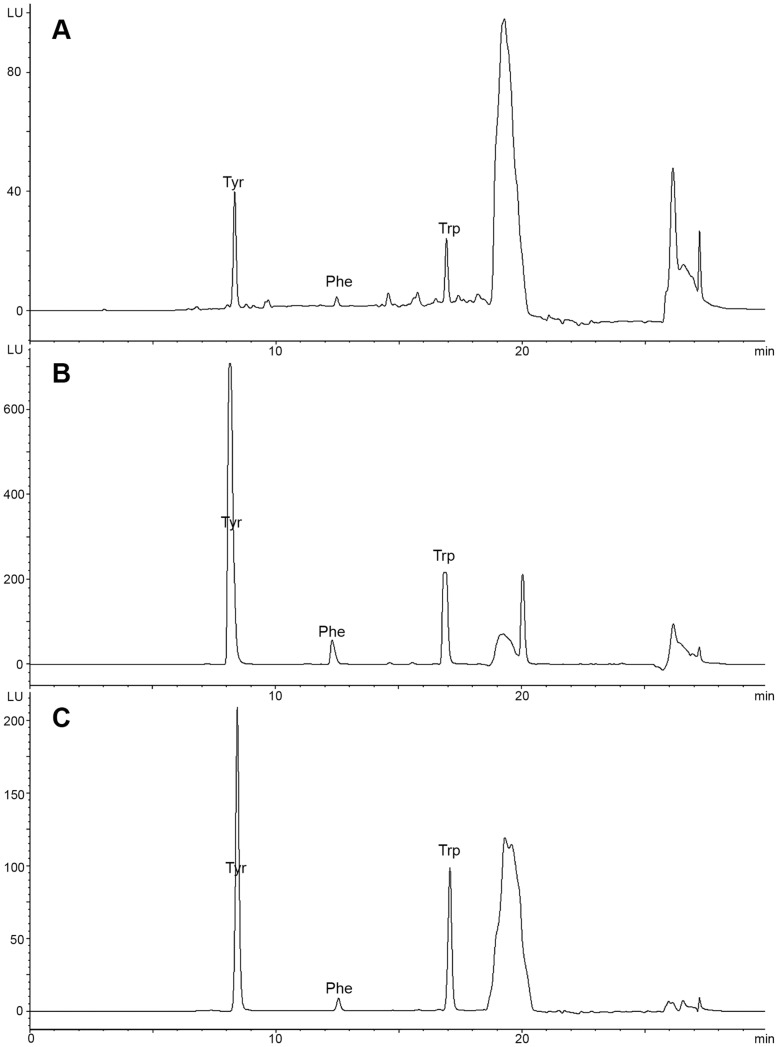
As shown in the fluorescence chromatogram (Figure 1), the three aromatic amino acids were completely separated. The retention times for each of the three amino acids were as follows: tyrosine, 8 min; phenylalanine, 12 min; and tryptophan, 16 min. The calibration curve, linearity and working ranges were as follows: tyrosine, Y = 0.32X+1.30, R2 = 0.9999, 0.5–20.0 µg/ml; phenylalanine, Y = 7.15X+1.03, R2 = 0.9999, 0.5–20.0 µg/ml; and tryptophan, Y = 0.25X+3.01, R2 = 0.9999, 0.5–20.0 µg/ml. The recoveries of tyrosine, phenylalanine and tryptophan were 100.5±2.7%, 101.3±3.4% and 99.6±1.6%, respectively. The precisions of tyrosine, phenylalanine and tryptophan were 100.1±0.5%, 100.0±1.8% and 98.7±1.5%, respectively. This experimental system was stable and reliable for the quantification of aromatic amino acids in gastric juice.
Figure 1. Fluorescence chromatograms of aromatic amino acids in gastric juice.
(A) NGD, (B) AGC and (C) EGC group. Tyr, tyrosine; Phe, phenylalanine; Trp, tryptophan.
Aromatic Amino Acids Assay
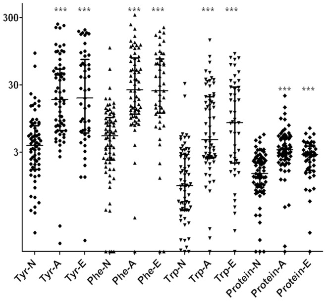
The median (25th to 75th percentile) values of tyrosine, phenylalanine and tryptophan were 3.8 (1.7–7.5) µg/ml, 5.3 (2.3–9.9) µg/ml and 1.0 (0.4–2.8) µg/ml in the NGD samples; 18.3 (6.4–52.3) µg/ml, 25.7 (11.1–76.1) µg/ml and 4.7 (2.5–20.3) µg/ml in the AGC samples; and 19.4 (5.8–72.4) µg/ml, 24.6 (11.5–73.7) µg/ml and 8.3 (2.1–28.0) µg/ml in the EGC samples (Table 3). The levels of aromatic amino acids in the gastric juice were significantly differently among the NGD, AGC and EGC groups (Kruskal-Wallis test, tyrosine χ2 = 49.847, phenylalanine χ2 = 62.397, tryptophan χ2 = 52.846, all P<0.0001). The levels of tyrosine, phenylalanine and tryptophan were significantly increased in the gastric juice obtained from both the AGC and EGC groups compared with those of the NGD group (Dunn's test, AGC vs. NGD, all P<0.0001; EGC vs. NGD, all P<0.0001) (Table 3 and Figure 2). After adjusting for the effect of the total protein levels in the gastric juice, significant associations still existed between the aromatic amino acid levels and the clinical diagnosis (Partial correlation, AGC vs. NGD: tyrosine, R = 0.334, P<0.001; phenylalanine, R = 0.257, P<0.001; and tryptophan, R = 0.312, P<0.001; EGC vs. NGD: tyrosine, R = 0.378, P<0.001; phenylalanine, R = 0.392, P<0.001; and tryptophan, R = 0.380, P<0.001). There were no significant differences in the levels of tyrosine, phenylalanine and tryptophan between the AGC and EGC gastric juice samples (Dunn's test, P = 0.811, P = 0.781 and P = 0.691, respectively) (Table 3). This result was consistent with previous findings [15] that indicated that no associations exist between Helicobacter pylori infection and the levels of aromatic amino acids in gastric juice (Mann-Whitney U test, tyrosine, P = 0.871; phenylalanine, P = 0.742; tryptophan, P = 0.913).
Figure 2. Scatter plot showing the levels of tyrosine, phenylalanine, tryptophan and total protein in gastric juice from NGD, AGC and EGC groups.
Tyr-N is the level of tyrosine in gastric juice from the NGD group. Tyr-A is the level of tyrosine in gastric juice from AGC group, and Tyr-E is the level of tyrosine in gastric juice from the EGC group. Phe-N indicates the level of phenylalanine in gastric juice from NGD group. Phe-A indicates the level of phenylalanine in gastric juice from AGC group and Phe-E indicates the level of phenylalanine in gastric juice from EGC group. Trp-N indicates the level of tryptophan in gastric juice from NGD group. Trp-A indicates the level of tryptophan in gastric juice from AGC group and Trp-E indicates the level of tryptophan in gastric juice from EGC group. Protein-N is the level of total protein in gastric juice from NGD group. Protein-A is the level of total protein in gastric juice from AGC group and Protein-E is the level of total protein in gastric juice from EGC group. ***: In comparison with NGD group, P value is less than 0.001. The median with interquartile range was shown in figure.
Validity of Aromatic Amino Acids for the Detection of AGC and EGC
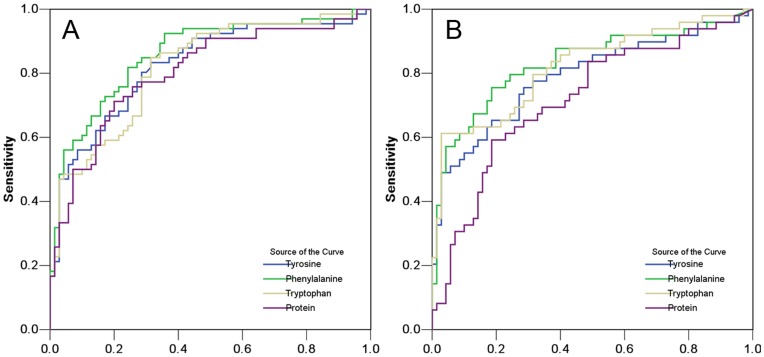
In the ROC curve analysis to distinguish between AGC and NGD, the areas under the curves (AUC) for tyrosine, phenylalanine, tryptophan and total protein were 0.824 [95% confidence interval (CI), 0.753–0.895, P<0.001], 0.858 (95% CI, 0.794–0.922, P<0.001), 0.818 (95% CI, 0.747–0.889, P<0.001) and 0.803 (95% CI, 0.728–0.878, P<0.001), respectively (Figure 3A and Table 4). According to the ROC curve analysis to distinguish between EGC and NGD, the AUCs for tyrosine, phenylalanine, tryptophan and total protein were 0.790 (95% CI, 0.703–0.877, P<0.001), 0.831 (95% CI, 0.750–0.911, P<0.001), 0.819 (95% CI, 0.739–0.900, P<0.001) and 0.719 (95% CI, 0.623–0.814, P<0.001), respectively (Figure 3B and Table 4). The optimized cut-off values to distinguish AGC or EGC from NGD were chosen by determining the maximum sums of sensitivity and specificity from the ROC curves (Table 4). After selecting the optimized values, the sensitivity, specificity and accuracy needed to distinguish between AGC and NGD were as follows: tyrosine, 83.3%, 68.6% and 75.7%; phenylalanine, 81.8%, 75.7% and 78.7%; tryptophan, 84.8%, 68.6% and 76.5%; and total protein, 75.8%, 74.3% and 75.0%. After selecting the optimized values, the sensitivity, specificity and accuracy needed to distinguish between EGC and NGD were as follows: tyrosine, 75.5%, 71.4% and 73.1%; phenylalanine, 75.5%, 81.4% and 79.0%; tryptophan, 61.2%, 97.1% and 82.4%; and total protein, 59.2%, 81.4% and 72.3% (Table 4). After selecting the optimized values reported in our previous study [12], [15], the sensitivity, specificity and accuracy for the detection of EGC were as follows: tyrosine, 51.0%, 94.3% and 76.5%; phenylalanine, 79.6%, 74.3% and 76.5%; tryptophan, 61.2%, 94.3% and 80.7%; and total protein, 83.7%, 62.9% and 71.4%. Using a different optimized value, we obtained similar results. Using a combination of tyrosine, phenylalanine and tryptophan did not significantly improve the detection of AGC or EGC because the concentrations of the three aromatic amino acids in the gastric juice samples were closely related to each other (tyrosine-phenylalanine, R = 0.871; tyrosine-tryptophan, R = 0.941; phenylalanine-tryptophan, R = 0.840, P<0.001 for each).
Figure 3. Predictive ability of gastric juice analysis for gastric cancer.
(A) ROC curves of tyrosine, phenylalanine, tryptophan and protein in gastric juice for detection of AGC. (B) ROC curves of tyrosine, phenylalanine, tryptophan and protein in gastric juice for detection of EGC.
Table 4. Performance of aromatic amino acids for detection of AGC or EGC.
| Marker | AUC (SE) | 95% CI | Cutoff value | Sensitivity | Specificity | Accuracy |
| Detection of AGC | ||||||
| Tyrosine | 0.824 (0.036) | 0.753–0.895 | 5.45 µg/ml | 83.3% (55/66) | 68.6% (48/70) | 75.7% (103/136) |
| Phenylalanine | 0.858 (0.033) | 0.794–0.922 | 9.68 µg/ml | 81.8% (54/66) | 75.7% (53/70) | 78.7% (107/136) |
| Tryptophan | 0.818 (0.036) | 0.747–0.889 | 1.94 µg/ml | 84.8% (56/66) | 68.6% (48/70) | 76.5% (104/136) |
| Protein | 0.803 (0.038) | 0.728–0.878 | 2.34 mg/ml | 75.8% (50/66) | 74.3% (52/70) | 75.0% (102/136) |
| Detection of EGC | ||||||
| Tyrosine | 0.790 (0.045) | 0.703–0.877 | 5.88 µg/ml | 75.5% (37/49) | 71.4% (50/70) | 73.1% (87/119) |
| Phenylalanine | 0.831 (0.041) | 0.750–0.911 | 11.74 µg/ml | 75.5% (37/49) | 81.4% (57/70) | 79.0% (94/119) |
| Tryptophan | 0.819 (0.041) | 0.739–0.900 | 5.67 µg/ml | 61.2% (30/49) | 97.1% (68/70) | 82.4% (98/119) |
| Protein | 0.719 (0.049) | 0.623–0.814 | 2.66 mg/ml | 59.2% (29/49) | 81.4% (57/70) | 72.3% (86/119) |
AUC, area under the ROC curve; SE, the standard error of AUC; Cutoff value, optimized cutoff points were the values yielding maximum sums of sensitivity and specificity from the ROC curves; AGC, advanced gastric cancer; EGC, early gastric cancer.
Logistic Regression Analysis
In the logistic regression analysis, the association between the levels of aromatic amino acids in the gastric juice and EGC was evaluated. Univariate logistic regression analyses were performed to evaluate the predictive values of candidate biomarkers for the detection of EGC. In these univariate analyses, the β coefficients (SE) of age, sex, Helicobacter pylori status, protein and elevated aromatic amino acid levels in gastric juice were calculated. Four variables (age and elevated tyrosine, phenylalanine and tryptophan levels in gastric juice) had a P<0.05 and were identified using multiple logistic regression analysis (Table 5). Because there were collinearities between tyrosine, phenylalanine and tryptophan (all R>0.8), we assessed the relationship between the levels of tyrosine, phenylalanine or tryptophan and EGC in three separate models and estimated the adjusted coefficients in each model. The elevated levels of these aromatic amino acids in the gastric juice were associated with EGC (tyrosine: adjusted β coefficient 1.801, SE 0.527; phenylalanine: adjusted β coefficient 2.555, SE 0.586; tryptophan: adjusted β coefficient 4.414, SE 0.945) (Table 5). Thus, elevated levels of aromatic amino acids in the gastric juice may be independent markers for the detection of EGC.
Table 5. Univariate and multivariate analyses of the predictive values of aromatic amino acids for EGC.
| Univariate analysis | Multivariate analysis | |||
| Factor | β Coefficient (SE)a | P valueb | Adjusted β Coefficient (SE)c | P valued |
| Model with tyrosine included | ||||
| Age | 0.032 (0.015) | 0.036 | 0.042 (0.018) | 0.021 |
| Sex (reference: male) | −1.012 (0.389) | 0.009 | −1.249 (0.472) | 0.008 |
| Helicobacter pylori status (reference: negative) | −0.322 (0.404) | 0.426 | −0.445 (0.486) | 0.360 |
| Protein | 0.586 (0.161) | <0.001 | 0.289 (0.189) | 0.127 |
| Tyrosinee (reference: <5.88 µg/ml) | 2.042 (0.425) | <0.0001 | 1.801 (0.527) | <0.001 |
| Model with phenylalanine included | ||||
| Age | 0.032 (0.015) | 0.036 | 0.047 (0.019) | 0.016 |
| Sex (reference: male) | −1.012 (0.389) | 0.009 | −1.179 (0.498) | 0.018 |
| Helicobacter pylori status (reference: negative) | −0.322 (0.404) | 0.426 | −0.482 (0.517) | 0.351 |
| Protein | 0.586 (0.161) | <0.001 | 0.137 (0.203) | 0.498 |
| Phenylalaninee (reference: <11.74 µg/ml) | 2.604 (0.453) | <0.0001 | 2.555 (0.586) | <0.0001 |
| Model with tryptophan included | ||||
| Age | 0.032 (0.015) | 0.036 | 0.055 (0.022) | 0.014 |
| Sex (reference: male) | −1.012 (0.389) | 0.009 | −0.947 (0.549) | 0.085 |
| Helicobacter pylori status (reference: negative) | −0.322 (0.404) | 0.426 | −1.298 (0.696) | 0.062 |
| Protein | 0.586 (0.161) | <0.001 | 0.187 (0.209) | 0.371 |
| Tryptophane (reference: <5.67 µg/ml) | 3.983 (0.775) | <0.0001 | 4.414 (0.945) | <0.0001 |
Coefficients for each factor were evaluated using univariate logistic regression;
Two-sided Chi-squared test for coefficients obtained from univariate logisitic regression model;
Adjusted coefficients for each covariate were evaluated using multivariate logistic regression with adjustment for age, sex, Helicobacter pylori status, and protein concentration;
Two-sided Chi-squared test for coefficients obtained from the multivariate logisitic regression model;
Because there were collinearities between tyrosine phenylalanine and tryptophan (all R>0.8 ), we assessed the relationship between the levels of tyrosine, phenylalanine or tryptophan and the occurrence of EGC in three separate models, and the adjusted coefficients were estimated in each model.
Discussion
In our previous studies, we have observed increased fluorescence of gastric juice in AGC and identified three fluorescence biomarkers for screening advanced gastric cancer [12], [15]. Whether these potential biomarkers are useful for the early detection of gastric cancer is uncertain. In our current investigation, we tested the validity of using aromatic amino acids in the gastric juice for the diagnosis of EGC. Our results show that high levels of aromatic amino acids are present in the gastric juice from patients with EGC compared to NGD patients. Thus, high levels of aromatic amino acids in the gastric juice may be valuable for early detection of EGC.
Because of the thesmall sample size, power analysis for Kruskal-Wallis test and ROC AUC were performed. To our knowledge, the formulas or procedures for determining the sample size necessary to test for differences in multiple-sample locations using nonparametric methods are much more complicated [19], and no power or sample size calculation method is currently available regarding the generic alternative hypothesis for the Kruskal-Wallis test [20]. Lachenbruch and Clements suggested that the Kruskal-Wallis test might have greater power than the F-test when the population distributions are not normal. They claimed that, compared with the F-test, the Kruskal-Wallis test was more robust against departures from the assumption of the equality of variance [21]. Thus, using the power of the F-test to approximate the power of the Kruskal-Wallis test may provide a poor lower bound for normal distributions. Following a rank transformation, the proximate powers (1 - β error) were calculated using the F-test (ANOVA) as implemented in SAS 8.1. All of the proximate powers are more than 0.99. Thus, we can reliably conclude that the levels of total protein and aromatic amino acids in gastric juice are significantly different among NGD, AGC and EGC. Power analyses for the ROC curves were performed using PASS 2008 software. None of the powers was less than 0.99. Therefore, it is acceptable to conclude that high levels of aromatic amino acids in the gastric juice may be valuable predictors for AGC or EGC.
The enhanced production of aromatic amino acids in EGC corresponds to the results for a previous study [15] and is consistent with early reports in other malignant diseases (blood [22], lung [23], [24], [25], breast [23], [24], bladder [26], etc.). We speculate that the following reasons may account for this phenomenon. First, tumour cells require sufficient aromatic amino acids to accumulate in the cancer foci to meet the sharply increased needs for protein synthesis to support rapid growth. A high expression of amino acids transporters (such as L-type amino-acid transporter 1) in various tumours [27], [28], [29], [30], [31], [32], [33], [34], [35] has been reported in many studies. Amino acid transporters may enhance the transport of amino acids and accumulate aromatic amino acids near cancer foci. The infiltration of excessive aromatic amino acids may significantly increase in gastric juice in EGC compared to NGD. Secondly, aberrant metabolism in cancer cells would precede the visible changes in stomach carcinogenesis [11]. It is known that the abnormalities of amino acid transport and metabolism in a cancerous region occur frequently in the early stage of gastric cancer. Gastric juice, which is generated by the gastric epithelium, contains a substantial amount of information about the metabolic status of the epithelium. Therefore, the information obtained from analysing the gastric juice may be useful in separating biomarkers for the early detection of gastric cancer. Finally, matrix metalloproteinases (MMPs), which are enzymes that degrade the extracellular matrix and basement membrane, play roles in the early phase of tumourigenesis [36], [37], [38], [39], [40]. The over-expression and activity of MMPs accelerates the degradation of extracellular matrix and basement membrane. The degradation of matrix generates aromatic amino acids in the gastric juice surrounding tumour cells. Additionally, blood aromatic amino acids could easily penetrate the impaired basement membrane and epithelia of carcinoma foci and enhance amino acid levels in gastric juice. These causes could explain the significant enhancement of aromatic amino acids in gastric juice from EGC patients.
The early phase of gastric cancer has few symptoms and can be easily missed by conventional endoscopy [6], [41]. These problems reduce the rate of early detection and effective treatment for gastric cancer. Abnormal levels of aromatic amino acids in the gastric juice may be used to monitor the occurrence of amino acid metabolism disorders and reveal early malignant transformations that occur in the stomach. Notably, our findings indicate that the levels of aromatic amino acids in the gastric juice can distinguish EGC from gastritis. This finding introduces the possibility that these biomarkers can be used for the mass screening of gastric cancer at an early stage. The measurement of aromatic amino acid levels in gastric juice samples may be an efficient, economical, rapid, and convenient method for detecting AGC or EGC in a population.
Although aromatic amino acids may be promising biomarkers for EGC, there are still drawbacks associated with this approach. The use of less invasive procedures would be beneficial for screening purposes. The gastric juices in our study were aspirated during endoscopy, which would severely limit the applicability of assessing gastric juice biomarkers for the detection of EGC. To increase the potential application of this finding, non-endoscopic means can be introduced for sampling gastric juice. An endogastric capsule [42], [43], [44] is a 45–50 cm length of nylon thread connected to a capsule contained absorbent paper. A capsule can make gastric juice sampling more acceptable, painless and convenient for patients compared to gastric juice collection by endoscopy. The levels of aromatic amino acids in the gastric juice can be influenced by the fluid and food consumed by the patient. Therefore, full fasting in patients with poor gastric motility is essential for enhancing the accuracy of biomarker assays and gastric cancer detection. Because the sample size in this study is small, larger studies are required to determine whether aromatic amino acids in gastric juice can help in screening early-stage gastric cancer. Additionally, further studies will identify the optimized boundary values for each marker. Once the diagnostic screening values are identified, new methods (such as mass spectrometry) could allow for the simple, rapid and high-throughput detection of aromatic amino acids in gastric juice.
In conclusion, significantly increased levels of aromatic amino acids were found in gastric juice from patients with late-stage or early-stage gastric cancer compared to the NGD group. An analysis of aromatic amino acids may be an efficient method for the early detection of gastric cancer. If validated in a large-scale study, aromatic amino acids in gastric juice would be useful as biomarkers for screening EGC in the general population.
Acknowledgments
The authors would like to thank to Qiuming Geng, Shigang Ding Qinqin Tao, Jingjing Lu, Yan Deng and all colleagues in the endoscopy rooms of Peking University Third Hospital for their assistance in gastric juice collection.
Funding Statement
This research was supported by the National Natural Science Foundation of China [Grant number: 3097415](URL: http://www.nsfc.gov.cn/e_nsfc/desktop/zn/0101.htm). The funders had no role in study design, data collection and analysis, decision to publish, or preparation of the manuscript.
References
- 1. Arvanitakis C, Nikopoulos A, Giannoulis E, Theoharidis A, Georgilas V, et al. (1992) The impact of early or late diagnosis on patient survival in gastric cancer in Greece. Hepatogastroenterology 39: 355–357. [PubMed] [Google Scholar]
- 2. Hartgrink HH, Jansen EP, van Grieken NC, van de Velde CJ (2009) Gastric cancer. Lancet 374: 477–490. [DOI] [PMC free article] [PubMed] [Google Scholar]
- 3. Cunningham SC, Kamangar F, Kim MP, Hammoud S, Haque R, et al. (2005) Survival after gastric adenocarcinoma resection: eighteen-year experience at a single institution. J Gastrointest Surg 9: 718–725. [DOI] [PubMed] [Google Scholar]
- 4. Itoh H, Oohata Y, Nakamura K, Nagata T, Mibu R, et al. (1989) Complete ten-year postgastrectomy follow-up of early gastric cancer. Am J Surg 158: 14–16. [DOI] [PubMed] [Google Scholar]
- 5. Pathirana A, Poston GJ (2001) Lessons from Japan–endoscopic management of early gastric and oesophageal cancer. Eur J Surg Oncol 27: 9–16. [DOI] [PubMed] [Google Scholar]
- 6. Yalamarthi S, Witherspoon P, McCole D, Auld CD (2004) Missed diagnoses in patients with upper gastrointestinal cancers. Endoscopy 36: 874–879. [DOI] [PubMed] [Google Scholar]
- 7. Amin A, Gilmour H, Graham L, Paterson-Brown S, Terrace J, et al. (2002) Gastric adenocarcinoma missed at endoscopy. J R Coll Surg Edinb 47: 681–684. [PubMed] [Google Scholar]
- 8. Duraker N, Naci CA, Gencler N (2002) The prognostic significance of gastric juice CA 19–9 and CEA levels in gastric carcinoma patients. Eur J Surg Oncol 28: 844–849. [DOI] [PubMed] [Google Scholar]
- 9. Miki K, Morita M, Sasajima M, Hoshina R, Kanda E, et al. (2003) Usefulness of gastric cancer screening using the serum pepsinogen test method. Am J Gastroenterol 98: 735–739. [DOI] [PubMed] [Google Scholar]
- 10. Miki K (2006) Gastric cancer screening using the serum pepsinogen test method. Gastric Cancer 9: 245–253. [DOI] [PubMed] [Google Scholar]
- 11. McDermott EW, Barron ET, Smyth PP, O'Higgins NJ (1990) Premorphological metabolic changes in human breast carcinogenesis. Br J Surg 77: 1179–1182. [DOI] [PubMed] [Google Scholar]
- 12. Zhou LY, Lin SR, Li Y, Geng QM, Ding SG, et al. (2011) The intrinsic fluorescence spectrum of dilute gastric juice as a novel diagnostic tool for gastric cancer. J Dig Dis 12: 279–285. [DOI] [PubMed] [Google Scholar]
- 13.Sun XN, Zhou LY, Lin SR, Zhao YM, Lv JQ (2004) Study on autofluorescence spectrum of gastric juice in clinical screening for gastric cancer. Chin J Dig : 10–13 (in Chinese).
- 14.Li Y, Lin SR, Zhou LY, Peng JR, Guo HL (2003) Preliminary identification of a specific fluorescent substance in gastric juice from patients with gastric carcinoma. Chin J Dig : 11–14 (in Chinese).
- 15. Deng K, Lin S, Zhou L, Geng Q, Li Y, et al. (2011) Three aromatic amino acids in gastric juice as potential biomarkers for gastric malignancies. Anal Chim Acta 694: 100–107. [DOI] [PubMed] [Google Scholar]
- 16. Lansdown M, Quirke P, Dixon MF, Axon AT, Johnston D (1990) High grade dysplasia of the gastric mucosa: a marker for gastric carcinoma. Gut 31: 977–983. [DOI] [PMC free article] [PubMed] [Google Scholar]
- 17. Deng K, Lin S, Zhou L, Geng Q, Li Y, et al. (2011) Three aromatic amino acids in gastric juice as potential biomarkers for gastric malignancies. Anal Chim Acta 694: 100–107. [DOI] [PubMed] [Google Scholar]
- 18. Lian W, Ma DJ, Xu X, Chen Y, Wu YL (2012) Rapid high-performance liquid chromatography method for determination of tryptophan in gastric juice. J Dig Dis 13: 100–106. [DOI] [PubMed] [Google Scholar]
- 19.Shein-Chung Chowm, Wang H, Shao J (2008) Sample Size Calculations in Clinical Research, 2nd edition. Boca Raton, Florida: Chapman &Hall/CRC.
- 20. Fan C, Zhang D, Zhang CH (2011) On sample size of the kruskal-wallis test with application to a mouse peritoneal cavity study. Biometrics 67: 213–224. [DOI] [PubMed] [Google Scholar]
- 21. Lachenbruch PA, Clements PJ (1991) Anova, kruskal-wallis, normal scores and unequal variance. Communications in Statistics - Theory and MethodsCommunications in Statistics - Theory and MethodsCommunications in Statistics - Theory and Methods 20: 107–126. [Google Scholar]
- 22. Muscaritoli M, Conversano L, Petti MC, Torelli GF, Cascino A, et al. (1999) Plasma amino acid concentrations in patients with acute myelogenous leukemia. Nutrition 15: 195–199. [DOI] [PubMed] [Google Scholar]
- 23. Cascino A, Muscaritoli M, Cangiano C, Conversano L, Laviano A, et al. (1995) Plasma amino acid imbalance in patients with lung and breast cancer. Anticancer Res 15: 507–510. [PubMed] [Google Scholar]
- 24. Laviano A, Cascino A, Muscaritoli M, Fanfarillo F, Rossi FF (2003) Tumor-induced changes in host metabolism: a possible role for free tryptophan as a marker of neoplastic disease. Adv Exp Med Biol 527: 363–366. [DOI] [PubMed] [Google Scholar]
- 25. Huang Z, McWilliams A, Lui H, McLean DI, Lam S, et al. (2003) Near-infrared Raman spectroscopy for optical diagnosis of lung cancer. Int J Cancer 107: 1047–1052. [DOI] [PubMed] [Google Scholar]
- 26. Draga RO, Grimbergen MC, Vijverberg PL, van SCF, Jonges TG, et al. (2010) In vivo bladder cancer diagnosis by high-volume Raman spectroscopy. Anal Chem 82: 5993–5999. [DOI] [PubMed] [Google Scholar]
- 27. Ganapathy V, Thangaraju M, Prasad PD (2009) Nutrient transporters in cancer: relevance to Warburg hypothesis and beyond. Pharmacol Ther 121: 29–40. [DOI] [PubMed] [Google Scholar]
- 28. Fuchs BC, Bode BP (2005) Amino acid transporters ASCT2 and LAT1 in cancer: partners in crime. Semin Cancer Biol 15: 254–266. [DOI] [PubMed] [Google Scholar]
- 29. Nawashiro H, Otani N, Shinomiya N, Fukui S, Ooigawa H, et al. (2006) L-type amino acid transporter 1 as a potential molecular target in human astrocytic tumors. Int J Cancer 119: 484–492. [DOI] [PubMed] [Google Scholar]
- 30. Kaira K, Oriuchi N, Imai H, Shimizu K, Yanagitani N, et al. (2008) Prognostic significance of L-type amino acid transporter 1 expression in resectable stage I-III nonsmall cell lung cancer. Br J Cancer 98: 742–748. [DOI] [PMC free article] [PubMed] [Google Scholar]
- 31. Yoon JH, Kim YB, Kim MS, Park JC, Kook JK, et al. (2004) Expression and functional characterization of the system L amino acid transporter in KB human oral epidermoid carcinoma cells. Cancer Lett 205: 215–226. [DOI] [PubMed] [Google Scholar]
- 32. Kobayashi H, Ishii Y, Takayama T (2005) Expression of L-type amino acid transporter 1 (LAT1) in esophageal carcinoma. J Surg Oncol 90: 233–238. [DOI] [PubMed] [Google Scholar]
- 33. Nakanishi K, Ogata S, Matsuo H, Kanai Y, Endou H, et al. (2007) Expression of LAT1 predicts risk of progression of transitional cell carcinoma of the upper urinary tract. Virchows Arch 451: 681–690. [DOI] [PubMed] [Google Scholar]
- 34.Kobayashi K, Ohnishi A, Promsuk J, Shimizu S, Kanai Y, et al. (2008) Enhanced tumor growth elicited by L-type amino acid transporter 1 in human malignant glioma cells. Neurosurgery 62: 493–503; discussion 503–504. [DOI] [PubMed]
- 35. Sakata T, Ferdous G, Tsuruta T, Satoh T, Baba S, et al. (2009) L-type amino-acid transporter 1 as a novel biomarker for high-grade malignancy in prostate cancer. Pathol Int 59: 7–18. [DOI] [PubMed] [Google Scholar]
- 36. Talvensaari A, Apaja-Sarkkinen M, Hoyhtya M, Westerlund A, Puistola U, et al. (1999) Matrix metalloproteinase 2 immunoreactive protein appears early in cervical epithelial dedifferentiation. Gynecol Oncol 72: 306–311. [DOI] [PubMed] [Google Scholar]
- 37. Rudolph-Owen LA, Chan R, Muller WJ, Matrisian LM (1998) The matrix metalloproteinase matrilysin influences early-stage mammary tumorigenesis. Cancer Res 58: 5500–5506. [PubMed] [Google Scholar]
- 38. Cai M, Onoda K, Takao M, Kyoko IY, Shimpo H, et al. (2002) Degradation of tenascin-C and activity of matrix metalloproteinase-2 are associated with tumor recurrence in early stage non-small cell lung cancer. Clin Cancer Res 8: 1152–1156. [PubMed] [Google Scholar]
- 39. Sier CF, Casetta G, Verheijen JH, Tizzani A, Agape V, et al. (2000) Enhanced urinary gelatinase activities (matrix metalloproteinases 2 and 9) are associated with early-stage bladder carcinoma: a comparison with clinically used tumor markers. Clin Cancer Res 6: 2333–2340. [PubMed] [Google Scholar]
- 40. Nosho K, Yamamoto H, Taniguchi H, Adachi Y, Yoshida Y, et al. (2004) Interplay of insulin-like growth factor-II, insulin-like growth factor-I, insulin-like growth factor-I receptor, COX-2, and matrix metalloproteinase-7, play key roles in the early stage of colorectal carcinogenesis. Clin Cancer Res 10: 7950–7957. [DOI] [PubMed] [Google Scholar]
- 41. Axon A (2006) Symptoms and diagnosis of gastric cancer at early curable stage. Best Pract Res Clin Gastroenterol 20: 697–708. [DOI] [PubMed] [Google Scholar]
- 42. Wang RT, Zao JS, Liu D, Peng BS, He PY (1991) The gastric cancer series tests with minimum volume of gastric juice collected by advanced micro device. J Beijing Med Univ 23: 9–11 (in Chinese).. [Google Scholar]
- 43. Muretto P, Ruzzo A, Pizzagalli F, Graziano F, Maltese P, et al. (2008) Endogastric capsule for E-cadherin gene (CDH1) promoter hypermethylation assessment in DNA from gastric juice of diffuse gastric cancer patients. Ann Oncol 19: 516–519. [DOI] [PubMed] [Google Scholar]
- 44. Muretto P, Graziano F, Staccioli MP, Barbanti I, Bartolucci A, et al. (2003) An endogastric capsule for measuring tumor markers in gastric juice: an evaluation of the safety and efficacy of a new diagnostic tool. Ann Oncol 14: 105–109. [DOI] [PubMed] [Google Scholar]


