Abstract
A variant of Bacillus thuringiensis subsp. kurstaki containing a single, stable copy of a uniquely amplifiable DNA oligomer integrated into the genome for tracking the fate of biological agents in the environment was developed. The use of genetically tagged spores overcomes the ambiguity of discerning the test material from pre-existing environmental microflora or from previously released background material. In this study, we demonstrate the utility of the genetically “barcoded” simulant in a controlled indoor setting and in an outdoor release. In an ambient breeze tunnel test, spores deposited on tiles were reaerosolized and detected by real-time PCR at distances of 30 m from the point of deposition. Real-time PCR signals were inversely correlated with distance from the seeded tiles. An outdoor release of powdered spore simulant at Aberdeen Proving Ground, Edgewood, MD, was monitored from a distance by a light detection and ranging (LIDAR) laser. Over a 2-week period, an array of air sampling units collected samples were analyzed for the presence of viable spores and using barcode-specific real-time PCR assays. Barcoded B. thuringiensis subsp. kurstaki spores were unambiguously identified on the day of the release, and viable material was recovered in a pattern consistent with the cloud track predicted by prevailing winds and by data tracks provided by the LIDAR system. Finally, the real-time PCR assays successfully differentiated barcoded B. thuringiensis subsp. kurstaki spores from wild-type spores under field conditions.
INTRODUCTION
The development of sensitive and unequivocal approaches for detecting and tracking highly pathogenic bacteria has traditionally relied upon the use of nonpathogenic spore-producing Bacillus species as model organisms or simulants, whose physical and biochemical properties mimic those of the threat agent. Bacillus anthracis is a proven biothreat agent (5, 14–15, 20, 24) due its high virulence and the ability to form hardy and persistent spores, which can persist for decades in certain environments (21). Historically, nonpathogenic spore-forming bacteria such as Bacillus atrophaeus subsp. globigii have been used as surrogate organisms to simulate B. anthracis (9, 11). The physical properties of Bacillus thuringiensis subsp. kurstaki and its close genetic relatedness to B. anthracis, most notably with regard to the presence of an exosporium, which is absent from B. atrophaeus subsp. globigii, have led to recent preference for the use of B. thuringiensis subsp. kurstaki over B. atrophaeus subsp. globigii (10). However, the use of B. atrophaeus subsp. globigii and B. thuringiensis subsp. kurstaki in test sites is complicated by the fact that both organisms occur naturally in the environment (18; see also the excellent review of environmental B. thuringiensis subsp. kurstaki prevalence by Van Cuyk et al. [27]). B. thuringiensis subsp. kurstaki has a long history of use as a biopesticide, dating back to 1929 studies in the northeastern United States that showed B. thuringiensis to be effective for insect control against the gypsy moth (17). Today the global use of B. thuringiensis subsp. kurstaki as a pesticide is widespread, with over 13,000 tons produced annually, and the U.S. Department of Agriculture (USDA) estimates that the worldwide market for B. thuringiensis subsp. kurstaki-based products for forestry and agriculture is greater than $80 million per year (1).
For the purposes of open-air release and tracking of biowarfare (BW) agent surrogate organisms, the natural occurrence and widespread industrial use of B. atrophaeus subsp. globigii and B. thuringiensis subsp. kurstaki make specific identification of newly disseminated isolates difficult (16, 27). Furthermore, once disseminated, spores can persist in the environment for years or even decades (22, 26), complicating analysis of subsequent field release tests. Repeated use of B. atrophaeus subsp. globigii spores at military proving grounds has saturated such areas with the accumulation of 75 years' worth of biological test materials (K. Kester, personal communication). Other studies have attempted to develop better BW simulants to replace B. atrophaeus subsp. globigii spores. One approach modified polystyrene beads to attach moieties that help track these synthetic products. The ability to detect and track the fate of the material is a valuable attribute, but because the simulants were built from synthetic particles, there are new questions about how well they mimic aerosolization and surface adhesion properties of a real BW agent (7). Another approach (3, 4) created plasmid vectors that housed multiple gene-based targets, but this simulant strategy was not developed for aerosolization testing purposes.
The accompanying paper reports a study in which a unique genetic “tag” was integrated into the bacterial genome for the purposes of developing a traceable simulant (2). Using genetic exchange technology for Bacillus group species and information gleaned from the newly sequenced genome of ATCC 33679, a well-characterized HD-1 biopesticide strain of B. thuringiensis subsp. kurstaki with an exemplary safety record (12, 13, 23, 25), we designed and introduced a series of 46-bp sequences into specific loci of the genome. These insertions lie within intergenic regions chosen to minimize the potential of disrupting protein-coding genes or regulatory sequences which could affect the phenotype of the organism. The 46-bp sequence constitutes a “barcode” which creates a lineage-specific locus that is paired with a chromosomal locus. The design allows for the use of a tailor-made real-time PCR (RT-PCR) assay for specific spore detection and differentiation.
The purpose of creating a traceable BW simulant was to provide a better way to validate urban threat models, perform tests in which one can sample and detect multiple strains in parallel from a single dissemination event, and reduce test costs by allowing data to be collected from multiple release tests in parallel. Two clear objectives of this study were (i) to demonstrate the suitability of barcoded B. thuringiensis subsp. kurstaki spores for tracking spore release in breeze tunnel and in open-air field release and (ii) to demonstrate reaerosolization of released spores in a controlled environment using tailored PCR assays. The results from the first indoor and outdoor field tests of barcoded strains correlated with the standard microbiological and PCR detection methods, and these are summarized in this paper.
MATERIALS AND METHODS
Large-scale production of B. thuringiensis subsp. kurstaki strains for dissemination.
Wild-type (WT) B. thuringiensis subsp. kurstaki (ATCC 33679) and barcoded B. thuringiensis subsp. kurstaki spores derived from ATCC 33679 (2) were grown in NZ-Amine A growth medium consisting of the following (in grams per liter unless other units noted): 10.0 glucose, 5.0 casein peptone type S, 1.0 yeast extract, 4.0 K2HPO4, 3.0 KH2PO4, 0.134 CaCl2 · 2H2O, 0.02 FeSO4 · 7H2O, 0.05 MgSO4 · 7H2O, 0.023 MnSO4 · H2O, and 0.02 ZnSO4 · 7H2O, and 0.05 ml antifoam 204 (Sigma-Aldrich) per liter of culture volume. All medium ingredients were autoclaved together with the exception of glucose and metals. Glucose filtered through 0.8/0.2-μm filters was aseptically added to the sterile media. The two metal solutions (one containing CaCl2 · 2H2O, FeSO4 · H2O, MgSO4 · H2O, and MnSO4 · H2O and the other containing the ZnSO4 · 7H2O) were separately prepared, filtered sterilized, and added to prevent precipitation of CaSO4. One-liter seed cultures in a 4-liter flask were prepared by inoculating with two vials of frozen stock and incubating (Innova 4300 shaking incubator; New Brunswick Scientific) at 30°C and 200 rpm until the optical density at 600 nm (Genesys 20 spectrophotometer; ThermoSpectronic) reached approximately 0.4 optical density units. The 1-liter seed cultures were aseptically transferred by a presterilized 2-liter transfer bottle into the 100-liter fermentor (IF 150; New Brunswick Scientific) containing the presterilized NZ-Amine A medium. The operating conditions for the 100-liter fermentor were controlled at 240 rpm, an airflow of 1 vvm (i.e., air volume per liquid volume per minute), 30°C, and pH 7.0 (with 3 M H3PO4 and 3 M NaOH). When the percentage of spores (as estimated by phase-contrast microscopy [microscope model BX51; Olympus]) exceeded 95%, the spore suspension was heat-shocked for 1 h at 70°C. After cooling down to 30°C, the spore suspension was diafiltrated (0.2-μm Pellicon TFF filters; Millipore) to approximately a 20-liter retentate volume, washed five times with deionized water, and concentrated to a final volume of approximately 10 liters. The washed and concentrated 10-liter spore suspension was spray dried (Niro) and subsequently milled (Sturtevant) with Aerosol 202 (Evonik). The spray-dried spore preparation was characterized in terms of particle size distribution (using a TSI, Inc., aerodynamic particle sizer [APS 3321]), moisture content (Karl Fischer C20 moisture analyzer; Mettler-Toledo), and viability (serial dilution and plate counting). The final dried and milled products contained 1.1 × 10¹¹ (WT B. thuringiensis subsp. kurstaki) and 2.6 × 10¹¹ (barcoded B. thuringiensis subsp. kurstaki) CFU/g. Analysis of a representative preparation using a Petroff-Hauser chamber revealed that approximately 90% of total spores were viable prior to dissemination.
Indoor reaerosolization.
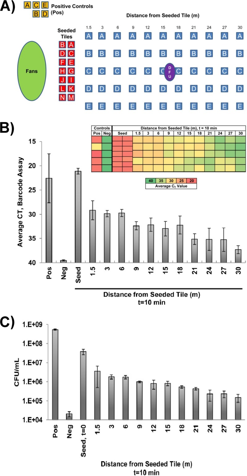
In order to test the ability of barcoded B. thuringiensis subsp. kurstaki spores to be reaerosolized, a test was conducted in a 61-m-long ambient breeze tunnel. This tunnel provided a controlled indoor environment that simulated outdoor wind conditions. The experimental setup is illustrated in Fig. 1A. Vinyl tiles were cleaned with 70% (vol/vol) ethanol-water and placed down the length of the ambient breeze tunnel at 3-m intervals to a distance of 30 m away from the contaminated tiles. At the front of the ambient breeze tunnel, a row of tiles enclosed in boxes were carefully seeded with 100 mg of the aerosolized barcoded B. thuringiensis subsp. kurstaki spores. Five of the seeded tiles were set aside and not subjected to a breeze to serve as the positive-control tiles. Midway down the tunnel, a single dry filter unit (DFU) air sampler (see below) was also situated to collect any spores that drifted down the tunnel. Prior to initiation of the test, five unseeded tiles were sampled as negative controls to ensure that the seeding process did not itself cause spores to drift in the sampling area. To begin the test, fans directed at the seeded tiles were turned on, creating a breeze that was allowed to blow air across the contaminated tiles for a period of 10 min. The velocity of the breeze was measured to be 3.8 m per s by anemometers positioned just above the seeded tiles. The fans were then turned off, and reaerosolized spores were allowed to settle overnight. In the morning, each tile was swabbed with a single sterilized 2-in2 nonwoven cotton fabric wipe (Dukal Corporation). The sponges were wetted with 5 ml of PBS from a 50-ml conical tube and then swabbed across the tile, covering the entire tile. The wipe was then folded inside out and used to swab the entire tile again in the other direction. The wipes were placed back into their respective 50-ml tubes and processed for analysis.
Fig 1.
Re-aerosolization in the ambient breeze tunnel tested by PCR. (A) Experimental design. Twenty tiles (1 ft2 each) were seeded with powdered barcoded spores (seed and positive controls). Seed tiles were subjected to 10 min of wind disturbance at 3.8 m per s for reaerosolization. The reaerosolized spores were allowed to settle on clean tiles arrayed at intervals down a 30-m section of a 61-m-long tunnel, followed by analysis by culture and PCR. The negative-control data represent a composite of clean tiles prior to reaerosolization, and values for samples indicated by their distance from the seeded tiles are the averages for five replicate tiles, with each PCR assay performed in triplicate. The dry filter unit was placed 15 m from the seeded tiles and was measured before application of the breeze (T = 0) and after (DFU following release). (B) Results of the reaerosolization test. The graph shows averages of all CT values determined for each distance. Results are presented as averages ± standard deviations. (Inset) Heat map of results from each tile. Each PCR assay was performed in triplicate. Results are averages for each tile. (C) Enumeration of viable colonies from each sample. Results are averages ± standard deviations for all five tiles in each distance range.
Outdoor dissemination and detection of B. thuringiensis subsp. kurstaki strains.
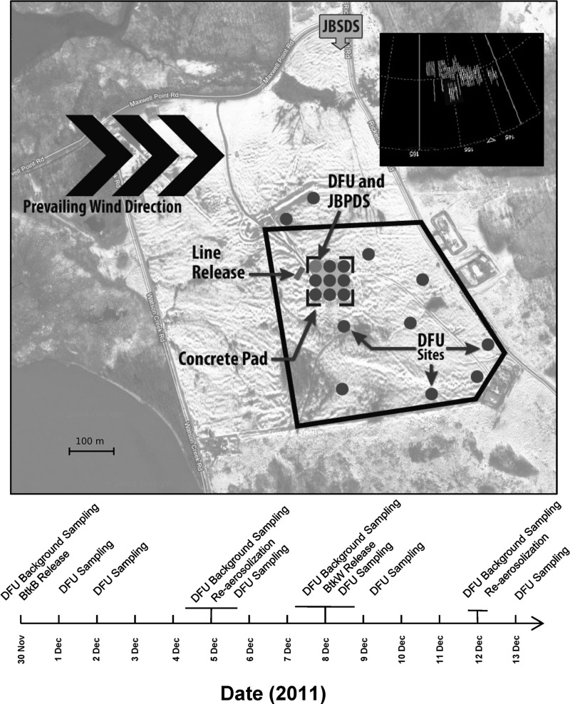
The outdoor dissemination took place at the Edgewood Chemical Biological Center's M Field test range, Aberdeen Proving Ground, MD. The M Field test range is an open field on the Edgewood Peninsula on which a large concrete pad is located to facilitate positioning of test equipment (Fig. 2). A test grid consisting of multiple dry filter unit (DFU) collectors and a single joint biological point detection system (see below) was arranged across the field downwind of the release point. The test plan was designed to release 100 g (∼1.1 × 10¹³ spores) of dried barcoded B. thuringiensis subsp. kurstaki spores on the first day and continue to sample the air for an additional 2 days. The layout of the test grid and timeline of dissemination and sampling events are shown in Fig. 2. On the third day after the initial release event, a team of two people used commercial blowers for a 15-min period to simulate air turbulence around a 30-m2 area near the site of the release. The air samples were monitored for an additional day to detect barcoded spores that might have been reaerosolized by this event. On the eighth day, a 100-g batch of wild-type (WT B. thuringiensis subsp. kurstaki; lacking the barcode) spores (approximately 2.6 × 10¹³ spores) was released in the identical manner, and the air was monitored. The goal of this second release was to demonstrate the selectivity of the detection method.
Fig 2.
Experimental design of outdoor dissemination test. The map shows a layout of the test range in detail, with a 100-m by 100-m concrete pad as the center point of the experiment. A line release was carried out from the position indicated to maximize the capture of agent by an array of DFU 2000 air samplers that were deployed based on recommendations by a sensor placement tool from Ensco. The location of the joint biological standoff detection system (JBSDS) LIDAR system is indicated. (Inset) LIDAR image of progress of the cloud of barcoded B. thuringiensis subsp. kurstaki spores over the test range. The satellite image was obtained from Google Earth (copyright 2012 DigitalGlobe, GeoEye, U.S. Geological Service, USDA Farm Service Agency).
Dissemination of B. thuringiensis subsp. kurstaki powder.
The dissemination platform for dispersing the spore preparations consisted of a Metronics model 10 Skilblower mounted on the bed of a high-mobility multipurpose wheeled vehicle (HMMWV; AM General LLC, South Bend, IN), better known as the Humvee. The spores were released at a point approximately 50 m west of the test grid, primarily targeting the northwest corner of the pad (Fig. 2). The Skilblower is a high-volume dissemination system specifically for dry powders. It utilizes a high-power motor blower to disseminate large volumes of powders to great distances, generates high shear forces for maximum breakup of particles, and incorporates a variable-feed-rate system to provide controlled amounts of powder from 2 to 40 g/min. The Skilblower was mounted at a height of approximately 1.5 m above the bed of the Humvee at an angle of 35 degrees from horizontal. The Humvee was driven at ∼3 km per h along the north/south line until the dissemination was complete (i.e., until all of the powder was disseminated). The powder was manually fed into the Skilblower to better control the timing of the release. The feeding of the biological material was done at a steady rate by holding the container of powder to the intake of the Skilblower at a 90° angle. The first release occurred on 30 November 2011 at 6:24 PM over a 5-min period and consisted of 100 g of barcoded B. thuringiensis subsp. kurstaki spores, which was calculated to consist of 3.3 × 10¹¹ colonies per gram and had a mass mean diameter of 3.39 μm and a moisture content of 8.85%. The second release occurred 8 days later at 7:24 PM over a 6.5-min period and consisted of 87 g of WT B. thuringiensis subsp. kurstaki spores, which was calculated to consist of 1.1 × 10¹¹ colonies per gram and had a mass mean diameter of 3.72 μm and a moisture content of 9.09%.
Standoff detection of B. thuringiensis subsp. kurstaki clouds.
To track the emerging barcoded B. thuringiensis subsp. kurstaki cloud during the dissemination, a joint biological standoff detection system (JBSDS) was fielded during the release. The JBSDS is a vehicle-mounted light detection and ranging (LIDAR) system that uses infrared and UV lasers to detect aerosol clouds out to 5 km and discriminate biological aerosol clouds from nonbiological aerosol clouds using the intrinsic fluorescence of biomolecules. The system was located approximately 800 m from the M Field concrete pad and scanned an azimuth of approximately 30°. The system display was remotely linked to the command module located off the M Field test grid. This information allowed the test team to deduce in real time that the aerosol cloud would follow the predicted model track and continue traveling northwest across the test grid, passing directly over the concrete test pad and the centrally located collection and detection systems.
Point detection of B. thuringiensis subsp. kurstaki clouds.
Detecting and identifying a cloud as it passes over a fixed location, such as the central concrete pad on the test field, is referred to as point detection, and for this purpose the Department of Defense's Joint biological point detection system (JBPDS) was fielded during this test. The JBPDS is a fully automated system capable of providing detection and presumptive identification of potential biological warfare aerosol agents. The JBPDS utilizes a UV fluorescence-based detector which samples and counts particles from the ambient air. When the algorithm detects a concentration of biological particles above the average measured background, it triggers the system to initiate aerosol collection, using a wetted wall cyclone impinger, into a liquid buffer solution. This liquid is then used to inoculate a flat cassette which houses 10 immunochromatographic assay test strips specific for biological threat agents. For the purposes of the November 30 test, one of the antibody-based test strips was assigned to provide real-time presumptive identification of the B. thuringiensis subsp. kurstaki spores.
Dry filter sampling.
An array of DFU 2000 samplers was fielded to collect the B. thuringiensis subsp. kurstaki spore release. The DFU is a high-volume biological sample collection system used by the military to collect air samples in the field. Twenty DFU 2000 samplers were positioned across the test grid. The DFU collects 1,000 liters of air per min and consists of a high-flow air-sampling pump that collects airborne particulates on 47-mm-diameter polyester (PEF-1) filters from an air intake mast 3 m in height. Over the course of the 12-day test, the units were run for a single 8-h period at various time points for 12 days, the collection filters were sterilely replaced, and the test filters were transported back to a lab for PCR analysis and microbial culture using the spiral plating method.
Microbiological culture analysis.
Culture analysis of the DFU and tile samples was performed using an Autoplate 4000 and QCounter (Spiral Biotech, Inc., Norwood, MA). Each DFU sample was spiral plated in triplicate. An additional 3-log dilution was prepared for samples expected to have high concentrations of B. thuringiensis subsp. kurstaki (i.e., the DFUs on the “pad” during the releases). The third serial dilution was also spiral plated in triplicate for these samples. Plates were incubated at 30°C for ∼16 h. The QCounter was used to count the colonies on the plate according to the manufacturer's instructions.
Sample resuspension and disruption.
Following sampling, the DFU filters were transported to the laboratory in 50-ml conical tubes and manually suspended in 10 ml phosphate-buffered saline (PBS) with 0.1% Triton X-100. A 1-ml aliquot was transferred from the 50-ml conical tube to a 2-ml screw-top tube containing 0.29 g (±0.18 g) of 0.1-mm glass beads (no. 11079110; BioSpec Products, Bartlesville, OK). Samples were processed in a mini-Beadbeater 96 (BioSpec Products) for 15 min. Processed samples were removed, and beads were allowed to settle for 2 min. The supernatant was removed, and 625 μl of the resulting liquid was processed by the DNA extraction method described below.
DNA extraction.
A BioMek FX laboratory automation workstation was employed for DNA extractions. A 625-μl aliquot of a sample was added to 625 μl blood lysis buffer (MagneSil One fixed-yield blood genomic system; catalog no. MD1392; Promega, Madison, WI) and shaken at 900 rpm for 1 min, followed by incubation at room temperature for 10 min. MagneSil red (60 μl, diluted 2:5; catalog no. A1641; Promega, Madison, WI) was added and shaken at 900 rpm for 1 min, followed by the addition of 250 μl isopropanol and mixing by pipetting on the Biomek for 5 min. The MagneSil particles were removed from the solution in two half-volume steps by 1-min incubations using a MagnaBot 96 magnetic separation device (catalog no. V8151; Promega, Madison, WI). The MagneSil particles were washed with an additional 250 μl of blood lysis buffer, shaken at 1,500 rpm for 1 min, and recovered with the MagnaBot for 1 min. The MagneSil particles were washed with 70% ethanol (200 μl 70% ethanol, shaken at 1,500 rpm for 1 min, recovered with MagnaBot magnetic separation device for 1 min) three times. Particles were dried for 10 to 15 min at room temperature. Purified DNA was eluted into water (150 μl water, incubated at room temperature for 1 min, shaken at 1,500 rpm for 1 min, heated at 65°C for 3 min, shaken at 1,500 rpm for 1 min, and MagnaBot device for 1 min), and transferred into a shallow 96-well plate.
Real-time PCR.
Amplification, data acquisition, and data analysis were carried out on an Applied Biosystems model 7900HT fast real-time PCR system (Applied Biosystems, Foster City, CA). Primers specific for the common tag of the two-component barcode module were as described in reference 2. PCRs were performed in 50-μl volumes in 96-well optical PCR plates (catalog no. 4306737; Applied Biosystems, Foster City, CA). Each reaction was set up using SYBR green PCR master mix (catalog no. 4309155; Applied Biosystems, Foster City, CA), 0.25 μM forward and 1 μM reverse primer, nuclease-free sterile water, and 1 μl extracted DNA product. The thermocycler conditions were as follows: 50°C for 2 min, 95°C for 10 min, 40 cycles of 95°C for 15 s, and 60°C for 1 min, followed by a disassociation stage of 95°C for 15 s, 60°C for 15 s, and 95°C for 15 s. For detection of all B. thuringiensis subsp. kurstaki strains, a nonspecific TaqMan fast-block RT-PCR assay for B. thuringiensis subsp. kurstaki was purchased from the Department of Defense Critical Reagents Program (http://www.jpeocbd.osd.mil/packs/Default.aspx?pg=1205). B. thuringiensis subsp. kurstaki PCRs were performed in 20-μl volumes in 384-well optical PCR plates (catalog no. 4309849; Applied Biosystems, Foster City, CA). Each reaction was set up using 14.75 μl of master mix, 0.25 μl of Taq polymerase, and 5 μl of extracted DNA product. The thermocycler conditions were as follows: 50°C for 2 min, 95°C for 20 s, 40 cycles of 95°C for 1 s, and 60°C for 20 s. Analysis for all assays was performed using Sequence Detection software, v.2.3.
RESULTS AND DISCUSSION
Demonstration of the utility of the genetically tagged spores for reaerosolization in controlled environments was the key objective of this study. Figure 1 summarizes the results of the detection for the reaerosolized spores in the ambient breeze tunnel based on the distance relative to the contaminated tiles. The data show that deposited spores were reaerosolized and deposited up to 30 m away following simulation of light wind. The average cycle threshold (CT) values increased with distance from the seeded tiles (Fig. 1B) and tracked with the counts of viable spores (Fig. 1C).
To demonstrate utility of barcoded B. thuringiensis subsp. kurstaki spores in open-air release studies, outdoor dissemination of both barcoded and WT B. thuringiensis subsp. kurstaki spores was monitored with a suite of detection equipment arrayed over a test grid (Fig. 2) to visualize the plume and collect samples downwind of a line-release for each spore type. A JBPDS unit positioned on the northwest corner of the concrete pad at the test field immediately visualized the released spores. Forty seconds after the long-distance JPSDS visualized the emerging cloud (Fig. 2, inset), the spore cloud passed over the concrete test pad as predicted. The JBPDS was set to register a positive response if biological particulate matter breached a threshold predetermined for the system. The unit triggered and radioed an alarm to the command center with maximum counts indicative of a dense cloud based on prior experience (data not shown). The immunochromatographic assays in the JBPDS provided a presumptive positive identification that B. thuringiensis subsp. kurstaki spores were present. The unit then archived a liquid sample which was later recovered and tested to prove that it did contain the uniquely tagged spores (data not shown).
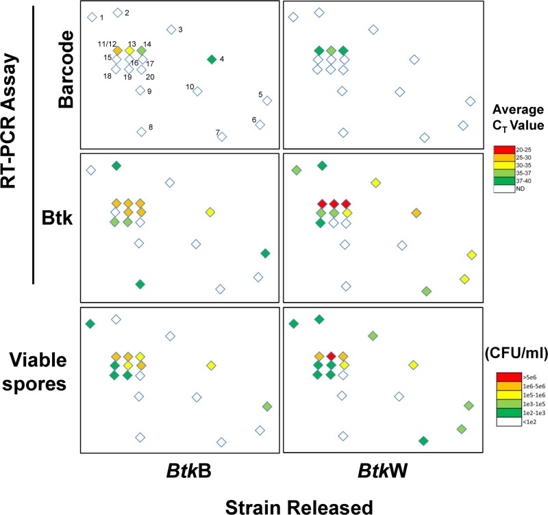
The results for the air sampling immediately following each of the release events are summarized in Fig. 3 (as analyzed by using DFU filter units). The CT values and CFU data are available as Table S1 in the supplemental material. The differences between the average CT values obtained for the barcode PCR assay and the B. thuringiensis subsp. kurstaki assay are probably due to the location of the PCR target on either chromosome (barcode) or a plasmid that is present in higher relative copy numbers (B. thuringiensis subsp. kurstaki). These results show unambiguous detection of WT B. thuringiensis subsp. kurstaki and barcoded B. thuringiensis subsp. kurstaki genome in both challenges by RT-PCR. The signals obtained by RT-PCR correlated with the total amount of viable material recovered from the DFUs. While both RT-PCR assays registered positive results in both challenges, the overall signal strength for the barcoded B. thuringiensis subsp. kurstaki assay was much lower than that for the WT B. thuringiensis subsp. kurstaki assay during the second challenge, with only one of the assays registering a CT value lower than 37, in contrast to the low CT values observed for the generic B. thuringiensis subsp. kurstaki PCR assay.
Fig 3.
Detection of barcoded strains in the field. Following the release, dry filter units were run for 8-h periods each day over a 12-day period, and the processed samples were subjected to PCR tests in triplicate. Resuspended material from the filters was also plated on culture medium to enumerate culturable material. Results are shown for the periods immediately following the release of barcoded (BtkB) and wild-type (BtkW) material.
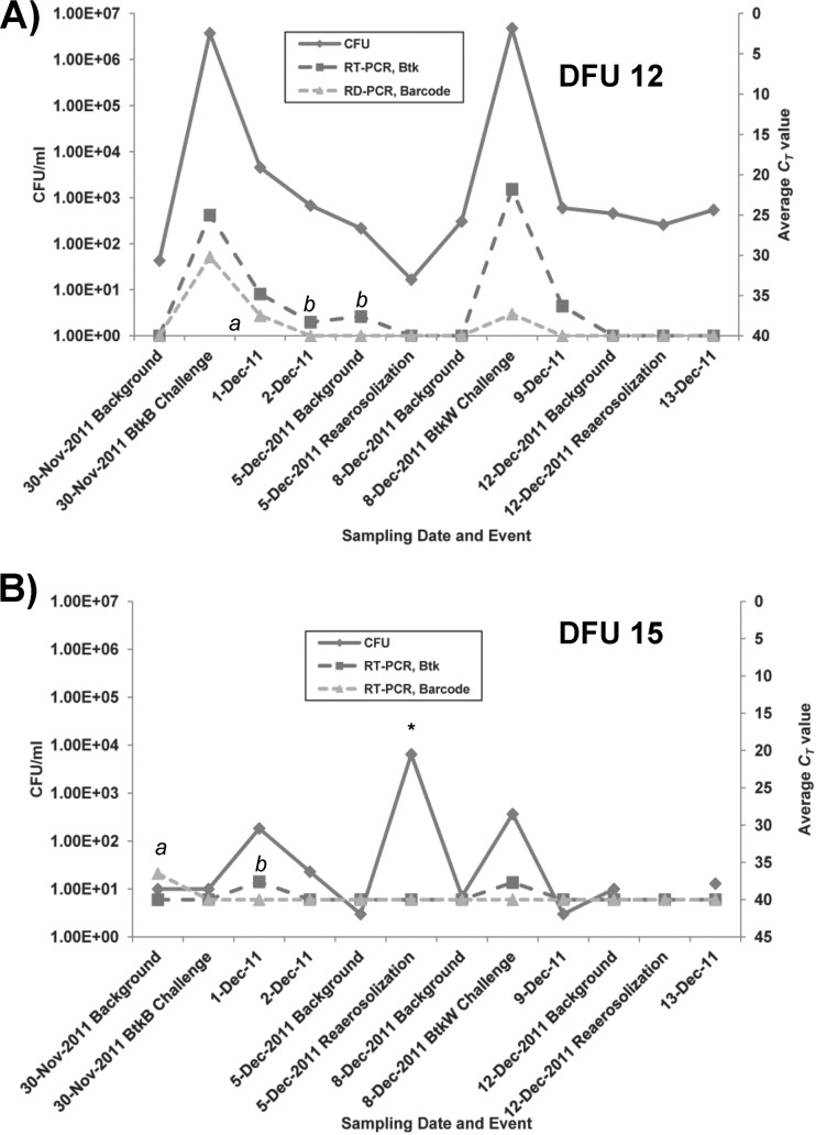
We monitored the ability to recover viable spores and generate RT-PCR signals over time following each release by activating each DFU for an 8-h period over subsequent days (Fig. 4). While the RT-PCR signals rapidly dropped to undetectable levels, viable material was recovered, though at levels near or below the detection limits for each of the assays. The complete CT values and detection data are found in the supplemental material. Immediately following the release, large amounts of viable material producing robust RT-PCR signals were recovered from several downwind DFUs (Fig. 3 and 4A). The amount of viable material dropped rapidly following each release, with an immediate 3-log drop on the day following the release and an approximately 1-log decrease per day thereafter. In contrast, very few, if any, viable spores were recovered from DFUs not directly downwind of the release point (Fig. 3 and 4B). DFU no. 15 registered a relatively large increase in culturable material on 5 December following an attempt to reaerosolize deposited barcoded B. thuringiensis subsp. kurstaki from the field (see below). This material exhibited colony morphologies that were easily distinguishable from that of B. thuringiensis subsp. kurstaki (data not shown) and therefore probably represented spores/cells of the environmental microbiome.
Fig 4.
Detection of B. thuringiensis subsp. kurstaki strains over time. Culture and RT-PCR results are shown from DFU no. 12 (A) and DFU no. 15 (B) (see Fig. 3 for locations of the DFUs on the test grid). a, one of three barcoded B. thuringiensis subsp. kurstaki assays was positive; b, one of three WT B. thuringiensis subsp. kurstaki assays was positive. The asterisk indicates that colony morphology was distinct from that of B. thuringiensis subsp. kurstaki.
The use of leaf blowers to disturb settled spores on the high grass of the test field on day 3 following the release did not result in any additional PCR detections. The failure of the leaf blowers to reaerosolize the spores is not surprising, given that the weather during the period of this test was not conducive to measurement of reaerosolization. The high humidity and noticeable amounts of early-morning dew resulted in significant wetting of the grassy areas surrounding the test field each morning. High humidity and surface moisture are known to reduce reaerosolization; both serve to increase adhesion between particles and surfaces (6, 8). Furthermore, wind speeds during the initial spore release were higher than expected, averaging 17 kph with occasional gusts up to 32 kph. Given that the milled dry powder released in this test had a mass mean diameter of 3.39 μm, the high wind speeds on the evening of the release likely resulted in the production of a long, sparse initial deposition area. While the amount of material reaerosolized does not scale directly with surface concentration, the concentration of reaerosolized material at any given location is a function of the surrounding surface concentration (19). If spores were reaerosolized in this experiment, the resultant concentrations were below the detection limits of the sampling techniques.
Surprisingly, on the eighth day of the outdoor field test, several collectors yielded weakly positive signals for the barcode assay following the release of WT B. thuringiensis subsp. kurstaki spores, which had been meant as negative-control release (Fig. 3 and 4A). We examined the possibility that the low-level barcoded B. thuringiensis subsp. kurstaki signal observed in the WT B. thuringiensis subsp. kurstaki release may have resulted from cross-reactivity of the PCR assay itself. Our laboratory results (2) showed no cross-reactivity between wild-type and barcoded strains under the assay conditions utilized in this study, which used 1 ng of purified genomic DNA and yielded CT values comparable to those obtained with samples derived from the barcoded B. thuringiensis subsp. kurstaki release. Furthermore, genomic DNA extracted from the WT B. thuringiensis subsp. kurstaki preparation utilized in this field test did not yield the barcoded B. thuringiensis subsp. kurstaki signal in real-time PCR assays (data not shown). We therefore believe it to be unlikely that the barcoded B. thuringiensis subsp. kurstaki signal observed during the WT B. thuringiensis subsp. kurstaki release was due to cross-reaction of the PCR assay with wild-type DNA.
As no PCR signal was observed for the barcoded B. thuringiensis subsp. kurstaki assay in the period between days 3 and 7, and low-level PCR activity began immediately following the wild-type release, the most likely possibility is that barcoded spores may have adsorbed to the intake ports of the DFUs during the initial release of barcoded B. thuringiensis subsp. kurstaki, which then may have been dislodged by the influx of WT B. thuringiensis subsp. kurstaki spores during the second challenge. Because neither the colony counts nor the PCR detection assay for WT B. thuringiensis subsp. kurstaki dropped to completely undetectable levels prior to the dissemination of WT B. thuringiensis subsp. kurstaki, we cannot definitively rule out the residual presence of barcoded B. thuringiensis subsp. kurstaki spores on surfaces of the detection equipment. Another possibility is that the surface of the Humvee may have been residually contaminated with barcoded B. thuringiensis subsp. kurstaki from the first test and that vibration during the test reaerosolized material adsorbed to the surface of the Humvee.
Our laboratory results show that the assays should be specific within the range of CT values observed in this study. However, because the construction of the first generation of barcoded spores utilized a small section of host genome sequence as a binding site for the reverse PCR primer, we cannot definitively rule out asymmetric amplification from wild-type DNA, leading to a spurious detection result. Therefore, modifications of the barcoded spore development strategy are under way to reduce the complexity of producing additional variants and to convert the embedded PCR assay from a SYBR green detection format to a TaqMan fluorescence resonance energy transfer (FRET)-based assay.
In conclusion, the present study and the data summarized here demonstrated that reaerosolization did occur in the ambient breeze tunnel. The outdoor test also demonstrated the need to thoroughly decontaminate all equipment used in outdoor field tests and showed that the current variant of the barcoded spore required changes in its construction. The insertion of genetic barcodes into biological simulants has great advantages for test and evaluation methods, and traceable unique biological materials can provide a better way to validate urban threat release models. In addition, the inclusion of test-specific barcodes introduces the possibility of the simultaneous sampling and detection of multiple strains in a single sample collection point. Use of genetically tagged BW simulants is predicted to simplify tracking and specific detection, which is critically needed for generating data to support diffusion models and reduce the test costs by allowing data to be collected from multiple tests with minimal interference from pre-existing spore populations.
Supplementary Material
ACKNOWLEDGMENTS
This study was made possible by ECBC internal funding. Creation of the barcoded strains was supported by the Defense Threat Reduction Agency project number CB3654 to H.S.G. and P.A.E.
Opinions presented in this work are those of the authors and do not represent the official policy of the Army, Department of Defense, or the U.S. Government. All information in this report has been cleared for public release.
Footnotes
Published ahead of print 21 September 2012
Supplemental material for this article may be found at http://aem.asm.org/.
REFERENCES
- 1. Anonymous 1999, posting date Bacillus thuringiensis. International Programme on Chemical Safety, United Nations Environment Programme and World Health Organization [Google Scholar]
- 2. Buckley P, et al. 2012. Genetic barcodes for improved environmental tracking of an anthrax simulant. Appl. Environ. Microbiol. 78:8272–8280 [DOI] [PMC free article] [PubMed] [Google Scholar]
- 3. Carrera M, Sagripanti JL. 2009. Artificial plasmid engineered to simulate multiple biological threat agents. Appl. Microbiol. Biotechnol. 81:1129–1139 [DOI] [PubMed] [Google Scholar]
- 4. Carrera M, Sagripanti JL. 2009. Non-infectious plasmid engineered to simulate multiple viral threat agents. J. Virol. Methods 159:29–33 [DOI] [PubMed] [Google Scholar]
- 5. Dando MR. 1999. Biohazard. Nature 400:632 [Google Scholar]
- 6. de Boer MP, de Boer PCT. 2007. Thermodynamics of capillary adhesion between rough surfaces. J. Colloid Interface Sci. 311:171–185 [DOI] [PubMed] [Google Scholar]
- 7. Farrell S, Halsall HB, Heineman WR. 2005. Bacillus globigii bugbeads: a model simulant of a bacterial spore. Anal. Chem. 77:549–555 [DOI] [PubMed] [Google Scholar]
- 8. Fecan F, Marticorena B, Bergametti G. 1999. Parameterisation of the increase of the aeolian erosion threshold wind friction velocity due to soil moisture for arid and semiarid areas. Ann. Geophys. 17:149–157 [Google Scholar]
- 9. Gibbons HS, et al. 2011. Genomic signatures of strain selection and enhancement in Bacillus atrophaeus var. globigii, a historical biowarfare simulant. PLoS One 6:e17836 doi:10.1371/journal.pone.0017836 [DOI] [PMC free article] [PubMed] [Google Scholar]
- 10. Greenberg DL, Busch JD, Keim P, Wagner DM. 2010. Identifying experimental surrogates for Bacillus anthracis spores: a review. Invest. Genet. 1:4. [DOI] [PMC free article] [PubMed] [Google Scholar]
- 11. Hayward AE, Marchetta JA, Hutton RS. 1946. Strain variation as a factor in the sporulating properties of the so-called Bacillus globigii. J. Bacteriol. 52:51–54 [DOI] [PubMed] [Google Scholar]
- 12. Human Health Surveillance Scientific Committee 1999. Human health surveillance during the aerial spraying for control of North American gypsy moth on southern Vancouver Island, British Columbia, 1999. Capital Health Region, Canada [Google Scholar]
- 13. Ibrahim MA, Griko N, Junker M, Bulla LA. 2010. Bacillus thuringiensis: a genomics and proteomics perspective. Bioeng. Bugs 1:31–50 [DOI] [PMC free article] [PubMed] [Google Scholar]
- 14. Jernigan DB, et al. 2002. Investigation of bioterrorism-related anthrax, United States, 2001: epidemiologic findings. Emerg. Infect. Dis. 8:1019–1028 [DOI] [PMC free article] [PubMed] [Google Scholar]
- 15. Kournikakis B, Ho J, Duncan S. 2010. Anthrax letters: personal exposure, building contamination, and effectiveness of immediate mitigation measures. J. Occup. Environ. Hyg. 7:71–79 [DOI] [PubMed] [Google Scholar]
- 16. Merrill L, Dunbar J, Richardson J, Kuske CR. 2006. Composition of bacillus species in aerosols from 11 U.S. cities. J. Forensic Sci. 51:559–565 [DOI] [PubMed] [Google Scholar]
- 17. Metalnikov S, Chorine V. 1929. On the infection of the gypsy moth and certain other insects with Bacillus thuringiensis. A preliminary report. Intern. Corn Borer Invest. Soc. Report. 2:60–61 [Google Scholar]
- 18. Nakamura LK. 1989. Taxonomic relationship of black-pigmented Bacillus subtilis strains and a proposal for Bacillus atrophaeus sp. nov. Int. J. Syst. Bacteriol. 39:295–300 [Google Scholar]
- 19. Nicholson KW. 1988. A review of particle resuspension. Atmosph. Environ. 22:2639–2651 [Google Scholar]
- 20. Regis E. 1999. The biology of doom: the history of America's secret germ warfare project. Henry Holt, New York, NY [Google Scholar]
- 21. Sinclair R, Boone SA, Greenberg D, Keim P, Gerba CP. 2008. Persistence of category A select agents in the environment. Appl. Environ. Microbiol. 74:555–563 [DOI] [PMC free article] [PubMed] [Google Scholar]
- 22. Turnbull PC, Lindeque PM, Le Roux J, Bennett AM, Parks SR. 1998. Airborne movement of anthrax spores from carcass sites in the Etosha National Park, Namibia. J. Appl. Microbiol. 84:667–676 [DOI] [PubMed] [Google Scholar]
- 23. U.S. Department of Agriculture 2008. Gypsy moth management in the United States: a cooperative approach. U.S. Department of Agriculture, Newtown Square, PA [Google Scholar]
- 24. U.S. Department of Justice 2010. Amerithrax investigative summary. U.S. Department of Justice, Washington, DC [Google Scholar]
- 25. Valadares de Amorim G, Whittome B, Shore B, Levin DB. 2001. Identification of Bacillus thuringiensis subsp. kurstaki strain HD1-like bacteria from environmental and human samples after aerial spraying of Victoria, British Columbia, Canada, with Foray 48B. Appl. Environ. Microbiol. 67:1035–1043 [DOI] [PMC free article] [PubMed] [Google Scholar]
- 26. Van Cuyk S, et al. 2011. Persistence of Bacillus thuringiensis subsp. kurstaki in urban environments following spraying. Appl. Environ. Microbiol. 77:7954–7961 [DOI] [PMC free article] [PubMed] [Google Scholar]
- 27. Van Cuyk S, et al. 2009. Bacillus thuringiensis var. kurstaki (Btk) agent fate characterization. 2008 final report. LA-UR-09-05794 Los Alamos National Laboratory, Los Alamos, NM [Google Scholar]
Associated Data
This section collects any data citations, data availability statements, or supplementary materials included in this article.


