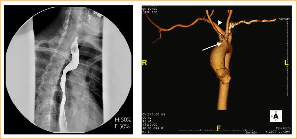
A 42-year-old woman was seen for evaluation of solid dysphagia immediately after the initial swallow. Her symptoms were tolerable with dietary modification. Her medical history included hypothyroidism, bilateral thumb surgery as a child, and an appendectomy. A barium esophagram (left panel)

showed posterior compression of the thoracic esophagus; a 13-mm barium tablet was held up at this level with reproduction of her symptoms. Reconstructed 3-D images (right panel) show an aberrant right subclavian artery (arrowhead) originating from the aortic arch distal to a common origin of the carotid arteries (arrow), the truncus bicaroticus, and the left subclavian artery.
In 1794, Bayford coined the term dysphagia lusus naturae (Latin for freak or jest of nature) to describe dysphagia caused by extrinsic compression from an aberrant right subclavian artery. This patient's congenital hand deformities illustrate the embryological origin of this vascular anomaly, which is the most common congenital aortic root abnormality and has a prevalence ranging from 0.5% to 1.8% in the general population.1 Barium esophagram is often suggestive, showing oblique compression of the esophagus running from left to right and inferior to superior. Noninvasive angiography (either computed tomography or magnetic resonance imaging) is essential for confirmation of the diagnosis and surgical planning. Surgical vascular reconstruction is reserved for highly symptomatic patients or those who do not respond to dietary modification.
This patient's symptoms were mild and controlled with dietary modifications. Thus, reconstructive vascular surgery was not recommended. She had no further progression of her symptoms at 6 month follow-up.
Reference
- 1.Levitt B., Richter J.E. Dysphagia lusoria: a comprehensive review. Dis Esophagus. 2007;20(6):455–460. doi: 10.1111/j.1442-2050.2007.00787.x. [DOI] [PubMed] [Google Scholar]


