Abstract
Objective
To improve the ability of primary care physicians to recognize the mechanisms and common presentations of low-velocity Lisfranc injuries (LFIs) and to impart an improved understanding of the role of imaging and principles of primary care in low-velocity LFIs.
Sources of information
A MEDLINE literature review was performed and the results were summarized, reviewing anatomy and mechanisms, clinical and imaging-based diagnoses, and management principles in the primary care setting.
Main message
Low-velocity LFIs result from various mechanisms and can have very subtle findings on clinical examination and imaging. A high degree of suspicion and caution are warranted when managing this type of injury.
Conclusion
Although potentially devastating if missed, if a few treatment principles for low-velocity LFIs are applied from the initial presentation onward, outcomes from this injury can be optimized.
Case
A 30-year-old woman presents to the emergency department after a fall from a height of roughly 1.2 m off her back deck. The patient landed awkwardly on her right foot. She was able to walk back to the house, but shortly thereafter was unable to walk on her right foot. The foot became progressively swollen and now she cannot fit it into her shoe. She must have her foot elevated or the pain and swelling are dramatically worsened.
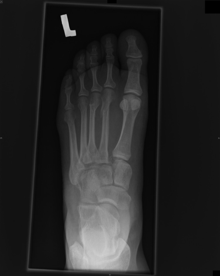
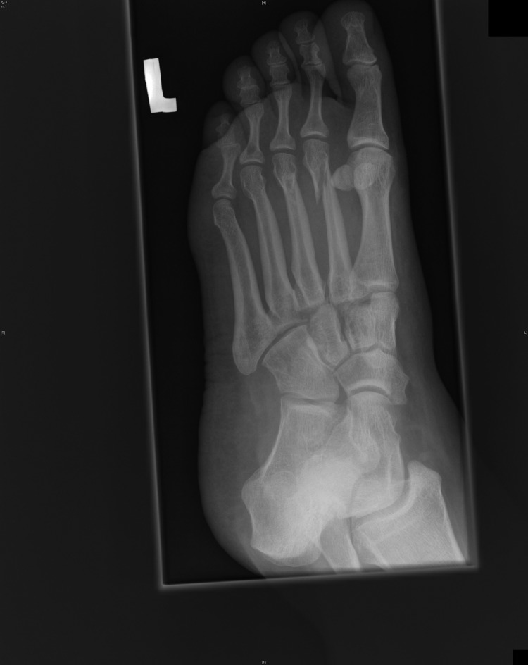
Examination of her right foot reveals that the foot and ankle are roughly normally aligned. There is a large amount of swelling in the area of her midfoot and forefoot. She does not allow examination of her midfoot because of pain that she rates as 8 out of 10 starting in her midfoot and shooting into her first, second, and third toes. She is uncomfortable on forefoot examination of the first, second, and third toes, secondary to referred pain up her foot to her ankle. She refuses to bear weight on the affected foot. Initial radiographs are shown in Figures 1 to 3.
Figure 1.
Anteroposterior view of the injured foot: Note the subtle shift of the second metatarsal base away from the medial cuboid bone, and the bony comminution that sits in the resultant space. The second metatarsal shaft fracture is a not-uncommon concomitant injury with this mechanism.
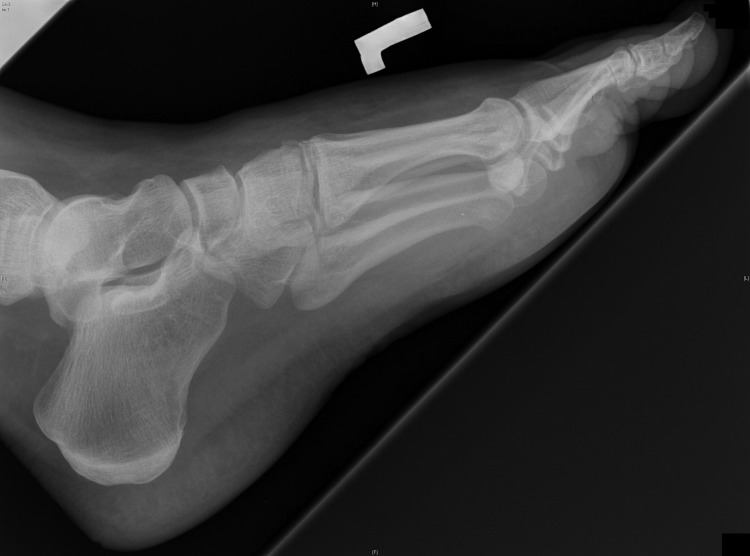
Figure 3.
Lateral view of the injured foot: Note the subtle shift of the second metatarsal base away from the medial cuboid bone, and the bony comminution that sits in the resultant space. The second metatarsal shaft fracture is a not-uncommon concomitant injury with this mechanism.
Sources of information
A MEDLINE literature review was performed and the results were summarized, reviewing anatomy and mechanisms, clinical and imaging-based diagnoses, and management principles for Lisfranc injury (LFI) in the primary care setting.
Main message
Substantial LFI, or tarsometatarsal joint injury, has been increasing in incidence.1 While representing a rare entity in the general population (0.2% of all orthopedic injuries),2 the injury is commonly encountered in certain subgroups; one case series documented an incidence of midfoot sprain in collegiate football players of 4% per year.3 Lisfranc injuries typically occur by 2 contrasting mechanisms. High-velocity LFIs typically involve dramatic mechanisms including high-velocity motor vehicle accidents and falls from a height. The forces involved often result in comminuted and displaced fractures, visible midfoot swelling, and ecchymoses; high-velocity LFIs rarely present a diagnostic dilemma.4 These injuries can represent a surgical emergency, as the neurovascular supply to the midfoot and forefoot might be in jeopardy, and disruption of the dorsalis pedis artery and soft-tissue swelling might result in development of compartment syndrome.5
Low-velocity LFIs are often caused by vague, seemingly trivial—although reproducible—mechanisms of injury such as walking off of a curb in an urban environment.2 Because of the trivial nature of the inciting trauma, low-velocity LFIs often present in a similar fashion to less substantial soft-tissue sprains to primary care physicians either in emergency departments or ambulatory clinics. Although exact rates are difficult to estimate, the subtle nature of the injury results in conservative estimates of missed diagnosis in 20% to 40% of cases,1 with some case series citing rates of missed injuries of up to 50%.6 Authors have reported LFI to be the injury most commonly missed in the emergency department.7,8
Anatomy
The tarsometatarsal, or Lisfranc joint—named for the Napoleonic surgeon who described osteotomy-free amputation of the forefoot through this articulation9—is composed of 3 joint cavities: the first, second, and third through fifth tarsometatarsal joints. Stability is imparted both through bony geometry and strong ligamentous complexes. The arch configuration of the joint complex and recessed second metatarsal base provide bony stability, while the second through fifth rays are bridged anteroposteriorly by dorsal and plantar tarsometatarsal ligaments, as well as laterally by dorsal, interosseous, and plantar intermetatarsal ligaments.
The first and second rays have unique ligamentous anatomy wherein no intermetatarsal ligaments exist, but extreme strength is imparted by dorsal, interosseous, and plantar bundles of ligament binding the lateral aspect of the medial cuneiform bone with the medial head of the second metatarsal bone—the Lisfranc ligamentous complex.
Injury to the Lisfranc ligamentous complex proceeds sequentially in a dorsal-plantar direction6 with increasing instability of the injury or failure of each bundle. Injured metatarsals “fall away” or displace laterally and plantarward (ie, traumatic flatfoot deformity) and account for classic radiographic findings. The intrinsic bony architecture of the midfoot when not bearing weight, however, has the ability to hold the foot in a relatively correct alignment. This can hide the deformity and mask the degree of soft-tissue or bony disruption on non–weight-bearing imaging, as discussed below.
Misdiagnosis and its consequences
This intrinsic midfoot stability leads to underestimation of injury and missed diagnoses in up to 20% to 50% of cases.1,6 In untreated cases following missed initial diagnoses, midfoot instability progresses until the patient develops a painful, rigid planovalgus deformity and rapidly progressive osteoarthritis.10 This clinical entity is often refractory to surgical management and, in most cases, leads to chronic pain, losses in working productivity, and large compensation claims.11 This is in contrast to the relatively acceptable outcomes12–14 that a patient can expect if timely diagnosis and surgical stabilization are achieved in less than 4 to 6 weeks. It is for this reason that medicolegal claims are also not uncommon when LFIs are missed.15
Clinical diagnosis
Lisfranc injuries are typically the result of 1 of 2 mechanisms: 1) a high-velocity mechanism such as motor vehicle trauma or 2) a low-velocity mechanism such as twisting or stepping off of a curb (dorsal fold-over injury).2 While high-velocity injuries can be missed, usually owing to a distracting injury from polytrauma, the focus of this article is to address the latter mechanism. Any low-energy injury resulting in an unanticipated twisting force on the stationary foot can result in insufficiency or complete failure of the Lisfranc joint, resulting in an unstable LFI. Typically, patients will present within 24 to 48 hours of what might have seemed to be a relatively innocuous injury.
On examination, the foot typically is swollen, particularly dorsally,2 with a dorsal focus of ecchymosis. Swelling typically becomes more diffuse and ecchymosis becomes apparent at the plantar aspect in the days following injury.9 The area of maximal tenderness usually corresponds to the dorsal-medial midfoot region, although some pain referral into the forefoot is also relatively common with attempted manipulation of the midfoot. Any pain noted in the area of the fifth metatarsal or hindfoot should be examined separately to rule out concomitant injuries.
The patient is typically unwilling or unable to effectively bear weight on the affected foot, although he or she will sometimes be able to offload the midfoot in a manner that allows antalgic or nonphysiologic weight-bearing. When forced to bear weight or walk normally, however, patients will be unable to do so. A distal neurovascular examination is essential to triaging any injury to the foot and ankle, although the findings are typically normal. Signs and symptoms of compartment syndrome should be assessed (ie, pain beyond what should be anticipated on assessment, pain on passive stretch, overly swollen foot) but are usually not present in this setting. Examination of the ankle and lower limb does not typically add any relevant data, but might reveal an associated injury at the level of the ankle and must be part of any assessment.
Radiography and computed tomography
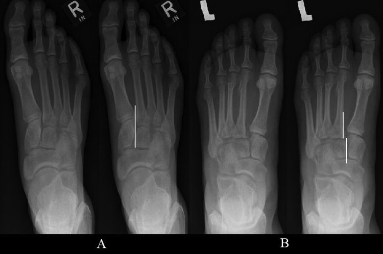
In the past, diagnostic imaging workup was initiated with plain-film radiographic examination including anteroposterior, lateral, and 30° internal oblique images of the affected foot.16,17 The cortex of the second metatarsal should form a line continuous with that of the medial cuneiform, and the intermetatarsal joint space should align with the intercuneiform joint space, measuring less than 2 mm transversely (Figure 4). On oblique films, the medial border of the third metatarsal and lateral cuneiform, and the fourth metatarsal and cuboid, should be aligned (Figure 5).
Figure 4.
Relationship of the metatarsal (MT) bones: A) The anteroposterior view of the foot displays the relationship of the first and second MTs to their corresponding medial and intermediate cuneiform bones. Specifically, the medial border of the second MT should align with the medial border of the intermediate cuneiform. B) The interruption of the second MT–intermediate cuneiform line is noted, signifying an unstable Lisfranc injury.
Figure 5.
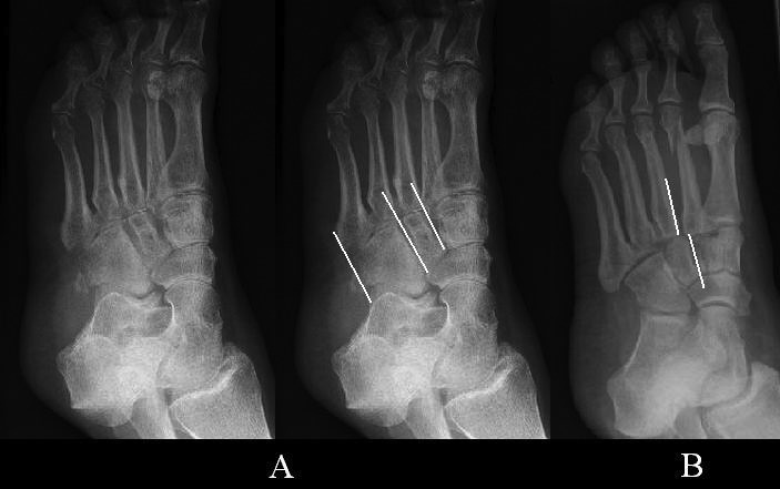
Contours of the tarsometatarsal joints: A) The oblique radiograph of the foot displays the normal contours (demarcated with white lines) of the third to fifth tarsometatarsal joints. These should be smooth and uninterrupted. B) There is an interruption of the normally smooth line between the third metatarsal and the lateral cuneiform bone. This signifies an unstable Lisfranc injury.
Abnormal alignment and the presence of the fleck sign—avulsion at the ligamentous insertion—are sufficient to confirm midfoot instability, and the patient should be referred for prompt surgical management (Figure 6). Weight-bearing radiographs are preferred, although they are notoriously difficult to obtain owing to patient discomfort. In one recent case series, not a single patient was able to comply with instructions to obtain adequate weight-bearing films owing to pain.18
Figure 6.
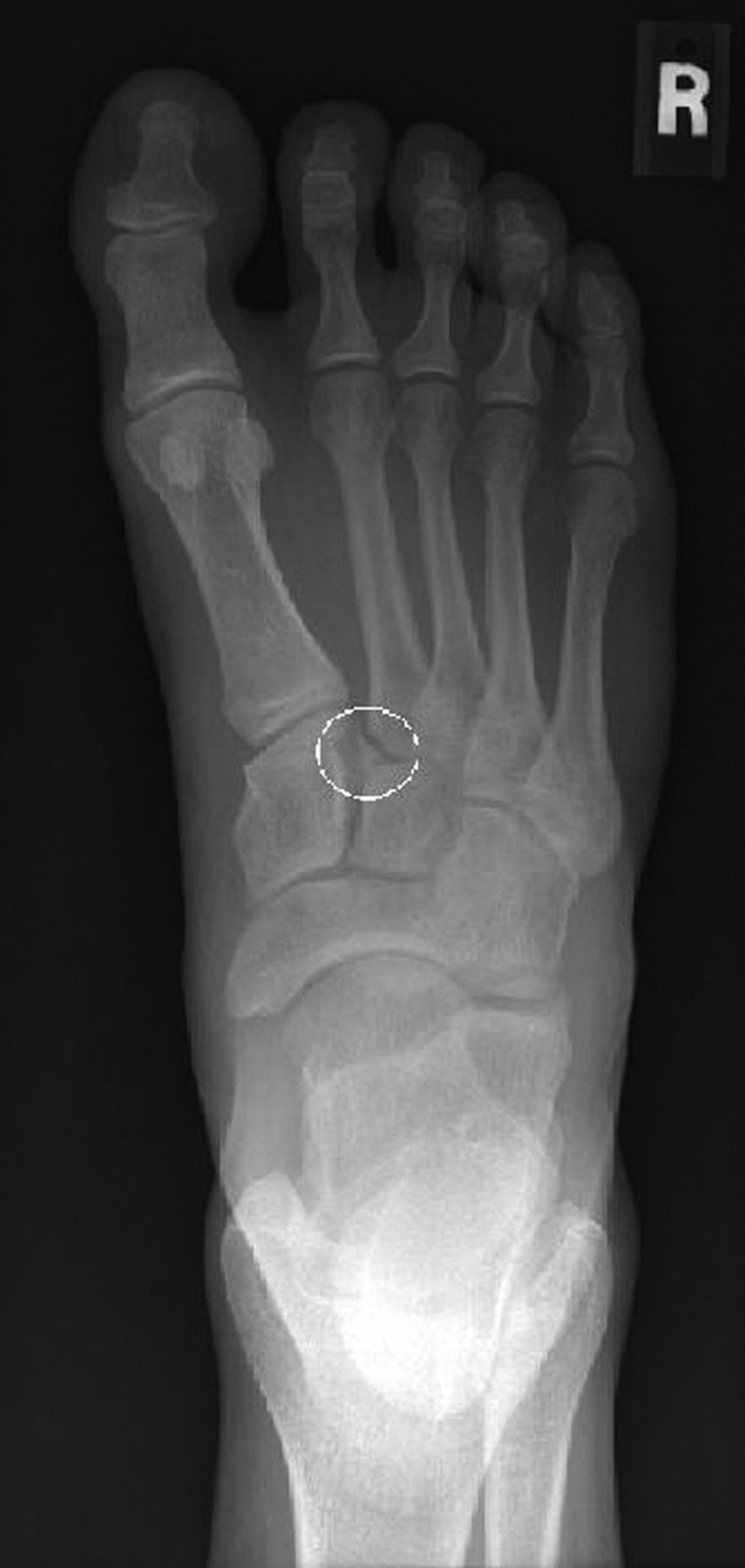
The fleck sign: Avulsion fracture of the base of the second metatarsal bone.
As an alternative to weight-bearing radiographs, assessment with fluoroscopy during manipulation under anesthesia (ie, “stress views”) has been advocated in cases of heightened clinical suspicion.19 This technique has the added benefit of allowing prompt progression to operative management in cases with positive or equivocal results. The risk conferred by the 10% to 20% of cases found to be normal intraoperatively was thought to be justified by the grave consequences of a missed diagnosis.16
Computed tomography (CT) adds sensitivity in assessing the integrity of bony structures, and vastly improves 3-dimensional spatial resolution in the modern era of multiplanar reconstruction. A recent case series using a multidetector CT protocol found 24% of Lisfranc fractures evident on CT were occult on radiographs.20 Further, CT adds sensitivity in assessing other structures of the midfoot; in the same case series, the sensitivity of radiography in detecting fractures of the other tarsal bones when compared with CT was from 24% to 33%.
These findings have led to the recommendation advocated by these authors—that in the presence of a suspicious mechanism of injury and clinical examination (even if radiographic examination has been performed and results interpreted as normal), CT should be considered the primary imaging modality and be performed at the time of presentation or as an urgent outpatient examination, with an appointment arranged before discharge.
Future role of magnetic resonance imaging (MRI) and ultrasound
A relatively extensive body of work has accumulated in recent years evaluating the role of MRI in LFIs. In the past, MRI has been validated as providing excellent correlation with anatomy on cadaveric series.21 More recently, MRI has been reported to have a sensitivity of up to 94% in demonstrating ligamentous disruption and from 70% to 90% accuracy in grading ligamentous injury in the absence of frank disruption as correlated with intraoperative findings.15,16
The potential role of ultrasound is under investigation. A recent case series has demonstrated that sonographic features, including widened first cuneiform to second metatarsal distance, widening with weight-bearing, and nonvisualization of the dorsal bundle of the Lisfranc complex, might prove successful in predicting unstable joints requiring intervention.21 While sonographic assessment at presentation and on follow-up is currently being employed in certain centres, this method has yet to be validated in larger case series.
Treatment
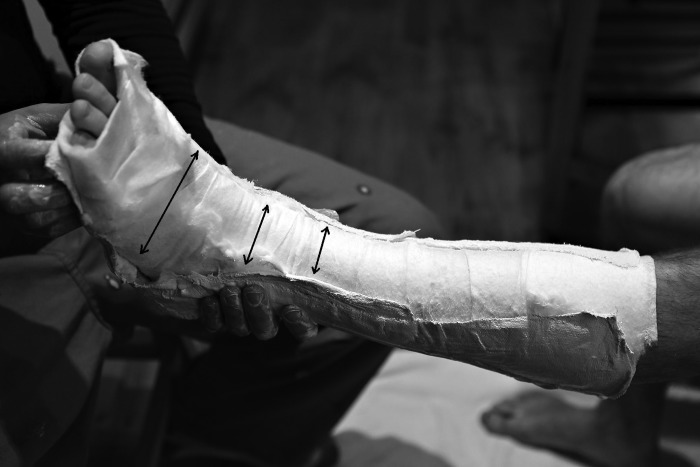
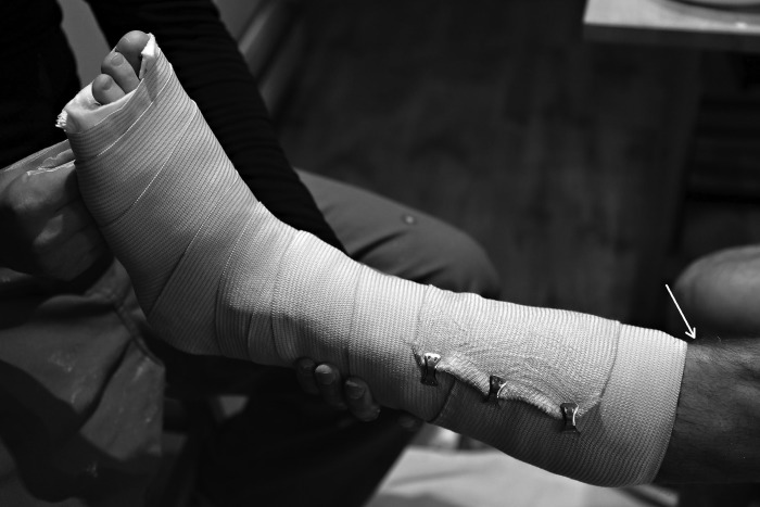
Once the diagnosis of LFI has been made, the patient must not bear weight on the affected foot. The foot should be placed in a below-knee, posterior plaster or fibreglass slab. This slab should be applied over generously and meticulously applied cotton under-cast padding, ensuring there are no wrinkles in the padding and no bare spots of skin. The slab should extend from the proximal calf to just beyond the toes, with the ankle in a position that is comfortable for the patient (Figure 7). The lower leg can then be overwrapped loosely with an elastic bandage (Figure 8). It is critical that this splint be made snug but not constrictive. The plaster or fibreglass should never touch anteriorly, so that an open corridor remains to allow for swelling. Patients should be referred urgently (ie, in < 7 days) to an orthopedic surgeon comfortable and experienced in managing the traumatized foot and ankle. Appropriate imaging should be made available to the patient and surgeon to prevent wasting of resources and further exposure to ionizing radiation.
Figure 7.
The 3-sided splint is applied in the following manner: All of the bony prominences (including the heel) are generously padded with cotton undercast padding. Then the plaster slabs are applied so that they never touch anteriorly along the distal tibia, and so that the foot, except for the plantar portion of the slab, is left relatively uncovered by plaster (black arrows). These features allow for the substantial swelling that can accompany foot trauma and Lisfranc injuries.
Figure 8.
Applying the final splint: The bandage covers from distal to the metatarsophalangeal joints (leaving the tips of the toes exposed for examination) to just distal to the tibial tubercle (white arrow) at the level of the calf.
Discharge instructions should include warnings about the symptoms of compartment syndrome of the foot. Patients should be told that if symptoms resembling compartment syndrome occur, they should return to the emergency department as soon as possible. Emergency department management in such cases would include loosening of the splint and emergency referral to an orthopedic surgeon. Appropriate analgesia is required, as well as routine instructions to not bear weight on the injured foot, apply ice if available, and elevate the foot whenever possible to minimize swelling.
Conclusion
Lisfranc injuries can be devastating. If missed, they can lead to substantial long-term morbidity and poor outcomes. If diagnosed and definitively treated in a timely fashion, acceptable outcomes can usually be expected. This highlights a need for prompt and efficient diagnosis in the emergency department. An approach to suspected LFI is summarized in Box 1.
Box 1. Summary of an approach to suspected Lisfranc injury.
-
History
-High-risk mechanism in a low-energy foot trauma setting (eg, low-pressure twisting, fall on plantar-flexed foot)
-
Physical examination
-Area of maximal tenderness around the medial midfoot
-Swollen midfoot
-Dorsal (acutely) and plantar (subacutely) bruising at the level of the midfoot
-Associated injuries to the foot and ankle should be ruled out
-
Imaging
-Anteroposterior radiograph: relationship of the first and second metatarsal bones to medial and intermediate cuneiform bones
-Oblique radiograph: relationship of the third metatarsal to the lateral cuneiform, and fourth metatarsal to the cuboid bone
-Importance of computed tomography if the history is appropriate and there is clinical suspicion, even in the context of normal radiographic examination findings. Magnetic resonance imaging and ultrasound performed depending on the centre
Urgent referral (in < 7 days) to an orthopedic surgeon; foot should not bear weight and should be encased in a posterior or 3-sided slab. Patients should be educated about the risk of developing compartment syndrome and directed to return to the emergency room should it arise
Figure 2.
Oblique view of the injured foot: Note the subtle shift of the second metatarsal base away from the medial cuboid bone, and the bony comminution that sits in the resultant space. The second metatarsal shaft fracture is a not-uncommon concomitant injury with this mechanism.
KEY POINTS
Low-velocity Lisfranc injuries (LFIs) are often caused by vague, seemingly trivial—although reproducible—mechanisms of injury such as walking off of a curb in an urban environment. Once the diagnosis of LFI has been made, the patient must not bear weight on the affected foot. The foot should be placed in a 3-sided, below-knee, posterior plaster or fibreglass slab. Patients should be told that if symptoms resembling compartment syndrome occur, they should return to the emergency department as soon as possible. If missed, LFIs can lead to substantial long-term morbidity and poor outcomes. If diagnosed and definitively treated in a timely fashion, acceptable outcomes can usually be expected.
Footnotes
This article has been peer reviewed.
This article is eligible for Mainpro-M1 credits. To earn credits, go to www.cfp.ca and click on the Mainpro link.
La traduction en français de cet article se trouve à www.cfp.ca dans la table des matières du numéro de novembre 2012 à la page e620.
Contributors
Dr D.J. Mayich and Dr M.S. Mayich were involved in the literature review and manuscript writing. Dr Daniels acted in an advisory role, assisting in the literature review, manuscript preparation, and editing of the manuscript.
Competing interests
None declared
References
- 1.Rammelt S, Schneiders W, Schikore H, Holch M, Heineck J, Zwipp H. Primary open reduction and fixation compared with delayed corrective arthrodesis in the treatment of tarsometarsal (Lisfranc) fracture dislocation. J Bone Joint Surg Br. 2008;90(11):1499–506. doi: 10.1302/0301-620X.90B11.20695. [DOI] [PubMed] [Google Scholar]
- 2.Sands AK, Grose A. Lisfranc injuries. Injury. 2004;35(Suppl 2):SB71–6. doi: 10.1016/j.injury.2004.07.014. [DOI] [PubMed] [Google Scholar]
- 3.Meyer SA, Callaghan JJ, Albright JP, Crowley ET, Powell JW. Midfoot sprains in collegiate football players. Am J Sports Med. 1994;22(3):392–401. doi: 10.1177/036354659402200316. [DOI] [PubMed] [Google Scholar]
- 4.Hardcastle PH, Reschauer R, Kutscha-Lissberg E, Schoffmann W. Injuries to the tarsometatarsal joint. Incidence, classification, and treatment. J Bone Joint Surg Br. 1982;64(3):349–56. doi: 10.1302/0301-620X.64B3.7096403. [DOI] [PubMed] [Google Scholar]
- 5.Lattermann C, Goldstein JL, Wukich DK, Lee S, Bach BR., Jr Practical management of Lisfranc injuries in athletes. Clin J Sport Med. 2007;17(4):311–5. doi: 10.1097/JSM.0b013e31811ed0ba. [DOI] [PubMed] [Google Scholar]
- 6.Nunley JA, Vertullo CJ. Classification, investigation, and management of midfoot sprains: Lisfranc injuries in the athlete. Am J Sports Med. 2002;30(6):871–8. doi: 10.1177/03635465020300061901. [DOI] [PubMed] [Google Scholar]
- 7.Preidler KW, Peicha G, Lajtai G, Seibert FJ, Fock C, Szolar DM, et al. Conventional radiography, CT, and MR imaging in patients with hyperflexion injuries of the foot: diagnostic accuracy in the detection of bony and ligamentous changes. AJR Am J Roentgenol. 1999;173(6):1673–7. doi: 10.2214/ajr.173.6.10584818. [DOI] [PubMed] [Google Scholar]
- 8.Englanoff G, Anglin D, Hutson HR. Lisfranc fracture-dislocation: a frequently missed diagnosis in the emergency department. Ann Emerg Med. 1995;26(2):229–33. doi: 10.1016/s0196-0644(95)70156-7. [DOI] [PubMed] [Google Scholar]
- 9.Hunter TB, Peltier LF, Lund PJ. Radiologic history exhibit. Musculoskeletal eponyms: who are those guys? Radiographics. 2000;20(3):819–36. doi: 10.1148/radiographics.20.3.g00ma20819. [DOI] [PubMed] [Google Scholar]
- 10.Crim J. MR imaging evaluation of subtle Lisfranc injuries: the midfoot sprain. Magn Reson Imaging Clin N Am. 2008;16(1):19–27. doi: 10.1016/j.mric.2008.02.007. [DOI] [PubMed] [Google Scholar]
- 11.Calder JD, Whitehouse SL, Saxby TL. Results of isolated Lisfranc injuries and the effect of compensation claims. J Bone Joint Surg Br. 2004;86(4):527–30. [PubMed] [Google Scholar]
- 12.Harper MC. Fractures and fracture-dislocations of the tarsometatarsal joint [Comment] J Bone Joint Surg Am. 1989;71(6):952–3. [PubMed] [Google Scholar]
- 13.Myerson MS, Fisher RT, Burgess AR, Kenzora JE. Fracture dislocations of the tarsometatarsal joints: end results correlated with pathology and treatment. Foot Ankle. 1986;6(5):225–42. doi: 10.1177/107110078600600504. [DOI] [PubMed] [Google Scholar]
- 14.Henning JA, Jones CB, Sietsema DL, Bohay DR, Anderson JG. Open reduction internal fixation versus primary arthrodesis for Lisfranc injuries: a prospective randomized study. Foot Ankle Int. 2009;30(10):913–22. doi: 10.3113/FAI.2009.0913. [DOI] [PubMed] [Google Scholar]
- 15.Gupta RT, Wadhwa RP, Learch TJ, Herwick SM. Lisfranc injury: imaging findings for this important but often-missed diagnosis. Curr Probl Diagn Radiol. 2008;37(3):115–26. doi: 10.1067/j.cpradiol.2007.08.012. [DOI] [PubMed] [Google Scholar]
- 16.Macmahon PJ, Dheer S, Raikin SM, Elias I, Morrison WB, Kavanagh EC, et al. MRI of injuries to the first interosseous cuneometatarsal (Lisfranc) ligament. Skeletal Radiol. 2009;38(3):255–60. doi: 10.1007/s00256-008-0613-6. Epub 2008 Dec 2. [DOI] [PubMed] [Google Scholar]
- 17.Hatem SF. Imaging of Lisfranc injury and midfoot sprain. Radiol Clin North Am. 2008;46(6):1045–60. vi. doi: 10.1016/j.rcl.2008.09.003. [DOI] [PubMed] [Google Scholar]
- 18.Raikin SM, Elias I, Dheer S, Besser MP, Morrison WB, Zoga AC. Prediction of midfoot instability in the subtle Lisfranc injury. Comparison of magnetic resonance imaging with intraoperative findings. J Bone Joint Surg Am. 2009;91(4):892–9. doi: 10.2106/JBJS.H.01075. [DOI] [PubMed] [Google Scholar]
- 19.Coss HS, Manos RE, Buoncristiani A, Mills WJ. Abduction stress and AP weight-bearing radiography of purely ligamentous injury in the tarsometatarsal joint. Foot Ankle Int. 1998;19(8):537–41. doi: 10.1177/107110079801900806. [DOI] [PubMed] [Google Scholar]
- 20.Haapamaki VV, Kiuru MJ, Koskinen SK. Ankle and foot injuries: analysis of MDCT findings. AJR Am J Roentgenol. 2004;183(3):615–22. doi: 10.2214/ajr.183.3.1830615. [DOI] [PubMed] [Google Scholar]
- 21.Woodward S, Jacobson JA, Femino JE, Morag Y, Fessell DP, Dong Q. Sonographic evaluation of Lisfranc ligament injuries. J Ultrasound Med. 2009;28(3):351–7. doi: 10.7863/jum.2009.28.3.351. [DOI] [PubMed] [Google Scholar]


