Abstract
Acute swelling of the parotid gland after general anesthesia (commonly known as anesthesia mumps or acute postoperative sialadenitis) is a rare but declared complication of anesthesia. The etiology is not clear, but some possible causes such as obstruction of glandular excretory ducts caused by patient position and increase in the viscosity of the saliva because of acute dehydratation and/or medications like atropin have been proposed. We report a swelling in the left preauricular and postauricular region extending to the angle of the mandibule in a 35-year-old patient after left lateral decubitus position for laparoscopic nephrectomy.
Keywords: Anesthesia, anesthesia mumps, salivary gland swelling
INTRODUCTION
Acute transient swelling of one or more salivary glands is a rare entity in anesthesia practice. When seen in the parotid gland, it is referred to as “anesthesia mumps” in the anesthesiology literature. While it can be both unilateral and bilateral, it is almost always painless and it resolves spontaneously over a period of hours or days, without leaving any sequela.[1–3] In this article, we present a unilateral parotid gland swelling after laparoscopic simple nephrectomy in the lateral decubitis position under general anesthesia.
CASE REPORT
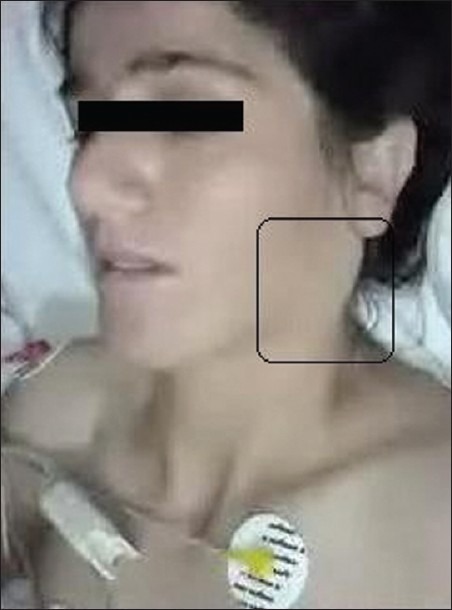
A 35-year-old, 60 kg female patient, with American Society of Anesthesiologists (ASA) physical status class II was scheduled for right laparoscopic simple nephrectomy for renal atrophy under general anesthesia. Her medical history and preoperative examination revealed no specific findings except for benign arterial hypertension. Upon arrival in the operating theater, an intravenous access was established and standard anesthesia monitoring (electrocardiogram, peripheral oxygen saturation, noninvasive blood pressure) was instituted. Before induction of anesthesia, midazolam 2 mg iv was administered as premedication. General anesthesia was induced with iv fentanyl 2 mcg/kg, propofol 2.5 mg/kg and vecuronium 0.1 mg/kg. After 3 min with mask ventilation, the patient was intubated with a 7.5 mm internal diameter (ID) tracheal tube and mechanical ventilation was performed with a tidal volume (VT) of 8 mL/kg, respiratory rate (RR) 10/min and positive end–expiratory pressure (PEEP) 7 cmH2O with volume-controlled ventilation (VCV) (Dräger Julian Plus Vitara 8060, ARRL- 0127, Germany). Fifteen minutes after endotracheal intubation, the patient was placed in the left lateral decubitus position. Her head was supported with a semi-soft bandage head ring used commonly for such positions under general anesthesia. Anesthesia was maintained with oxygen 1 L/min, air 1 L/min and sevoflurane with 1.7% end-tidal concentration, while the muscle relaxant and fentanyl were administered intermittently. During the operation, fluid replacement was maintained by crystaloid and colloids and monitored with 0.5–1 mL/kg urine output. The duration of the surgery was about 3.5 h, with a total blood loss of 650 mL. At the end of surgery, the patient was turned to the supine position and extubated. A swelling on the preauricular and postauricular region of the down-facing (left) side of her head extending to the angle of the mandibula was noted [Figure 1]. There were no local crepitations by palpation, no reddish lesions or a local increase in temperature over the swollen area. Postoperatively, the patient was transferred to the post-anesthesia care unit (PACU) for overnight postoperative care. In the PACU, portable ultrasonographic examination of the parotid gland showed hyperechogenic view and slight parotid duct dilatation on the left parotid gland compared with the right gland. The patient did not complain about pain after recovery. Eight milligrams of Dexamethasone (iv) and a nonsteroidal antiinflammatory drug (Diclofenac sodium 75 mg, im) were administrated. The size of the swelling diminished, and it disappeared after 24 h. The patient was discharged from the hospital on the second postoperative day.
Figure 1.

The similar swelling at the left parotis gland
DISCUSSION
Swelling of the salivary glands, either bilateral or unilateral, can be observed during surgery or in the early postoperative period, and spontaneously resolve over a period of a few hours/days, with no sequelae.[1–4] The etiology is yet to be understood, although several theories were put forward. The retrograde flow of air through the Stenon's orifice during straining and coughing under anesthesia, retention of secretions causing occlusion of the salivary ducts, perioperative use of various drugs like atropine, succinylcholine and morphine, and systemic dehydration are the most commonly accused factors.[3,5–8]
Pirat et al. reported bilateral submandibular salivary gland swelling following regional anesthesia for hip replacement. They hypothesized that it could be due to the sympathetic stimulation caused by perioperative use of vasopressors or hypovolemia.[4]
The other possible explanation is glandular ischemia and, as a result, ischemic sialadenitis. Compression of the arterial or venous vasculature may deteroriate the blood supply of the gland, which may lead to ischemia and, consecutively, to ischemic sialadenitis. Ischemic sialadenitis presents as a painful swelling usually unilateral in origin. Red hemorrhagic spots on the skin over the swollen area frequently associates the clinical picture.[3,9]
The position of the head during a prolonged surgical prodecure may also play a role in etiology. Several cases of salivary gland swelling have been reported, especially after long-lasting procedures in the sitting or prone position.[1,2,10] Obstruction of the Stenon duct by compression pressure during the long-lasting procedures in lateral decubitis position may cause obstructive acute transient sialadenopathy.
In our case, stagnation in the Stenon duct derived by dehydratation was not considered as a possible cause of swelling because intraoperative hydration of the patient was adequate. Also, there were no signs of inflammation, infection or crepitation, which is a finding usually related to subcutaneous emphysema on palpation of the swollen area. There were not any medications with a known relation to swelling in the salivary glands throughout the anesthesia course. There were no known diseases in our case, and the induction of anesthesia course was not problematic. Ischemic sialedenitis was not considered because swelling was painless and not hemorrhagic. In this case, we considered acute obstructive transient parotitis related to direct compression because of patient position. The swollen area was in the down-facing side of the head, which directly contacted the surface; painless swelling started to improve immediately after extubation and disappeared without a sequelae in 24 h.
In conclusion, acute salivary gland swelling may occur following either laparoscopic or open procedures performed in the lateral decubitis position. To prevent this complication in long-duration surgery, we suggest the use of adaptive-shaped soft pads conservatively and, sometimes, to change the head and neck position to avoid mechanical compression of the parotid gland and ducts. When we encounter such a clinical situation, it is important for the anesthesist, surgeon and the patient to know that this swelling has no clinical significance and will resolve spontaneously with sympthomatic care.
Footnotes
Source of Support: Nil
Conflict of Interest: None declared.
REFERENCES
- 1.Serin S, Kaya S, Kara CO, Baser S. A case of anesthesia mumps. Anesth Analg. 2007;104:1005. doi: 10.1213/01.ane.0000258824.59125.f8. [DOI] [PubMed] [Google Scholar]
- 2.Baykal M, Karapolat S. A case of anesthesia mumps after general anesthesia. Acta Anaesthesiol Scand. 2009;5:138. doi: 10.1111/j.1399-6576.2008.01809.x. [DOI] [PubMed] [Google Scholar]
- 3.Narang D, Trikha A, Chandralekha C. Anethesia mumps and Morbit obesity. Acta Anaesthesiol Belg. 2010;6:83–85. [PubMed] [Google Scholar]
- 4.Pirat A, Camkiran A, Balci ST, Zeyneloglu P, Kayhan Z. Bilateral submandibular salivary gland swelling following regional anesthesia for hip replacement. Acta Anaesthesiol Scand. 2009;53:272. doi: 10.1111/j.1399-6576.2008.01790.x. [DOI] [PubMed] [Google Scholar]
- 5.Brodie HA, Chole RA. Recurrent pneumosialadenitis: A case presentation and new surgical intervention. Otolaryngol Head Surg. 1988;98:350–3. doi: 10.1177/019459988809800416. [DOI] [PubMed] [Google Scholar]
- 6.Tefler MR, Irvine GH. Pneumoparotitis. Br J Surg. 1989;76:978. doi: 10.1002/bjs.1800760936. [DOI] [PubMed] [Google Scholar]
- 7.Mandel L, Surattanont F. Bilateral parotid swelling: A review. Oral Surg Oral Med Oral Pathol Oral Radiol Endod. 2002;93:221–37. doi: 10.1067/moe.2002.121163. [DOI] [PubMed] [Google Scholar]
- 8.Akcaboy EY, Akcaboy ZN, Alkan H, Gogus N. “Anesthesia mumps” after electroconvulsive therapy anesthesia. J ECT. 2011;27:e21–2. doi: 10.1097/YCT.0b013e3181df4ed0. [DOI] [PubMed] [Google Scholar]
- 9.Duncan IC, Spiro FI, van Standen D. Acute ischemic sialadenitis following facial artery embolization. Cardio-Vasc Intervent Radiol. 2004;27:300–2. doi: 10.1007/s00270-003-0217-4. [DOI] [PubMed] [Google Scholar]
- 10.Lui FC, Liou JT, Li AH, Chiou H, Jr, Day YJ. Acute unilateral parotid glands enlargement following endotrachealgeneral anesthesia:report of two cases. Chang Gung Med J. 2007;30:453–7. [PubMed] [Google Scholar]


