

Abstract
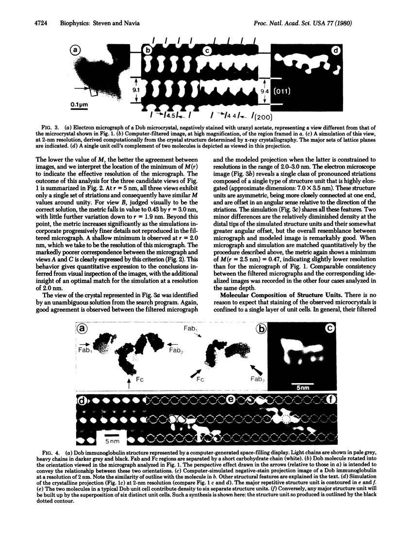
We have investigated the fidelity of structure representation in electron micrographs of negatively stained proteins by conducting a systematic evaluation of such micrographs in terms of a known molecular structure, solved by x-ray crystallography. Microcrystals of immunoglobulin G Dob were used as specimens in this comparison between micrograph images, optimized by computer image processing, and reference images derived computationally from the crystal structure. To an effective resolution of 2 nm, we observed a remarkably good correlation between the experimental images and their idealized counterparts, which are unaffected by those factors--electron irradiation and dehydration--that are thought to be primarily responsible for perturbation of protein structure during electron microscopy. Separate structural features resolved in these micrographs do not, in general, correspond to specific components of individual molecules but arise instead from complex superpositions involving several overlapping molecules.
Full text
PDF


Images in this article


Selected References
These references are in PubMed. This may not be the complete list of references from this article.
- Aebi U., Smith P. R., Dubochet J., Henry C., Kellenberger E. A study of the structure of the T-layer of Bacillus brevis. J Supramol Struct. 1973;1(6):498–522. doi: 10.1002/jss.400010606. [DOI] [PubMed] [Google Scholar]
- Amzel L. M., Poljak R. J. Three-dimensional structure of immunoglobulins. Annu Rev Biochem. 1979;48:961–997. doi: 10.1146/annurev.bi.48.070179.004525. [DOI] [PubMed] [Google Scholar]
- BRENNER S., HORNE R. W. A negative staining method for high resolution electron microscopy of viruses. Biochim Biophys Acta. 1959 Jul;34:103–110. doi: 10.1016/0006-3002(59)90237-9. [DOI] [PubMed] [Google Scholar]
- Beer M., Frank J., Hanszen K. J., Kellenberger E., Williams R. C. The possibilities and prospects of obtaining high-resolution information (below 30 A) on biological material using the electron microscope. Some comments and reports inspired by an EMBO workshop held at Gais, Switzerland, October 1973. Q Rev Biophys. 1974 May;7(2):211–238. doi: 10.1017/s0033583500001414. [DOI] [PubMed] [Google Scholar]
- Fuller S. D., Capaldi R. A., Henderson R. Structure of cytochrome c oxidase in deoxycholate-drived two-dimensional crystals. J Mol Biol. 1979 Oct 25;134(2):305–327. doi: 10.1016/0022-2836(79)90037-8. [DOI] [PubMed] [Google Scholar]
- Labaw L. W., Davies D. R. An electron microscopic study of human gamma Gl immunoglobulin crystals. Preliminary results. J Biol Chem. 1971 Jun 10;246(11):3760–3762. [PubMed] [Google Scholar]
- Labaw L. W., Davies D. R. The molecular outline of human gamma G1 immunoglobulin from an EM study of crystals. J Ultrastruct Res. 1972 Aug;40(3):349–365. doi: 10.1016/s0022-5320(72)90106-2. [DOI] [PubMed] [Google Scholar]
- Labaw L. W., Padlan E. A., Segal D. M., Davies D. R. An em study of phosphorylcholine-binding fab immunoglobulin fragment crystals. J Ultrastruct Res. 1975 Jun;51(3):326–339. doi: 10.1016/s0022-5320(75)80097-9. [DOI] [PubMed] [Google Scholar]
- Ohlendorf D. H., Wrenn R. F., Banaszak L. J. Three-dimensional structure of the lipovitellin-phosvitin complex from amphibian oocytes. Nature. 1978 Mar 2;272(5648):28–32. doi: 10.1038/272028a0. [DOI] [PubMed] [Google Scholar]
- Sarma V. R., Silverton E. W., Davies D. R., Terry W. D. The three-dimensional structure at 6 A resolution of a human gamma Gl immunoglobulin molecule. J Biol Chem. 1971 Jun 10;246(11):3753–3759. [PubMed] [Google Scholar]
- Silverton E. W., Navia M. A., Davies D. R. Three-dimensional structure of an intact human immunoglobulin. Proc Natl Acad Sci U S A. 1977 Nov;74(11):5140–5144. doi: 10.1073/pnas.74.11.5140. [DOI] [PMC free article] [PubMed] [Google Scholar]
- Smith P. R., Aebi U. The determination of the helical screw angle of a helical particle from its diffraction pattern. J Mol Biol. 1976 Sep 15;106(2):271–275. doi: 10.1016/0022-2836(76)90084-x. [DOI] [PubMed] [Google Scholar]
- Smith P. R. An integrated set of computer programs for processing electron micrographs of biological structures. Ultramicroscopy. 1978;3(2):153–160. doi: 10.1016/s0304-3991(78)80021-7. [DOI] [PubMed] [Google Scholar]
- Steven A. C., Aebi U., Showe M. K. Folding and capsomere morphology of the P23 surface shell of bacteriophage T4 polyheads from mutants in five different head genes. J Mol Biol. 1976 Apr 15;102(3):373–400. [PubMed] [Google Scholar]
- Steven A. C., Carrascosa J. L. Proteolytic cleavage and structural transformation: their relationship in bacteriophage T4 capsid maturation. J Supramol Struct. 1979;10(1):1–11. doi: 10.1002/jss.400100102. [DOI] [PubMed] [Google Scholar]
- Steven A. C., Couture E., Aebi U., Showe M. K. Structure of T4 polyheads. II. A pathway of polyhead transformation as a model for T4 capsid maturation. J Mol Biol. 1976 Sep 5;106(1):187–221. doi: 10.1016/0022-2836(76)90307-7. [DOI] [PubMed] [Google Scholar]
- Terry W. D., Matthews B. W., Davies D. R. Crystallographic studies of a human immunoglobulin. Nature. 1968 Oct 19;220(5164):239–241. doi: 10.1038/220239a0. [DOI] [PubMed] [Google Scholar]
- Unwin P. N. Electron microscopy of the stacked disk aggregate of tobacco mosaic virus protein. II. The influence of electron irradiation of the stain distribution. J Mol Biol. 1974 Aug 25;87(4):657–670. doi: 10.1016/0022-2836(74)90076-x. [DOI] [PubMed] [Google Scholar]