Abstract
Context
Most researchers investigating soccer injuries have studied elite athletes because they have greater athletic-exposure hours than other athletes, but most youth participate at the recreational level. If risk factors for injury vary by soccer level, then recommendations generated using research with elite youth soccer players might not generalize to recreational players.
Objective
To examine injury risk factors of strength and jump biomechanics by soccer level in female youth athletes and to determine whether research recommendations based on elite youth athletes could be generalized to recreational players.
Design
Cross-sectional study.
Setting
Seattle Youth Soccer Association.
Patients or Other Participants
Female soccer players (N = 92) aged 11 to 14 years were recruited from 4 randomly selected elite (n = 50; age = 12.5 years, 95% confidence interval [95% CI]) = 12.3, 12.8 years; height = 157.8 cm, 95% CI = 155.2, 160.3 cm; mass = 49.9 kg, 95% CI = 47.3, 52.6 kg) and 4 randomly selected recreational (n = 42; age = 13.2 years, 95% CI = 13.0, 13.5 years; height = 161.1 cm, 95% CI = 159.2, 163.1 cm; mass = 50.6 kg, 95% CI = 48.3, 53.0 kg) soccer teams.
Main Outcome Measure(s)
Players completed a questionnaire about demographics, history of previous injury, and soccer experience. Physical therapists used dynamometry to measure hip strength (abduction, adduction, extension, flexion) and knee strength (flexion, extension) and Sportsmetrics to measure vertical jump height and jump biomechanics. We compared all measurements by soccer level using linear regression to adjust for age and mass.
Results
Elite players were similar to recreational players in all measures of hip and knee strength, vertical jump height, and normalized knee separation (a valgus estimate generated using Sportsmetrics).
Conclusions
Female elite youth players and recreational players had similar lower extremity strength and jump biomechanics. This suggests that recommendations generated from research with elite youth soccer players could be generalized to recreational players.
Key Words: muscle strength, children, adolescents, risk factors, athletic injuries
Key Points
Lower extremity strength and jump biomechanics were similar for elite and recreational female soccer players aged 11 to 14 years.
Recommendations from research on elite youth soccer players should generalize to recreational players.
Soccer is the fifth most popular sport in the United States,1 with an estimated 8.6 million players from 6 to 11 years of age and 5 million players from 12 to 17 years of age.2 Most of these players participate at the recreational (nonelite) level, but most researchers studying injuries have focused on elite athletes.3–7 Researchers more commonly study elite athletes because they participate year-round and thus have greater athletic-exposure hours and more opportunity for injury. If elite and recreational players differ in terms of potential risk factors for injury, recommendations developed using research with elite athletes might not generalize to recreational players.
Data have suggested that adult elite soccer players are stronger than nonelite players.8,9 Cometti et al8 reported greater knee-flexion (hamstrings) strength in elite than nonelite soccer players, and Oberg et al9 reported that elite players were stronger in both knee flexion and extension. These differences in strength could result in altered risk of injury because hamstrings weakness is thought to increase susceptibility to anterior cruciate ligament (ACL) tears.10,11 However,12,13 researchers evaluating strength by soccer level in youth have not found differences in lower extremity strength. Whereas Hansen et al12 found differences by soccer level in hand grip and back strength of youth players, they did not find differences in quadriceps strength. In a similar study, le Gall et al13 reported no differences in quadriceps or hamstrings strength by soccer level in youth. To our knowledge, no one has compared strength by soccer level in female youth athletes.
Jump biomechanics, particularly valgus knee alignment on landing, have been shown to be a risk factor for knee injury because they place undue stress on the ACL.14 Investigators15 have suggested that poor jump biomechanics could contribute to the increased risk of ACL injury in female athletes, who more commonly exhibit valgus knee alignment during jumping maneuvers than male athletes. Researchers16–19 have evaluated vertical jump height by soccer level, but to our knowledge, none have examined differences in valgus alignment by soccer level in the adult or youth population.
Although researchers have found that adult soccer players designated as elite and recreational differ in strength and jump biomechanics,8,9,16–19 few have compared these characteristics by soccer level in the youth population, and to our knowledge, none have studied female youth. Therefore, the purpose of our study was to examine strength and jump biomechanics by soccer level in female youth athletes to determine whether research recommendations based on elite youth athletes could be generalized to recreational players.
METHODS
We conducted a cross-sectional study comparing lower extremity strength, vertical jump height, and normalized knee separation between elite and recreational female soccer players aged 11 to 14 years. These evaluations were part of a prospective cohort study, which has been described elsewhere.20
Participants
We recruited athletes from female elite (also called premier) and recreational teams in the Seattle Youth Soccer Association (SYSA). Elite teams in the SYSA require tryouts and have games throughout the calendar year, whereas recreational teams do not require tryouts and play for only 10 weeks in the fall. We randomly selected 4 elite and 4 recreational teams of girls aged 11 to 14 years from the 17 elite and 59 recreational teams in the SYSA. All selected teams agreed to participate in the study. From these teams, 78% (50/64) of the elite players and 71% (42/59) of the recreational players chose to participate, for an overall participation rate of 75%. Team membership and age from 11 to 14 years were the only requirements for inclusion in the study. Girls were excluded if they planned to move out of Seattle during the study year. All participants who were enrolled completed the study. All participants and their parents provided written assent and consent, respectively. The study was approved by the University of Washington Institutional Review Board.
Testing Procedures
We scheduled baseline evaluations to coincide with the start of the players' season in 2006: March for the elite players and September for the recreational players. If athletes were injured on the day of testing, the testing was delayed until they had recovered. With the assistance of their parents, participants completed a questionnaire about demographic information and soccer history. This questionnaire included information about years of soccer experience, previous injuries, menarchal status, and days of exercise per week. Information on menarchal status was relevant because the onset of puberty has been associated with changes in strength and jump biomechanics.21–23
Six experienced physical therapists (average time in practice = 7.7 years; range, 3.5–18 years) from the University of Washington measured the athletes' height and mass and completed testing of muscle strength and jump biomechanics. They were given detailed written instructions and a 2-hour training session. Each soccer athlete was tested for knee strength, hip strength, and jump biomechanics 3 times during the day of testing. The order in which these batteries of tests were performed was randomized for each instance of testing. Participants were evaluated twice by the same physical therapist and once by a different physical therapist. The mean of the 3 measurements was calculated for each player for each performance measure. The resulting single value for each player for each performance measure was used to analyze the average differences between the recreational and elite groups.
Knee Strength
Knee-extension and knee-flexion concentric torques were measured isokinetically using a dynamometer (model Pro 3; Biodex Medical Systems, Inc, Shirley, NY). Isokinetic dynamometry is considered to be one of the most accurate means for assessing muscle strength,24 and researchers have reported good reliability using these methods to measure knee strength in youth (intraclass correlation coefficient [ICC] range, 0.80–0.86).25,26 The dynamometer was calibrated according to the manufacturer's protocol before each testing session. The physical therapist provided a brief orientation to the Biodex testing system, positioned the participant in a seated position, aligned her joint axis with the axis of the dynamometer, and stabilized her with straps. Each participant performed a standardized warm-up of the right lower extremity of 3 submaximal concentric contractions followed by 10 maximal-effort repetitions at 180°/s. The greatest value of the quadriceps and hamstrings measurements was recorded as peak torque. Participants then repeated the procedure on the same limb at 300°/s. The athletes rested for 5 minutes between trials to reduce fatigue. We chose 180°/s and 300°/s because researchers have suggested that better differentiation exists between similar athletes when assessing strengths at higher velocities.21,23 The testing procedure was repeated on the left lower extremity. Both limbs were tested 3 times during the testing day, and the results for each performance measure for each limb were averaged.
Hip Strength
Maximal isometric hip strength was measured with a handheld dynamometer (HHD) (model microFET2; Hoggan Health Industries, Inc, Draper, UT) using the “break” test. In the break test, the examiner gradually increases the resistance to the participant's contraction until she overcomes it, recording the force required. Handheld dynamometry using the break test has been tested extensively27–32 and has been found31 to be a reasonable technique for measuring strength, with good reliability in youth (ICC for hip abduction = 0.76, hip flexion = 0.83, hip extension = 0.83). The physical therapist instructed the participant to perform 1 practice break trial for familiarization and then began the testing. The order of testing for the hip musculature was determined by convenience for the participant: hip flexion (seated), hip abduction (lying on the side), hip adduction (lying on the side), and hip extension (prone).
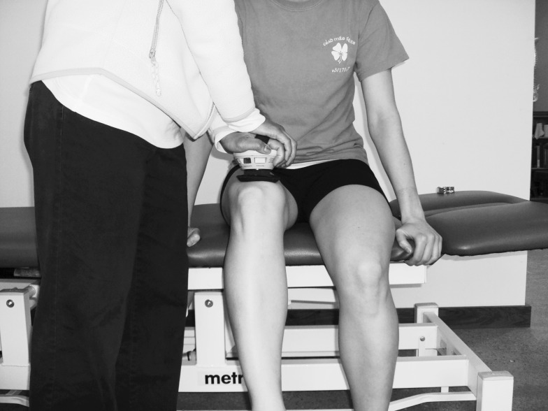
To measure hip flexion, participants sat on the edge of the therapy table with their legs hanging over the edge of the table and stabilized their trunks by grasping the edge of the table. The physical therapist placed the HHD on the distal femur just proximal to the patella and instructed participants to flex their hips 2 in (5.08 cm) off the table while the physical therapist applied resistance toward the floor (Figure 1).
Figure 1.
Hip-flexor testing using handheld dynamometry with the break test.
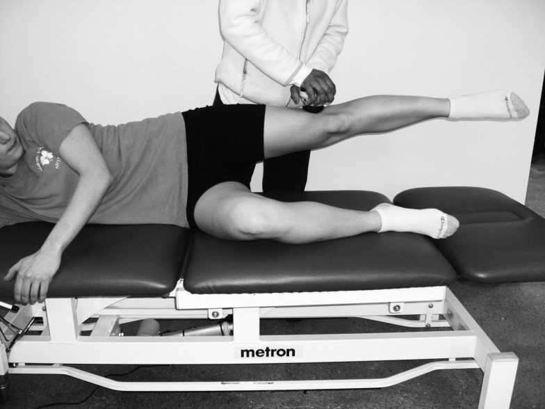
For hip abduction, participants lay on their sides with the lower extremity to be tested positioned straight and above the opposite limb, which was flexed to 90° at the hip and knee for stabilization. The physical therapist placed the HHD on the distal femur just proximal to the lateral knee and instructed participants to raise their test limbs 2 in (5.08 cm) while the physical therapist directed resistance to the floor (Figure 2).
Figure 2.
Hip-abductor testing using handheld dynamometry with the break test.
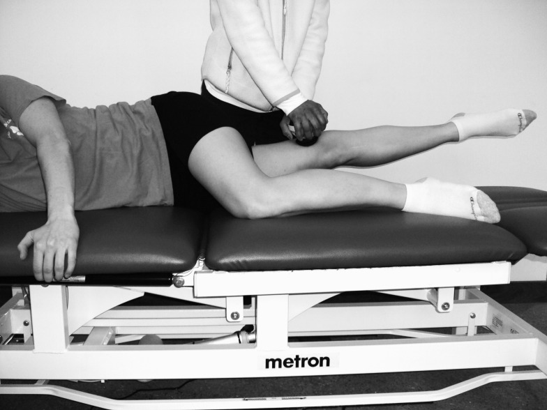
For hip adduction, participants lay on their sides with the lower extremity to be tested positioned straight and below the opposite limb, which was flexed at the hip and knee and supported by the table. The physical therapist placed the HHD on the medial surface of the distal femur just proximal to the medial side of the knee and instructed participants to raise their test limbs 2 in (5.08 cm) while the physical therapist directed resistance to the floor (Figure 3).
Figure 3.
Hip-adductor testing using handheld dynamometry with the break test.
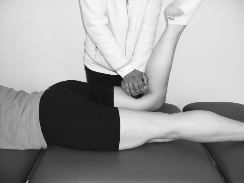
For hip extension, participants lay prone on the therapy table with their upper extremities overhead and the knee of the leg to be tested flexed to 90°. The physical therapist placed the HHD on the volar aspect of the distal femur just proximal to the popliteal fossa and instructed participants to extend their hips 2 in (5.08 cm) off the table while the physical therapist directed resistance toward the floor (Figure 4).
Figure 4.
Hip-extensor testing using handheld dynamometry with the break test.
The physical therapists conducted a videographic drop jump using the Sportsmetrics standardized video-analysis technique (Sportsmetrics, Inc, Cincinnati, OH). The Sportsmetrics system was developed by the Cincinnati Sports Medicine Research and Education Foundation and has been described extensively in their research, with excellent reliability reported (ICC > 0.90).26,33 The physical therapist positioned a Canon digital camcorder (model ZR850; Canon USA, Inc, Lake Success, NY) 12 ft (3.6 m) from a 31-cm-tall box, placed a calibrating placard next to the box, and loaded software onto a laptop computer to analyze the data. The physical therapist then placed reflective markers on the greater trochanter, the center of each patella, and the lateral malleolus of right and left legs of the athletes. All athletes wore Lycra (Invista, Wichita, KS) shorts and low-cut athletic shoes during the testing procedure. The athletes stepped on the box with their right sides facing the video camera and straightened their knees. Next, they faced the video camera, jumped off the box, landed, and immediately performed a maximum-effort vertical jump. The physical therapist reviewed the tape and captured images of (1) prelanding (toes just touching the ground), (2) landing (deepest point of flexion), (3) takeoff (beginning of the jump), and (4) maximal height. The Sportsmetrics software calculated the distances between participants' hips, knees, and ankles and the height of the vertical jump. Each participant completed the drop jump 3 times during the course of the testing day, and the results for each performance measure were averaged.
Statistical Analysis
A preliminary power analysis indicated that the sample size chosen (50 elite athletes, 42 recreational athletes) would provide adequate power (80%) to detect a moderate effect size (0.50) with a 2-sided equal-variance t test. We first examined demographic variables between the groups to determine whether any of these might confound the relationship between soccer level and strength or jump biomechanics. We compared age, years played, mass, height, body mass index, and days of exercise per week (continuous variables) by soccer level using the t test. We compared history of ever being injured, history of 3 or more previous injuries, position played, and menarchal status (categorical variables) by soccer level using Pearson χ2. Strength and jump biomechanics were both measured 3 times during the testing day (2 times by one physical therapist and 1 time by another physical therapist), and these measurements were averaged for each player (for each performance measure) before analysis. We used linear regression modeling to compare strength by soccer level while controlling for potential confounding variables. We generated separate linear regression models with each measure of strength as the dependent variable and a binary variable indicating soccer level as the predictor. We then assessed age, mass, and height as potential confounders in each model. Adjusting for mass in the linear regression analysis did not change the coefficients for the strength outcomes by more than 10% after adjusting for age, but it substantially improved the fit of the model, so mass was retained in the final model. Height was not different in any model, and including it in the regression negligibly changed both the fit of each model and the coefficient estimates. Therefore, we adjusted for mass and age only. We used the regression equations to generate age-adjusted and mass-adjusted mean strength measurements for recreational and elite players. All strength measures were adjusted to the mean age (12.8 years) and mean mass (50.2 kg) of the entire group. For each strength measurement, the statistical significance of soccer level (elite versus recreational) was based on the null hypothesis of a coefficient of zero for soccer level in the linear regression model that included age and mass. Effect sizes were calculated by dividing the difference in the adjusted means by the standard error of the estimate (root mean square error) from the regression model. This computation of an effect size is similar in principle to the effect size calculated for a comparison of means between 2 groups, where the difference in means is divided by the pooled standard deviation. We explored a potential team effect (clustering by team) by examining ICC for each outcome variable. All these ICCs were very small (range, 0.006–0.17), and thus we did not adjust for team in the analysis. We conducted all analyses using STATA (version 10; Statacorp, College Station, TX) with the α level set a priori at .05.
We analyzed the Sportsmetrics videographic jump data using the methods described by Noyes et al.33 They reported that one can reliably estimate valgus angle by using their software to capture videographic data, measuring the distance between the knees, and normalizing this distance to hip distance. We compared these estimates of valgus alignment at prelanding, landing, and takeoff by soccer level using a method similar to that used for the strength measurements, developing age-adjusted and mass-adjusted linear regression models to test for a nonzero coefficient of soccer level and generate adjusted means to use in calculating effect sizes.
RESULTS
Recreational players were slightly older (difference in mean age = 0.7 years; t = 3.67, P = .004) and taller (difference in mean height = 3.3 cm; t = 2.07, P = .04) than their elite counterparts, but they did not differ in years of soccer experience, mass, body mass index, average days of exercise per week, history of having been injured, menarchal status, or distribution of soccer positions (Table 1).
Table 1.
Demographic and Soccer Characteristics of Elite and Recreational Female Soccer Players Aged 11 to 14 Years in Seattle, 2006–2007a
| Characteristic |
Elite Players |
Recreational Players |
| Number of athletes | 50 | 42 |
| Mean age, y (95% CI)b | 12.5 (12.3, 12.8) | 13.2 (13.0, 13.5) |
| Mean height, cm (95% CI)c | 157.8 (155.2, 160.3) | 161.1 (159.2, 163.1) |
| Mean time played, y (95% CI) | 6.9 (6.4, 7.4) | 7.6 (6.9, 8.2) |
| Mean mass, kg (95% CI) | 49.9 (47.3, 52.6) | 50.6 (48.3, 53.0) |
| Mean body mass index, kg/m2 (95% CI) | 20.0 (19.0, 20.9) | 19.4 (18.7, 20.1) |
| Mean time exercised, d/wk (95% CI) | 4.6 (4.2, 5.0) | 4.6 (3.9, 5.2) |
| Ever injured, n (%) | 26 (52) | 20 (47) |
| ≥3 previous injuries, n (%) | 15 (30) | 11 (26) |
| Postmenarchal, n (%) | 31 (62) | 26 (62) |
| Primary position, n (%) | ||
| Defender | 16 (32) | 10 (24) |
| Forward | 15 (30) | 12 (29) |
| Midfielder | 17 (34) | 19 (45) |
| Goalkeeper | 2 (4) | 1 (2) |
Indicates 95% confidence intervals (CIs) were determined using the t test.
Indicates P < .01.
Indicates P < .05.
We used regression modeling to calculate age-adjusted and mass-adjusted means for each strength outcome. Hip-abduction strength was greater in elite than recreational players when adjusting for age alone (t = 2.12, P = .04), but it was not different after adjusting for mass (t = 1.87, P = .07). Elite players were similar to recreational players in other measures of hip strength (flexion, extension, adduction) and knee strength (flexion, extension) (Table 2). Elite and recreational players also had similar vertical jump heights and estimates of knee valgus alignment during prelanding, landing, and takeoff phases (normalized knee-separation distances) (Table 3).
Table 2.
Lower Extremity Strength Among Elite and Recreational Female Soccer Players Aged 11 to 14 Years in Seattle, 2006–2007,a Meanb (95% Confidence Interval)
| Measurement |
Elite Players |
Recreational Players |
Effect Size |
t |
P |
| Hip strength, N | |||||
| Flexion dominant | 42.0 (36.7, 47.4) | 40.0 (38.0, 42.1) | 0.26 | 1.24 | .22 |
| Flexion nondominant | 39.3 (34.6, 44.0) | 38.3 (36.5, 40.1) | 0.14 | 0.67 | .50 |
| Extension dominant | 39.5 (34.8, 44.3) | 39.5 (37.6, 41.4) | 0.003 | 0.01 | .99 |
| Extension nondominant | 38.7 (33.6, 43.7) | 37.4 (35.4, 39.4) | 0.18 | 0.83 | .41 |
| Abduction dominant | 39.9 (34.5, 45.4) | 36.8 (34.7, 39.0) | 0.41 | 1.87 | .07 |
| Abduction nondominant | 37.6 (32.8, 42.5) | 35.4 (33.4, 37.3) | 0.33 | 1.53 | .13 |
| Adduction dominant | 38.1 (33.6, 42.6) | 37.0 (35.3, 38.8) | 0.26 | 0.80 | .21 |
| Adduction nondominant | 37.1 (35.3, 38.9) | 37.1 (35.3, 38.9) | 0.17 | 1.27 | .43 |
| Knee strength, Nm | |||||
| Extension | |||||
| 180°/s dominant | 49.2 (42.6, 55.8) | 51.4 (48.8, 54.1) | −0.28 | −1.13 | .26 |
| 180°/s nondominant | 51.6 (45.7, 57.3) | 51.7 (49.4, 54.0) | −0.02 | −0.08 | .93 |
| 300°/s dominant | 38.8 (33.8, 43.8) | 38.9 (36.9, 40.9) | 0.02 | −0.06 | .95 |
| 300°/s nondominant | 39.3 (34.3, 44.3) | 39.5 (37.4, 41.6) | −0.04 | −0.16 | .87 |
| Flexion | |||||
| 180°/s dominant | 37.5 (33.9, 41.1) | 37.1 (35.7, 38.6) | 0.08 | 0.34 | .73 |
| 180°/s nondominant | 36.6 (33.0, 40.2) | 36.3 (34.8, 37.8) | 0.06 | 0.27 | .79 |
| 300°/s dominant | 37.1 (31.0, 42.2) | 35.6 (33.6, 37.7) | 0.24 | 0.94 | .35 |
| 300°/s nondominant | 35.1 (30.9, 39.3) | 33.7 (32.0, 35.5) | 0.23 | 1.00 | .28 |
Lower extremity dominance was defined as the extremity used to kick a ball.
Indicates all mean estimates were adjusted to the average age (12.8 years) and average mass (50.2 kg) of the entire group using linear regression.
Table 3.
Sportsmetricsa Knee Valgus Estimations and Vertical Jump Heights Among Elite and Recreational Female Soccer Players Aged 11 to 14 Years in Seattle, 2006–2007, Meanb (95% Confidence Interval)
| Measurement |
Elite Players |
Recreational Players |
Effect Size |
t |
P |
| Knee valgus estimate, knee | |||||
| distance/hip distance, ° | |||||
| Prelanding | 0.51 (0.44, 0.59) | 0.51 (0.48, 0.55) | 0.04 | 0.19 | .85 |
| Landing | 0.37 (0.29, 0.45) | 0.37 (0.33, 0.41) | 0.03 | −0.12 | .91 |
| Takeoff | 0.38 (0.30, 0.46) | 0.38 (0.35, 0.42) | 0.02 | 0.11 | .92 |
| Vertical jump, cm | 24.2 (20.2, 28.2) | 25.1 (23.5, 26.8) | 0.18 | −0.79 | .43 |
Sportsmetrics, Inc, Cincinnati, OH.
Indicates all mean estimates were adjusted to the average age (12.8 years) and average mass (50.2 kg) of the entire group using linear regression.
DISCUSSION
We conducted a cross-sectional study of female youth soccer players to determine whether lower extremity strength and jump biomechanics differed between elite and recreational players, because researchers have shown these variables are risk factors for injury in other populations. We found a trend for greater hip strength in elite than recreational athletes, but all other measures of hip and knee strength were similar. Athletes were similar in terms of jump biomechanics, years of soccer experience, and history of previous injury.
Hip-abductor strength was not different between groups, which is interesting since hip-abductor strength is thought to play a role in chronic knee injury, such as patellofemoral pain syndrome.34,35 In addition to affecting chronic knee injury, researchers have suggested that hip-abductor strength may affect jump biomechanics,36 and jump biomechanics have been associated with risk for ACL injury.14 Most investigators comparing strength between recreational and elite athletes have focused on quadriceps and hamstrings rather than hip abductors,8,9,12,13 but Thorborg et al32 found greater hip-abductor strength in elite soccer players than in a control group of recreational athletes. They hypothesized that hip abductors were important for kicking, accelerating, and sudden changes of direction, which are necessary skills for soccer.
One notable finding was that strength measurements were similar for all measures of hip strength in this study population (flexion, extension, abduction, adduction). In older participants, hip flexors and extensors are generally stronger than hip abductors and adductors,37 but we could not find any normative data on hip strength in female youth for comparison. Differentiations between muscle groups possibly are not yet prominent in this younger age group.
In contrast to the findings reported by researchers studying adult players,8,9 we did not find any differences in hamstrings or quadriceps strength by soccer level in our youth sample. However, our results are in agreement with those reported in studies of male youth athletes.12,13 In their study of soccer players aged 10 to 14 years, Hansen et al12 found differences between elite and subelite athletes in back and hand-grip strength, but they did not find differences in quadriceps strength after adjusting for age and mass. They did not evaluate hamstrings strength or hip strength. Le Gall et al13 found no differences in hamstrings or quadriceps strength by soccer level in their study of male soccer players aged 14 to 17 years.
We undertook this study to understand more about elite and recreational athletes in the youth population. We had observed that despite the barrier of tryouts and the differing time commitments, girls from elite and recreational teams in the SYSA seemed quite similar, and our data supported this observation. Playing elite soccer in the SYSA requires extensive financial and time commitments from both parents and athletes, and girls who play at this level likely are more motivated to become better soccer players. However, our data did not suggest that they differ in ways that affect their risks of injury. This finding is important because it suggests that clinicians could apply recommendations from research with elite youth athletes to their recreational-athlete patients. The data also suggest that hip-abductor strength might differentiate more-skilled athletes from their less-skilled counterparts, but future studies are needed to determine whether this difference in strength translates into true differences in injury risk.
One of the limitations of our study was the difference in mean age between elite and recreational players. Although the age difference was small (0.7 years), small differences can be correlated with increases in strength due to the proximity to puberty.38,39 We controlled for the age discrepancy using regression analysis; however, residual confounding might exist, and an ideal sample would have been age matched. Another limitation was the potential for measurement error in the strength and jump biomechanics measures. Researchers have reported good reliability using these measures,25,26,27–32 but any measurement error could obscure differences between the groups. We also had a relatively small sample of elite and recreational players, and more subtle differences possibly would be detected with a larger sample size. However, subtle differences in strength or jump biomechanics are unlikely to be relevant to injury risk. Finally, we studied youth soccer players in Seattle, and we cannot speculate about how these athletes compare with youth soccer players in other geographic locations.
CONCLUSIONS
We found that 11- to 14-year-old elite and recreational female soccer players in the SYSA were similar in terms of lower extremity strength and jump biomechanics, both of which have been suggested to be risk factors for injury in other populations. These results supported our hypothesis that recommendations from research on elite youth soccer players should generalize to recreational players.
ACKNOWLEDGMENTS
This study was supported by grant R21AR053371 from the National Institute of Arthritis and Musculoskeletal and Skin Diseases (Dr Schiff).
We thank the players and parents in the Seattle Youth Soccer Association for their participation in this study.
REFERENCES
- 1.National Federation of State High School Associations. 2008–09 High school athletics participation survey results. http://www.nfhs.org/content.aspx?id=3282&linkidentifier=id&itemid=3282. Accessed July 27, 2010. [Google Scholar]
- 2.Sporting Goods Manufacturer's Association. Teens & sports participation in America 2001. 2010 http://www.sgma.com/reports/153_Teens-%26-Sports-Participation-in-America-2001. Accessed July 29. [Google Scholar]
- 3.Brink MS, Visscher C, Arends S. Monitoring stress and recovery: new insights for the prevention of injuries and illnesses in elite youth soccer players. Br J Sports Med. 2010;44(11):809–815. doi: 10.1136/bjsm.2009.069476. [DOI] [PubMed] [Google Scholar]
- 4.Henderson G, Barnes CA, Portas MD. Factors associated with increased propensity for hamstring injury in English Premier League soccer players. J Sci Med Sport. 2010;13(4):397–402. doi: 10.1016/j.jsams.2009.08.003. [DOI] [PubMed] [Google Scholar]
- 5.Johnson A, Doherty PJ, Freemont A. Investigation of growth, development, and factors associated with injury in elite schoolboy footballers: prospective study. BMJ. 2009 doi: 10.1136/bmj.b490. 338:b490. [DOI] [PMC free article] [PubMed] [Google Scholar]
- 6.Le Gall F, Carling C, Reilly T. Injuries in young elite female soccer players: an 8-season prospective study. Am J Sports Med. 2008;36(2):276–284. doi: 10.1177/0363546507307866. [DOI] [PubMed] [Google Scholar]
- 7.Lehance C, Binet J, Bury T, Croisier JL. Muscular strength, functional performances and injury risk in professional and junior elite soccer players. Scand J Med Sci Sports. 2009;19(2):243–251. doi: 10.1111/j.1600-0838.2008.00780.x. [DOI] [PubMed] [Google Scholar]
- 8.Cometti G, Maffiuletti NA, Pousson M, Chatard JC, Maffulli N. Isokinetic strength and anaerobic power of elite, subelite and amateur French soccer players. Int J Sports Med. 2001;22(1):45–51. doi: 10.1055/s-2001-11331. [DOI] [PubMed] [Google Scholar]
- 9.Oberg B, Möller M, Gillquist J, Ekstrand J. Isokinetic torque levels for knee extensors and knee flexors in soccer players. Int J Sports Med. 1986;7(1):50–53. doi: 10.1055/s-2008-1025735. [DOI] [PubMed] [Google Scholar]
- 10.Söderman K, Alfredson H, Pietilä T, Werner S. Risk factors for leg injuries in female soccer players: a prospective investigation during one outdoor season. Knee Surg Sports Traumatol Arthrosc. 2001;9(5):313–321. doi: 10.1007/s001670100228. [DOI] [PubMed] [Google Scholar]
- 11.Myer GD, Ford KR, Barber Foss KD, Liu C, Nick TG, Hewett TE. The relationship of hamstrings and quadriceps strength to anterior cruciate ligament injury in female athletes. Clin J Sport Med. 2009;19(1):3–8. doi: 10.1097/JSM.0b013e318190bddb. [DOI] [PMC free article] [PubMed] [Google Scholar]
- 12.Hansen L, Bangsbo J, Twisk J, Klausen K. Development of muscle strength in relation to training level and testosterone in young male soccer players. J Appl Physiol. 1999;87(3):1141–1147. doi: 10.1152/jappl.1999.87.3.1141. [DOI] [PubMed] [Google Scholar]
- 13.le Gall F, Carling C, Williams M, Reilly T. Anthropometric and fitness characteristics of international, professional and amateur male graduate soccer players from an elite youth academy. J Sci Med Sport. 2010;13(1):90–95. doi: 10.1016/j.jsams.2008.07.004. [DOI] [PubMed] [Google Scholar]
- 14.Hewett TE, Myer GD, Ford KR et al. Biomechanical measures of neuromuscular control and valgus loading of the knee predict anterior cruciate ligament injury risk in female athletes: a prospective study. Am J Sports Med. 2005;33(4):492–501. doi: 10.1177/0363546504269591. [DOI] [PubMed] [Google Scholar]
- 15.Beutler A, de la Motte S, Marshall S, Padua D, Boden B. Muscle strength and qualitative jump-landing differences in male and female military cadets: the Jump-ACL study. J Sports Sci Med. 2009;8:663–671. [PMC free article] [PubMed] [Google Scholar]
- 16.Gissis I, Papadopoulos C, Kalapotharakos VI et al. Strength and speed characteristics of elite, subelite, and recreational young soccer players. Res Sports Med. 2006;14(3):205–214. doi: 10.1080/15438620600854769. [DOI] [PubMed] [Google Scholar]
- 17.Malina RM. Maturity status and injury risk in youth soccer players. Clin J Sport Med. 2010;20(2):132. doi: 10.1097/01.jsm.0000369404.77182.60. [DOI] [PubMed] [Google Scholar]
- 18.Sedano S, Vaeyens R, Philippaerts RM, Redondo JC, Cuadrado G. Anthropometric and anaerobic fitness profile of elite and non-elite female soccer players. J Sports Med Phys Fitness. 2009;49(4):387–394. [PubMed] [Google Scholar]
- 19.Smith R, Ford KR, Myer GD et al. Biomechanical and performance differences between female soccer athletes in National Collegiate Athletic Divisions I and III. J Athl Train. 2007;42(4):470–476. [PMC free article] [PubMed] [Google Scholar]
- 20.Schiff MA, Mack CD, Polissar NL et al. Soccer injuries in female youth players: comparison of injury surveillance by certified athletic trainers and Internet. J Athl Train. 2010;45(3):238–242. doi: 10.4085/1062-6050-45.3.238. [DOI] [PMC free article] [PubMed] [Google Scholar]
- 21.Hewett TE, Myer GD, Ford KR. Decrease in neuromuscular control about the knee with maturation in female athletes. J Bone Joint Surg Am. 2004;86(8):1601–1608. doi: 10.2106/00004623-200408000-00001. [DOI] [PubMed] [Google Scholar]
- 22.Hewett TE, Myer GD, Zazulak BT. Hamstrings to quadriceps peak torque ratios diverge between sexes with increasing isokinetic angular velocity. J Sci Med Sport. 2008;11(5):452–459. doi: 10.1016/j.jsams.2007.04.009. [DOI] [PMC free article] [PubMed] [Google Scholar]
- 23.Quatman CE, Ford KR, Myer GD, Hewett TE. Maturation leads to gender differences in landing force and vertical jump performance: a longitudinal study. Am J Sports Med. 2006;34(5):806–813. doi: 10.1177/0363546505281916. [DOI] [PubMed] [Google Scholar]
- 24.Wiggin M, Wilkinson K, Habetz S, Chorley J, Watson M. Percentile values of isokinetic peak torque in children six through thirteen years old. Pediatr Phys Ther. 2006;18(1):3–18. doi: 10.1097/01.pep.0000202097.76939.0e. [DOI] [PubMed] [Google Scholar]
- 25.Pierce SR, Lauer RT, Shewokis PA, Rubertone JA, Orlin MN. Test-retest reliability of isokinetic dynamometry for the assessment of spasticity of the knee flexors and knee extensors in children with cerebral palsy. Arch Phys Med Rehabil. 2006;87(5):697–702. doi: 10.1016/j.apmr.2006.01.020. [DOI] [PubMed] [Google Scholar]
- 26.Barber-Westin SD, Noyes FR, Galloway M. Jump-land characteristics and muscle strength development in young athletes: a gender comparison of 1140 athletes 9 to 17 years of age. Am J Sports Med. 2006;34(3):375–384. doi: 10.1177/0363546505281242. [DOI] [PubMed] [Google Scholar]
- 27.Bohannon RW, Andrews AW. Interrater reliability of hand-held dynamometry. Phys Ther. 1987;67(6):931–933. doi: 10.1093/ptj/67.6.931. [DOI] [PubMed] [Google Scholar]
- 28.Bohannon RW. Hand-held compared with isokinetic dynamometry for measurement of static knee extension torque (parallel reliability of dynamometers) Clin Phys Physiol Meas. 1990;11(3):217–222. doi: 10.1088/0143-0815/11/3/004. [DOI] [PubMed] [Google Scholar]
- 29.Fulcher ML, Hanna CM. Raina Elley C. Reliability of handheld dynamometry in assessment of hip strength in adult male football players. J Sci Med Sport. 2010;13(1):80–84. doi: 10.1016/j.jsams.2008.11.007. [DOI] [PubMed] [Google Scholar]
- 30.Martin HJ, Yule V, Syddall HE, Dennison EM, Cooper C, Aihie Sayer A. Is hand-held dynamometry useful for the measurement of quadriceps strength in older people? A comparison with the gold standard Biodex dynamometry. Gerontology. 2006;52(3):154–159. doi: 10.1159/000091824. [DOI] [PubMed] [Google Scholar]
- 31.Merlini L, Massone ES, Solari A, Morandi L. Reliability of hand-held dynamometry in spinal muscular atrophy. Muscle Nerve. 2002;26(1):64–70. doi: 10.1002/mus.10166. [DOI] [PubMed] [Google Scholar]
- 32.Thorborg K, Couppé C, Petersen J, Magnusson SP, Hömlich P. Eccentric hip adduction and abduction strength in elite soccer players and matched controls: a cross-sectional study. Br J Sports Med. 2011;45(1):10–13. doi: 10.1136/bjsm.2009.061762. [DOI] [PubMed] [Google Scholar]
- 33.Noyes FR, Barber-Westin SD, Fleckenstein C, Walsh C, West J. The drop-jump screening test: difference in lower limb control by gender and effect of neuromuscular training in female athletes. Am J Sports Med. 2005;33(2):197–207. doi: 10.1177/0363546504266484. [DOI] [PubMed] [Google Scholar]
- 34.Baldon Rde M, Nakagawa TH, Muniz TB, Amorim CF, Maciel CD, Serrão FV. Eccentric hip muscle function in females with and without patellofemoral pain syndrome. J Athl Train. 2009;44(5):490–496. doi: 10.4085/1062-6050-44.5.490. [DOI] [PMC free article] [PubMed] [Google Scholar]
- 35.Dolak KL, Silkman C, Medina McKeon J et al. Hip strengthening prior to functional exercises reduces pain sooner than quadriceps strengthening in females with patellofemoral pain syndrome: a randomized clinical trial. 41(9):700] J Orthop Sports Phys Ther. J Orthop Sports Phys Ther. 2011. 2011;41(8):560–570. doi: 10.2519/jospt.2011.3499. published correction appears in. [DOI] [PubMed] [Google Scholar]
- 36.Jacobs CA, Uhl TL, Mattacola CG, Shapiro R, Rayens WS. Hip abductor function and lower extremity landing kinematics: sex differences. J Athl Train. 2007;42(1):76–83. [PMC free article] [PubMed] [Google Scholar]
- 37.Stockton KA, Wrigley TV, Mengersen KA et al. Test-retest reliability of hand-held dynamometry and functional tests in systemic lupus erythematosus. Lupus. 2011;20(2):144–150. doi: 10.1177/0961203310388448. [DOI] [PubMed] [Google Scholar]
- 38.Taeymans J, Clarys P, Abidi H, Hebbelinck M, Duquet W. Developmental changes and predictability of static strength in individuals of different maturity: a 30-year longitudinal study. J Sports Sci. 2009;27(8):833–841. doi: 10.1080/02640410902874711. [DOI] [PubMed] [Google Scholar]
- 39.Forbes H, Sutcliffe S, Lovell A, McNaughton LR, Siegler JC. Isokinetic thigh muscle ratios in youth football: effect of age and dominance. Int J Sports Med. 2009;30(8):602–606. doi: 10.1055/s-0029-1202337. [DOI] [PubMed] [Google Scholar]


