

Abstract
Diarrheal disease remains one of the top 2 causes of young child mortality in the developing world. Whereas improvements in water/sanitation infrastructure and hygiene can diminish transmission of enteric pathogens, vaccines can also hasten the decline of diarrheal disease morbidity and mortality. From 1980 through approximately 2004, various case/control and small cohort studies were undertaken to address the etiology of pediatric diarrhea in developing countries. Many studies had methodological limitations and came to divergent conclusions, making it difficult to prioritize the relative importance of different pathogens. Consequently, in the first years of the millennium there was no consensus on what diarrheal disease vaccines should be developed or implemented; however, there was consensus on the need for a well-designed study to obtain information on the etiology and burden of more severe forms of diarrheal disease to guide global investment and implementation decisions. Accordingly, the Global Enteric Multicenter Study (GEMS) was designed to overcome drawbacks of earlier studies and determine the etiology and population-based burden of pediatric diarrheal disease. GEMS, which includes one of the largest case/control studies of an infectious disease syndrome ever undertaken (target approximately 12 600 analyzable cases and 12 600 controls), was rolled out in 4 sites in sub-Saharan Africa (Gambia, Kenya, Mali, Mozambique) and 3 in South Asia (Bangladesh, India, Pakistan), with each site linked to a population under demographic surveillance (total approximately 467 000 child years of observation among children <5 years of age). GEMS data will guide investment and help prioritize strategies to mitigate the morbidity and mortality of pediatric diarrheal disease.
In the 55 years between the end of World War II and the close of the 20th century, developing countries, including many newly established nations that emerged from the dissolution of colonial empires, grappled with growing their economies and improving the health of their people. While progressive economic development ensued in many countries (and was impressive in some), others countries notably lagged. By the late 1990s, the United Nations (UN) categorized a subset of approximately 43–50 as the “least developed countries,” many of which were located in sub-Saharan Africa and some parts of Asia [1]. These least developed countries, in particular, were characterized by extremely low gross national income per capita, high young child (<age 5 years) mortality, low adult (particularly female) literacy, and abbreviated adult life expectancy [2, 3]. Diarrheal diseases, pneumonia, measles, and malaria were typically among the top causes of young child mortality. In general, the higher the infant and young child mortality rate, the larger the fraction of mortality attributed to diarrheal diseases. Estimates of global young child (<age 5 years) mortality suggest that in the early years of the millennium an estimated 10.6 million young child deaths occurred annually [4, 5], with approximately 17%–21% of deaths due to diarrheal disease [4, 6, 7] and approximately 70% of all diarrheal mortality localized in 15 countries in Africa and South and Southeast Asia. Addressing the main causes of young child mortality in developing countries, including diarrheal diseases, became a global priority [8].
MOBILIZATION IN THE NEW MILLENNIUM
Circa 2000, 3 new entities came on the scene that rapidly interrelated in a synergistic way to offer extraordinary potential to accelerate the decline of young child mortality in developing countries, and particularly the component due to diarrheal diseases. In 2000, the 55th Session of the UN General Assembly adopted the UN Millennium Declaration [9], committing the countries of the world to mobilize resources to reduce poverty and improve health and education by 2015, with progress judged by whether or not certain specific goals were attained. One of these, Millennium Development Goal #4, aims to reduce young child mortality by 67% by 2015, compared to the 1990 baseline.
Second, in 1999 the nascent Bill & Melinda Gates Foundation entered the arena of global health and brought zeal, commitment, and passionate advocacy, as well as substantial new financial resources, to improve the survival of young children in developing countries. Finally, at the World Economic Forum in February 2000, the Global Alliance for Vaccines and Immunization (GAVI, now called the GAVI Alliance) was launched. The GAVI Alliance is a consortium that consists of UN agencies (World Health Organization [WHO], United Nations Children's Fund [UNICEF], World Bank) involved with immunization, vaccine supply, and vaccine financing; developing and donor countries; the vaccine industry (in both industrialized and developing countries); technical and research institutes; civil society; and the Bill & Melinda Gates Foundation and other private philanthropic foundations. In its decade of existence, GAVI has been highly successful in strengthening the delivery of immunization services and in introducing life-saving new vaccines into the Expanded Programme on Immunization of many of the poorest countries of the world, including those in sub-Saharan Africa. In 2002, GAVI established and funded 2 Accelerated Development and Introduction Plans (ADIPs), one for rotavirus vaccine and the other for pneumococcal conjugate vaccines. The fundamental aims of the rotavirus ADIP were (1) to provide information (eg, documentation of the safety, immunogenicity, and efficacy of rotavirus vaccines in infants in developing countries) that enables evidence-based decisions regarding the use of rotavirus vaccines, and (2) to accelerate the availability of new rotavirus vaccines appropriate for use in developing countries.
GEOGRAPHIC FOCUS
In order to intervene in a strategic way to accelerate the decline of young child mortality globally, efforts must be concentrated in 2 main geographic areas: sub-Saharan Africa, where 33 of the 35 countries with world's highest young child mortality rates are located [2, 3, 10–12], and South Asia, where the size of the young child population is enormous, leading to a large number of deaths, despite the mortality rates being lower than in sub-Saharan Africa [2, 3, 10–12].
CLINICAL SYNDROMES OF PEDIATRIC DIARRHEAL DISEASE IN DEVELOPING COUNTRIES
As seen by clinical health providers at fixed healthcare facilities in developing countries, almost all cases of pediatric diarrheal illness can be conveniently characterized as falling into 1 of 5 clinical syndromes [13] (Table 1). Approximately 80%–85% of patient episodes present as “simple gastroenteritis” with the subject passing loose or watery stool (often with mucus but not with blood), low-grade fever, occasional vomiting, anorexia, and apparent malaise (Figure 1). Approximately 5%–15% of children present with overt dysentery (gross blood in the diarrheic stools) (Figure 2), often accompanied by fever (sometimes high); many dysenteric patients appear clinically toxic. A small proportion of cases in older children present with profuse watery diarrhea, passing such voluminous “rice water” stools that even older children can rapidly become severely dehydrated (Figure 3). Another few percent of pediatric cases present with a history of apparent simple gastroenteritis that began 14 or more days previously but did not abate [14]; this defines “persistent diarrhea,” a syndrome that particularly can have adverse nutritional consequences [15]. Finally, a few percent of children are brought by caretakers for care because of vomiting rather than diarrhea as the main complaint. Few reports have described expanded etiologic analyses in relation to these clinical syndromes.
Table 1.
The 5 Main Clinical Syndromes of Diarrheal Disease Seen Among Infants and Young Children Presenting to Health Centers and Hospitals in Developing Countries
| Clinical Syndrome | Characteristic Signs and Symptoms | Proportion of Pediatric Diarrhea Patients Presenting to Health Facilities Who Exhibit This Syndrome | Some Etiologic Agents Associated With This Syndrome |
|---|---|---|---|
| Simple gastroenteritis | Loose stools (often with mucus but no blood), occasional vomiting, anorexia, low-grade fever, malaise | 80%–85% | Rotavirus, ETEC, EPEC, Cryptosporidium, norovirus GI and II, enteric adenovirus types 40 & 41 |
| Dysentery | Gross blood in loose stools (that may be quite scanty), fever (sometimes high), abdominal cramps, tenesmus; many dysentery patients appear clinically toxic | 5%–15% | Shigella, Campylobacter jejuni, Entamoeba histolytica, nontyphoidal Salmonella |
| Profuse purging | Passage of copious watery stools that may resemble “rice water”; fever, if present, is typically low grade; signs of overt dehydration (diminished skin turgor, sunken eyes, dry mucous membranes) may be present | <5% | Vibrio cholerae O1 & O139, ETEC |
| Persistent diarrhea | The same signs and symptoms as simple gastroenteritis but diarrhea continues unabated for at least 14 days | <5% | Giardia lamblia, EPEC |
| Acute vomiting | Acute onset of nausea and repetitive vomiting with little or no diarrhea | ∼1%–2% | Norovirus |
See Kotloff et al in this supplement for precise clinical definitions used in the GEMS study.
Abbreviations: EPEC, enteropathogenic Escherichia coli; ETEC, enterotoxigenic Escherichia coli; GEMS, Global Enteric Multicenter Study.
Figure 1.

An infant who presented with diarrheal dehydration consequent to simple gastroenteritis that was not treated promptly or effectively. Loss of turgor of skin over the abdomen is visible as “tenting,” following pinching. Simple gastroenteritis caused by many etiologic agents in young infants in developing countries can lead to dehydration. The fundamental reason is that per kilogram of body weight, the daily water and electrolyte requirements of young infants are substantially greater than those of older children. Thus, abnormal losses from diarrhea, vomiting, and fever, accompanied by inadequate fluid intake and lack of prompt and appropriate replacement (as with glucose/electrolyte oral rehydration solution), can lead to moderate and severe dehydration and death. This photograph was kindly provided by Dr Dipika Sur of the National Institute of Cholera and Enteric Diseases, Kolkata, India.
Figure 2.
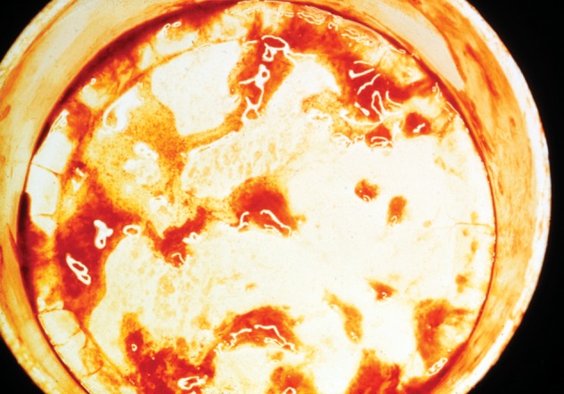
Dysentery is diagnosed clinically as the presence of gross blood in diarrheal stools. Dysentery stools can be quite scanty and composed mainly of mucus and blood (shown here). Bacillary dysentery is typically preceded by 18–24 hours of watery diarrhea, accompanied by high fever and toxemia, before the loose stools become scanty and bloody. Dysentery indicates substantial damage to the mucosa of the colon and terminal ileum.
Figure 3.
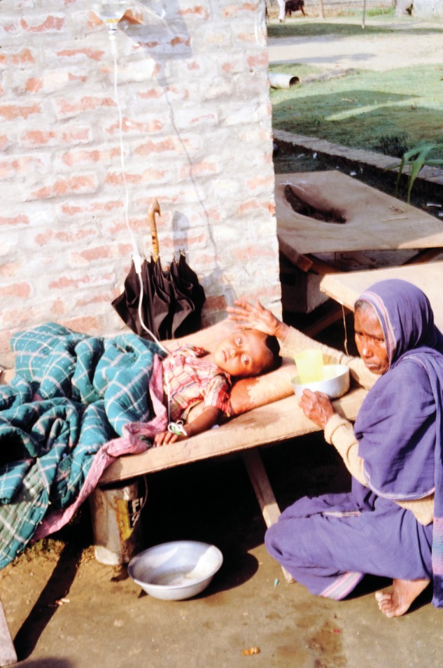
A Bangladeshi child with cholera is shown who experienced copious purging of rice water stools prior to presenting with severe dehydration. The child, with deeply sunken eyes, is lying on a cholera cot with his watery stools being collected in a bucket for measurement of volume (to guide replacement therapy). After rapid replacement of the child's fluid and electrolyte deficits with intravenous fluids, the health worker is attempting to transition the child to oral rehydration fluids administered by his caretaker, under supervision.
THE INTERRELATIONSHIP BETWEEN NUTRITIONAL STATE AND DIARRHEAL DISEASE
It has long been recognized that there is an intimate relationship between diarrheal disease and undernutrition in pediatric populations in developing countries [16, 17]. Diarrheal disease, with its injury to the gut, can lead children to fall off their growth curve. Conversely, more extreme forms of chronic malnutrition predispose young children to diarrhea-related mortality. For example, moderate and severe stunting is a strong risk factor for death from diarrheal disease [18].
LESSONS FROM THE EARLY 20TH CENTURY IN NORTH AMERICA AND EUROPE
Mortality from diarrheal disease is currently extremely low in industrialized countries, but it was a vexing public health problem a century ago when populations in current industrialized countries lived in conditions resembling those endured by people in developing countries today [19–23]. In fact, wherever populations live in crowded conditions marked by widespread fecal contamination, lack of treated water supplies and sanitation to remove human fecal waste, and lack of refrigeration to preserve food, the transmission of bacterial, viral, and protozoal enteric pathogens is enhanced and pediatric diarrheal disease can rage rampant. A shared vision of the Millennium Declaration is that all countries will undergo accelerated development such that with improved housing, provision of sanitation and safe water, enhanced food safety, and access to primary health care, diarrheal disease and pneumonia mortality will plummet. While that is the ultimate aim, it may be possible to accelerate markedly the decline in diarrheal disease mortality by certain cross-cutting general interventions (such as improved treatment of diarrhea and focused water/sanitation/hygiene improvements) and by immunizing infants and young children against the major etiologic agents responsible for clinically severe and fatal forms of diarrheal disease.
In the early years of the millennium, other than vaccines against rotavirus, there was not a broad consensus on what other diarrheal disease vaccines should be high priority for development and accelerated introduction, given the limited resources and supply issues pertinent at the global level. One must also recognize that for rotavirus vaccines there were mature industrialized country markets waiting to reward companies that invested in rotavirus vaccines and achieved licensure for their products in North America, Europe, and Australia. This guaranteed the development of these vaccines, a situation not operative for pathogens prevalent in developing countries but uncommon in industrialized countries (eg, Shigella species, enterotoxigenic Escherichia coli [ETEC]).
WHY KNOWLEDGE OF THE SPECIFIC ETIOLOGY OF PEDIATRIC DIARRHEAL DISEASE IN DEVELOPING COUNTRIES IS IMPORTANT
In developing country pediatric populations, it has long been recognized that there is a striking association between measles, diarrhea, and mortality [24, 25], and measles vaccine in such populations has been referred to as the first diarrheal disease vaccine [26]. The impressive and precipitous decline of measles as a cause of global young child mortality consequent to repetitive mass immunization campaigns with measles vaccine [27], particularly in sub-Saharan Africa, has led many to hypothesize that a sizable reduction of diarrheal disease mortality might be achievable if the specific major offending diarrheal pathogens were clearly elucidated and if vaccines existed and could be delivered to populations at high risk. And if licensed vaccines against those pathogens did not exist, advocacy could be undertaken to accelerate or initiate their development. Regrettably, as discussed below, as of the first years of the millennium, these data were not available with the precision necessary to drive investment decisions and to establish implementation priorities. In the 1980s and 1990s, in the absence of a robust evidence base, a Steering Committee on Diarrheal Diseases of WHO, following Delphian deliberations, proposed that the highest priority vaccines needed to prevent diarrheal diseases in developing countries were ones against rotavirus, ETEC, Shigella species, and Vibrio cholerae O1.
EARLY STUDIES INVESTIGATING THE ETIOLOGY OF SEVERE AND FATAL FORMS OF DIARRHEAL DISEASE IN YOUNG CHILDREN IN DEVELOPING COUNTRIES
Studies attempting to define the etiology of pediatric diarrheal disease in developing countries have been carried out for many decades. In the 1950s and early 1960s these studies were hampered by the fact that only a relatively few diarrheal pathogens were recognized and they were recovered from only a small proportion of diarrhea cases [28–33]. Thus, in that period the urgent need was to identify the etiologic agents. The 1970s and 1980s ushered in an age in which a plethora of new enteric pathogens were described including ETEC, rotavirus, Campylobacter jejuni, enteric adenovirus serotypes 40 and 41, what came to be known as noroviruses, astrovirus, enteroaggregative E. coli (EAEC), enteroinvasive E. coli (EIEC), enterohemorrhagic E. coli (EHEC), diffuse adherence E. coli, and Cryptosporidium species, to name some. In early studies, some of these agents were detected in a proportion of cases of pediatric diarrhea in developing countries.
For some years practical, robust, economical tests to detect even relatively common etiologic agents, such as ETEC, enteropathogenic E. coli (EPEC), rotavirus, EAEC, and norovirus, remained unavailable. Thus, for some agents, animal models [34], electron microscopy [35], laborious fecal concentration followed by acid fast, Giemsa, or fluorescent staining and direct examination by a skilled light microscopist [36–38], or complicated competitive enzyme-linked immunosorbent assays [39] were required, making large-scale comparisons impractical. However, with time, improved (often commercial) diagnostics became available to detect some of these pathogens with a high degree of standardization, thus enabling comparisons of etiology across geographic sites and over time. In particular, the advent of nucleic acid-based testing revolutionized the landscape, initially with DNA hybridization probes [40–43], then with iterations of polymerase chain reaction (PCR) (including multiplex techniques) [44–46; Panchalingam et al, this supplement] and quantitative reverse transcriptase PCR (to detect RNA viruses). Advances were also made in diagnostic methods to detect protozoal enteropathogens such as Cryptosporidium species and Entamoeba histolytica, including highly standardized, practical commercial immunoassay kits [47, 48].
MODERN STUDIES OF THE ETIOLOGY OF DIARRHEA IN YOUNG CHILDREN IN DEVELOPING COUNTRIES
By reviewing studies carried out since 1980, one identifies a number that employed tests for many of the “modern” etiologic agents. One might assume, therefore, that one can derive a clear landscape of the major enteric pathogens responsible for diarrheal disease of a severity that might lead to death in the geographic areas of highest mortality risk for young children. In fact, while there are indeed reports, most have notable shortcomings that limit their utility to address the question at a global level. For example, while there have been many studies of the etiology of pediatric diarrhea, relatively few have been performed in sites with very high or high young child mortality [49–78], as defined by UNICEF [79]. In particular, very few studies were carried out in sub-Saharan Africa [51–54, 57–59, 64–67]. Although a number of studies enrolled subjects at several sites within a single country, a multinational study such as that sponsored by WHO and reported by Huilan et al was a rare exception [55]. Most studies examining the etiology of pediatric diarrhea limited enrollment to infants and toddlers <24 months of age [61, 66, 74, 75, 80–84] or occasionally to children up to 35 months of age [49, 55, 56, 85]. Few studies enrolled children through 59 months of age, which could capture pathogens such as V. cholerae O1 or O139, which are more heavily represented in older preschool children with severe diarrhea (who comprise a potential target group for prevention).
Because the transmission of many diarrheal pathogens is highly seasonal and since there may also be considerable year-to-year variation in the relative frequency with which they circulate, it is important that studies of the etiology of pediatric diarrhea take this into account and be performed over a period of at least 2 and preferably 3 years. Some studies enrolled for <6 calendar months [57, 65, 66, 69, 82, 86], others for 6–24 months [50–56, 58–60, 62, 63, 67, 68, 71–75, 80, 81, 84, 87–97]. A few studies proceeded for 24–36 months [49, 81, 83, 85, 98–100] and 3 studies enrolled for >36 calendar months [70, 74, 97].
Approximately one-half of the studies investigating the etiology of pediatric diarrhea in developing countries mentioned above also sought pathogens in matched or relevant control subjects [49, 50, 52, 55, 57–60, 62, 63, 65, 68, 69, 73, 82, 84, 85, 87–97, 100, 101]. Some case/control studies were linked to a large defined population that had undergone a detailed recent census or that was under prospective demographic surveillance so that population-wide incidence rates could potentially be calculated; some cohort studies were also nested within such defined populations to allow potential extrapolations of incidence to the larger population. However, no case/control study recorded a baseline survey to estimate the healthcare seeking patterns and preferences of the larger population served by the hospitals, health centers, or other sites where enrollment of patients was carried out.
Most studies looked for an array of enteric pathogens that by that time were widely regarded as being associated with pediatric diarrhea in developing countries, such as rotavirus, ETEC, EPEC, EAEC, Shigella species, nontyphoidal Salmonella, C. jejuni, V. cholerae (usually in Asian studies), Cryptosporidium species, and Giardia species. In addition, some tested for 1 or more of the following: EIEC, diffusely adherent E. coli, EHEC, Aeromonas hydrophila, Plesiomonas shigelloides, enterotoxigenic Bacteroides fragilis, Clostridium difficile toxin, noroviruses, enteric adenoviruses, E. histolytica, Cyclospora cayetanensis, Strongyloides stercoralis. Many studies characterized ETEC isolates by toxin types, that is, those that elaborate heat-stable or heat-labile enterotoxin only, or those that produce both; a proportion of studies serogrouped Shigella isolates. However, few reports characterized ETEC by the fimbrial colonization factors that they express or fully serotyped Shigella isolates. Such information is important to guide vaccine development.
Among the post-1980 case/control reports of the broad etiology of pediatric diarrhea in developing countries, none related etiology to the different clinical syndromes of diarrheal disease and none described a follow-up visit (or visits) after a period of 1–2 months to ascertain whether the child was still alive and whether overt sequelae were evident. Few studies enrolled enough subjects to assure reasonable statistical power to detect significant differences in the rate of isolation in cases versus controls and to allow the calculation of odds ratios to assess the degree of pathogenicity by the strength of association.
THE GENESIS OF THE GEMS
Despite the many publications on the etiology of pediatric diarrheal disease, the recognition in the first years of the millennium of the existence of so many different potential diarrheal pathogens, the limitations of most of the studies and the great variation in results and conclusions made it impossible to set priorities on what enteric vaccines or other specific interventions were most needed to control morbidity and mortality in developing countries. A consensus emerged in the enteric disease research and disease control communities on the need for a definitive multicenter study that would attempt to address all the limitations of previous studies. An exhortation was made to design, organize, and undertake a large, well-powered, case/control study of the etiology and burden of pediatric diarrheal disease in multiple sites of high mortality, particularly in sub-Saharan Africa and South Asia [13]. It was proposed that the study should use state-of-the-art microbiological methods to detect a wide array of pathogens in patients whose clinical syndromes of presentation were carefully documented [13] and to perform the study in a defined population. It was also urged that a novel design be utilized that included a follow-up visit to case and control households 1–2 months after enrollment to ascertain whether there was mortality that occurred beyond the peri-enrollment period [13].
In 2004 the Bill & Melinda Gates Foundation made a strategic decision to expand its portfolio of projects in the area of enteric diseases, recognizing that these illnesses were one of the major killers of young children. At the behest of the Foundation, the Center for Vaccine Development of the University of Maryland School of Medicine submitted a proposal to undertake a definitive, multicenter, 3-year, highly-powered case/control study to determine the diarrheal pathogens that exact the highest burden of morbidity, mortality, and nutritional consequences in 3 different pediatric age strata (0–11, 12–23, and 24–59 months) in multiple sites in sub-Saharan Africa and South Asia (Table 2), with each site linked to a defined population under ongoing demographic surveillance (total of approximately 467 000 child-years of observation over 36 calendar months among the 7 sites) so that population-based incidence rates could be calculated, and to link etiology to clinical syndrome. The project, which was funded in 2006, would utilize optimal clinical and laboratory methods standardized across the different study sites. Officially designated “Diarrheal Disease in Infants and Young Children in Developing Countries,” the project came to be known as the Global Enteric Multicenter Study (GEMS). The keystone component of GEMS is one of the largest case/control studies ever carried out of an infectious disease syndrome, with a target enrollment of 600 analyzable cases of moderate-to-severe diarrhea (defined by Kotloff et al in this supplement) per each of 3 age strata (0–11, 12–23, and 24–59 months), per each of 7 sites, over 3 years (total of approximately 12 600 analyzable cases) and a similar number of matched controls. Additional subaims of the GEMS include the identification of water/sanitation/hygiene risk factors for specific pathogens, quantification of the economic burden of pediatric diarrhea on poor households in sub-Saharan Africa and Asia, full serotyping of Shigella isolates, elucidation of the fimbrial colonization factor antigen types of ETEC strains, and genotypic or further characterization of other major enteropathogens identified. This initial 3-year case/control study of moderate-to-severe diarrhea has been coined GEMS-1. A subsequent 1-year follow-on study in the same 7 sites that is investigating the etiology of less severe diarrhea, as well as moderate-to-severe diarrhea, is referred to as GEMS-1A.
Table 2.
Several Salient Features of the 7 Field Sites of the Global Enteric Multicenter Study
| Country | Collaborating Institution | Field Site | Setting | Annual Young Child (<5 y)Population UnderDemographic Surveillancea |
|---|---|---|---|---|
| The Gambia | Medical Research Council Unit, The Gambia | Basse (Upper River Division) | Rural | 29 076 |
| Kenya | CDC/Kenya Medical Research Institute (KEMRI) Research Station | Nyanza Province | Rural | 21 603 |
| Mali | Centre pour le Développment des Vaccins du Mali (CVD-Mali) | Djikoroni Para and Banconi quartiers, Bamako | Urban | 31 768 |
| Mozambique | Centro de Investigação em Saúde de Manhiça (CISM) | Manhiça District | Rural | 15 380 |
| Bangladeshm | International Center for Diarrheal Disease Research, Bangladesh (ICDDR,B) | Mirzapur Sub-District, Tangail District | Rural | 25 560 |
| India | National Institute of Cholera and Enteric Diseases (NICED) | Wards 14, 31, 34, 58, & 59 | Urban | 13 416 |
| Pakistan | Aga Khan University | Coastal settlements 20 km south of Karachi | Periurban | 25 659 |
a Median No. of children <5 years of age in the population at each GEMS site based on multiple rounds of demographic surveillance.
In this supplement, contributing papers describe basic assumptions that guided the GEMS-1 study design (Farag et al); the selection of the 7 GEMS-1 sites and the clinical and epidemiologic methods (Kotloff et al); the biostatistical strategies to analyze the data (Blackwelder et al); the data management methods needed to handle the enormous quantities of data (Biswas et al); and an innovative approach that uses the cohorts of cases and controls prospectively followed for approximately 60 days after enrollment into GEMS-1 and weighted generalized linear model regression to estimate the association between exposures recorded during the case/control component and outcomes detected during the follow-up (Sommerfelt et al). Additional papers provide a detailed review of the published literature accompanied by meta-analyses to examine the association between Giardia lamblia and acute and persistent diarrhea (Muhsen and Levine); the standardized laboratory methods used to identify diarrheal pathogens (Panchalingam et al); factors that explain the excretion of enteric pathogens by persons without diarrhea (Levine and Robins-Browne); laboratory diagnostic challenges in case/control studies of diarrhea in developing countries (Robins-Browne and Levine); and analyses of the economic burden of diarrheal disease at 6 of the 7 GEMS sites in Africa and Asia just prior to initiation of the case/control studies (Rheingans et al).
It is anticipated that the GEMS data will help to guide investment and implementation decisions in the area of diarrheal diseases on the global level. The GEMS consortium can also serve as a platform in the future to evaluate various interventions against diarrheal diseases (vaccines, water/sanitation hygiene improvements, novel therapies, diagnostics) at multiple sites, simultaneously. In this way the time required to obtain definitive answers can be diminished.
Notes
Acknowledgments. The authors thank Drs Thomas Brewer, Regina Rabinovich, Jan Agosti, and Niranjan Bose and Abby Dain of the Bill & Melinda Gates Foundation for their steadfast support and encouragement. Thanks are also due to Gloria Smedley, Jean Flanders Armstrong, and Dottie Small of the Center for Vaccine Development for their tireless and invaluable administrative support. Finally, we greatly appreciate the encouragement, sage advice, and collegial friendship of Professors George Griffin (St George's Hospital, London), Fred Binka (University of Ghana, Accra), and Zulfiqar Bhutta (Aga Khan University, Karachi), co-chairs of the GEMS International Strategic Advisory Committee.
Financial support. This work was supported by the Bill & Melinda Gates Foundation (grant number 38874).
Supplement sponsorship. This article was published as part of the supplement entitled “The Global Enteric Multicenter Study (GEMS),” sponsored by the Bill & Melinda Gates Foundation.
Potential conflicts of interest. All authors: No reported conflicts.
All authors have submitted the ICMJE Form for Disclosure of Potential Conflicts of Interest. Conflicts that the editors consider relevant to the content of the manuscript have been disclosed.
References
- 1.Geneva, Switzerland: United Nations; 2002. United Nations Conference on Trade and Development Secretariat. The least developed countries report 2002. [Google Scholar]
- 2.United Nations Children's Fund. New York, NY: UNICEF; 2000. State of the World's Children 2000. [Google Scholar]
- 3.United Nations Children's Fund. New York, NY: UNICEF; 2002. State of the world's children 2002. Leadership. [Google Scholar]
- 4.Bryce J, Boschi-Pinto C, Shibuya K, Black RE. WHO estimates of the causes of death in children. Lancet. 2005;365:1147–52. doi: 10.1016/S0140-6736(05)71877-8. [DOI] [PubMed] [Google Scholar]
- 5.Black RE, Cousens S, Johnson HL, et al. Global, regional, and national causes of child mortality in 2008: a systematic analysis. Lancet. 2010;375:1969–87. doi: 10.1016/S0140-6736(10)60549-1. [DOI] [PubMed] [Google Scholar]
- 6.Parashar UD, Hummelman EG, Bresee JS, Miller MA, Glass RI. Global illness and deaths caused by rotavirus disease in children. Emerg Infect Dis. 2003;9:565–72. doi: 10.3201/eid0905.020562. [DOI] [PMC free article] [PubMed] [Google Scholar]
- 7.Boschi-Pinto C, Velebit L, Shibuya K. Estimating child mortality due to diarrhoea in developing countries. Bull World Health Organ. 2008;86:710–7. doi: 10.2471/BLT.07.050054. [DOI] [PMC free article] [PubMed] [Google Scholar]
- 8.World Health Organization. Removing obstacles to healthy development. 1999 Report on infectious diseases Report No. WHO/CDS/99.1. [Google Scholar]
- 9.United Nations General Assembly; 2000. United Nations Millennium Declaration, A/RES/55/2. [Google Scholar]
- 10.United Nations Children's Fund. State of the world's children 2011. Adolescence, an age of opportunity. New Jersey: Hatteras Press; 2011. pp. 1–128. [Google Scholar]
- 11.United Nations Children's Fund. State of the world's children 2008. Child survival. New York: Hatteras Press; 2007. [Google Scholar]
- 12.United Nations Children's Fund. State of the world's children 2009. In: Moccia P, editor. Maternal and Newborn Health. Sterling, VA: Colorcraft of Virginia; 2008. [Google Scholar]
- 13.Levine MM. Enteric infections and the vaccines to counter them: future directions. Vaccine. 2006;24:3865–73. doi: 10.1016/j.vaccine.2006.03.039. [DOI] [PubMed] [Google Scholar]
- 14.Lanata CF, Black RE, Gilman RH, Lazo F, Del AR. Epidemiologic, clinical, and laboratory characteristics of acute vs persistent diarrhea in periurban Lima, Peru. J Pediatr Gastroenterol Nutr. 1991;12:82–8. doi: 10.1097/00005176-199101000-00017. [DOI] [PubMed] [Google Scholar]
- 15.Cruz JR, Pareja G, Caceres P, Cano F, Chew F. Acute and persistent diarrheal disease and its nutritional consequences in Guatemalan infants. Arch Latinoam Nutr. 1989;39:263–77. [PubMed] [Google Scholar]
- 16.Mata LJ. The children of Santa Maria Cauque. Cambridge, MA: MIT Press; 1978. [Google Scholar]
- 17.Scrimshaw NS. Historical concepts of interactions, synergism and antagonism between nutrition and infection. J Nutr. 2003;133:316S–21S. doi: 10.1093/jn/133.1.316S. [DOI] [PubMed] [Google Scholar]
- 18.Black RE, Allen LH, Bhutta ZA, et al. Maternal and child undernutrition: global and regional exposures and health consequences. Lancet. 2008;371:243–60. doi: 10.1016/S0140-6736(07)61690-0. 19. [DOI] [PubMed] [Google Scholar]
- 19.Stern A. Infant mortality in summer: how to conduct a successful campaign against it. Arch Pediatr. 1911;28:287–94. [Google Scholar]
- 20.Levine MM, Robins-Browne R. Vaccines, global health and social equity. Immunol Cell Biol. 2009;87:274–8. doi: 10.1038/icb.2009.15. [DOI] [PubMed] [Google Scholar]
- 21.Levine MM, Edelman R. Acute diarrheal infections in infants. I. Epidemiology, treatment and immunoprophylaxis. Hospital Practice. 1979;14:89–100. doi: 10.1080/21548331.1979.11707662. [DOI] [PubMed] [Google Scholar]
- 22.Levine MM, Losonsky G, Herrington D, et al. Pediatric diarrhea: the challenge of prevention. Pediatric Infectious Disease. 1986;5:S29–43. [PubMed] [Google Scholar]
- 23.Meyer EC. Infant mortality in New York City. A study of the results accomplished by infant-life saving agencies. New York, NY: The Rockefeller Foundation International Health Board; 1921. [Google Scholar]
- 24.Morley DC. Measles in the developing world. Proc R Soc Med. 1974;67:1112–5. [PMC free article] [PubMed] [Google Scholar]
- 25.Koster FT, Curlin GC, Aziz KM, Haque A. Synergistic impact of measles and diarrhoea on nutrition and mortality in Bangladesh. Bull World Health Organ. 1981;59:901–8. [PMC free article] [PubMed] [Google Scholar]
- 26.Feachem RG, Koblinsky MA. Interventions for the control of diarrhoeal diseases among young children: measles immunization. Bull World Health Organ. 1983;61:641–52. [PMC free article] [PubMed] [Google Scholar]
- 27.Zarocostas J. Mortality from measles fell by 91% in Africa from 2000 to 2006. BMJ. 2007;335:1173. doi: 10.1136/bmj.39419.393275.DB. [DOI] [PMC free article] [PubMed] [Google Scholar]
- 28.Ramos-Alvarez M, Sabin AB. Enteropathogenic viruses and bacteria; role in summer diarrheal diseases of infancy and early childhood. J Am Med Assoc. 1958;167:147–56. doi: 10.1001/jama.1958.02990190001001. [DOI] [PubMed] [Google Scholar]
- 29.Olarte J, Ramos-Alvarez M, Galindo E. Isolation of Shigella, Salmonella & coli enteropathogens in rectal swabs of 802 cases of sporadic diarrhea. Bol Med Hosp Infant Mex. 1957;14:257–62. [PubMed] [Google Scholar]
- 30.Moore Ha, Delacruz E, Perez Fi. Prevalence of enteropathogenic bacteria in Costa Rica. Bol Oficina Sanit Panam. 1964;56:564–73. [PubMed] [Google Scholar]
- 31.Malherbe H, Roux P, Kahn E. The role of enteropathogenic bacteria and viruses in acute diarrhoeal disorders of infancy and childhood in Johannesburg. ii. “Non-specific” gastro-enteritis. S Afr Med J. 1963;37:259–61.. [PubMed] [Google Scholar]
- 32.Roux P, Kahn E, Malherbe H, Cassel R. The role of enteropathogenic bacteria and viruses in acute diarrhoeal disorders of infancy and childhood in Johannesburg. i. Summer diarrhoea. S Afr Med J. 1963;37:256–9. [PubMed] [Google Scholar]
- 33.Hodes HL. The etiology of infantile diarrhea. Adv Pediatr. 1956;8:13–52. [PubMed] [Google Scholar]
- 34.Dean AG, Ching YC, Williams RG, Harden LB. Test for Escherichia coli enterotoxin using infant mice: application in a study of diarrhea in children in Honolulu. J Infect Dis. 1972;125:407–11. doi: 10.1093/infdis/125.4.407. [DOI] [PubMed] [Google Scholar]
- 35.Ryder RW, Sack DA, Kapikian AZ, et al. Enterotoxigenic Escherichia coli and reovirus-like agent in rural Bangladesh. Lancet. 1976;1:659–63. doi: 10.1016/s0140-6736(76)92776-8. [DOI] [PubMed] [Google Scholar]
- 36.Tee GH, Moody AH, Cooke AH, Chiodini PL. Comparison of techniques for detecting antigens of Giardia lamblia and Cryptosporidium parvum in faeces. J Clin Pathol. 1993;46:555–8. doi: 10.1136/jcp.46.6.555. [DOI] [PMC free article] [PubMed] [Google Scholar]
- 37.Moodley D, Jackson TF, Gathiram V, van den Ende J. A comparative assessment of commonly employed staining procedures for the diagnosis of cryptosporidiosis. S Afr Med J. 1991;79:314–7. [PubMed] [Google Scholar]
- 38.Shahid NS, Rahman AS, Anderson BC, Mata LJ, Sanyal SC. Cryptosporidiosis in Bangladesh. Br Med J (Clin Res Ed) 1985;290:114–5. doi: 10.1136/bmj.290.6462.114. [DOI] [PMC free article] [PubMed] [Google Scholar]
- 39.Thompson MR, Jordan RL, Luttrell MA, et al. Blinded, two-laboratory comparative analysis of Escherichia coli heat-stable enterotoxin production by using monoclonal antibody enzyme-linked immunosorbent assay, radioimmunoassay, suckling mouse assay, and gene probes. J Clin Microbiol. 1986;24:753–8. doi: 10.1128/jcm.24.5.753-758.1986. [DOI] [PMC free article] [PubMed] [Google Scholar]
- 40.Lanata CF, Kaper JB, Baldini MM, Black RE, Levine MM. Sensitivity and specificity of DNA probes with the stool blot technique for detection of Escherichia coli enterotoxins. J Infect Dis. 1985;152:1087–90. doi: 10.1093/infdis/152.5.1087. [DOI] [PubMed] [Google Scholar]
- 41.Levine MM, Prado V, Robins-Browne R, et al. Use of DNA probes and HEp-2 cell adherence assay to detect diarrheagenic Escherichia coli. J Infect Dis. 1988;158:224–8. doi: 10.1093/infdis/158.1.224. [DOI] [PubMed] [Google Scholar]
- 42.Gicquelais KG, Baldini MM, Martinez J, et al. Practical and economical method for using biotinylated DNA probes with bacterial colony blots to identify diarrhea-causing Escherichia coli. J Clin Microbiol. 1990;28:2485–90. doi: 10.1128/jcm.28.11.2485-2490.1990. [DOI] [PMC free article] [PubMed] [Google Scholar]
- 43.Levine MM, Ferreccio C, Prado V, et al. Epidemiologic studies of Escherichia coli infections in a low socioeconomic level periurban community in Santiago, Chile. Am J Epidemiol. 1993;138:849–69. doi: 10.1093/oxfordjournals.aje.a116788. [DOI] [PubMed] [Google Scholar]
- 44.Vidal M, Kruger E, Duran C, et al. Single multiplex PCR assay to identify simultaneously the six categories of diarrheagenic Escherichia coli associated with enteric infections. J Clin Microbiol. 2005;43:5362–5. doi: 10.1128/JCM.43.10.5362-5365.2005. [DOI] [PMC free article] [PubMed] [Google Scholar]
- 45.Vidal R, Vidal M, Lagos R, Levine M, Prado V. Multiplex PCR for diagnosis of enteric infections associated with diarrheagenic Escherichia coli. J Clin Microbiol. 2004;42:1787–9. doi: 10.1128/JCM.42.4.1787-1789.2004. [DOI] [PMC free article] [PubMed] [Google Scholar]
- 46.Rappelli P, Folgosa E, Solinas ML, et al. Pathogenic enteric Escherichia coli in children with and without diarrhea in Maputo, Mozambique. FEMS Immunol Med Microbiol. 2005;43:67–72. doi: 10.1016/j.femsim.2004.07.006. [DOI] [PubMed] [Google Scholar]
- 47.Newman RD, Jaeger KL, Wuhib T, Lima AA, Guerrant RL, Sears CL. Evaluation of an antigen capture enzyme-linked immunosorbent assay for detection of Cryptosporidium oocysts. J Clin Microbiol. 1993;31:2080–4. doi: 10.1128/jcm.31.8.2080-2084.1993. [DOI] [PMC free article] [PubMed] [Google Scholar]
- 48.Youn S, Kabir M, Haque R, Petri WA., Jr Evaluation of a screening test for detection of Giardia and Cryptosporidium parasites. J Clin Microbiol. 2009;47:451–2. doi: 10.1128/JCM.01736-08. [DOI] [PMC free article] [PubMed] [Google Scholar]
- 49.Mubashir M, Khan A, Baqai R, et al. Causative agents of acute diarrhoea in the first 3 years of life: hospital-based study. J Gastroenterol Hepatol. 1990;5:264–70. doi: 10.1111/j.1440-1746.1990.tb01627.x. [DOI] [PubMed] [Google Scholar]
- 50.Chatterjee BD, Thawani G, Sanyal SN. Etiology of acute childhood diarrhoea in Calcutta. Trop Gastroenterol. 1989;10:158–66. [PubMed] [Google Scholar]
- 51.Casalino M, Yusuf MW, Nicoletti M, et al. A two-year study of enteric infections associated with diarrhoeal diseases in children in urban Somalia. Trans R Soc Trop Med Hyg. 1988;82:637–41. doi: 10.1016/0035-9203(88)90542-1. [DOI] [PubMed] [Google Scholar]
- 52.Simango C, Dindiwe J. The aetiology of diarrhoea in a farming community in Zimbabwe. Trans R Soc Trop Med Hyg. 1987;81:552–3. doi: 10.1016/0035-9203(87)90403-2. [DOI] [PubMed] [Google Scholar]
- 53.Nathoo KJ, Mason PR, Trijssenaar FJ, Lyons NF, Tswana SA. Microbial pathogens associated with diarrhoea in children admitted to Harare Hospital for rehydration therapy. Cent Afr J Med. 1986;32:118–23. [PubMed] [Google Scholar]
- 54.Mackenjee MK, Coovadia YM, Coovadia HM, Hewitt J, Robins-Browne RM. Aetiology of diarrhoea in adequately nourished young African children in Durban, South Africa. Ann Trop Paediatr. 1984;4:183–7. doi: 10.1080/02724936.1984.11755417. [DOI] [PubMed] [Google Scholar]
- 55.Huilan S, Zhen LG, Mathan MM, et al. Etiology of acute diarrhoea among children in developing countries: a multicentre study in five countries. Bull World Health Organ. 1991;69:549–55. [PMC free article] [PubMed] [Google Scholar]
- 56.Khan MM, Iqbal J, Ghafoor A, Burney MI. Aetiologic agents of diarrhoeal diseases in hospitalised children in Rawalpindi, Pakistan. J Diarrhoeal Dis Res. 1988;6:228–31. [PubMed] [Google Scholar]
- 57.Gascon J, Vargas M, Schellenberg D, et al. Diarrhea in children under 5 years of age from Ifakara, Tanzania: a case-control study. J Clin Microbiol. 2000;38:4459–62. doi: 10.1128/jcm.38.12.4459-4462.2000. [DOI] [PMC free article] [PubMed] [Google Scholar]
- 58.Ogunsanya TI, Rotimi VO, Adenuga A. A study of the aetiological agents of childhood diarrhoea in Lagos, Nigeria. J Med Microbiol. 1994;40:10–4. doi: 10.1099/00222615-40-1-10. [DOI] [PubMed] [Google Scholar]
- 59.Loening WE, Coovadia YM, van den Ende J. Aetiological factors of infantile diarrhoea: a community-based study. Ann Trop Paediatr. 1989;9:248–55. doi: 10.1080/02724936.1989.11748641. [DOI] [PubMed] [Google Scholar]
- 60.Haque R, Mondal D, Karim A, et al. Prospective case-control study of the association between common enteric protozoal parasites and diarrhea in Bangladesh. Clin Infect Dis. 2009;48:1191–7. doi: 10.1086/597580. [DOI] [PMC free article] [PubMed] [Google Scholar]
- 61.Hasan KZ, Pathela P, Alam K, et al. Aetiology of diarrhoea in a birth cohort of children aged 0–2 year(s) in rural Mirzapur, Bangladesh. J Health Popul Nutr. 2006;24:25–35. [PubMed] [Google Scholar]
- 62.Baqui AH, Sack RB, Black RE, et al. Enteropathogens associated with acute and persistent diarrhea in Bangladeshi children <5 years of age. J Infect Dis. 1992;166:792–6. doi: 10.1093/infdis/166.4.792. [DOI] [PubMed] [Google Scholar]
- 63.Albert MJ, Faruque AS, Faruque SM, Sack RB, Mahalanabis D. Case-control study of enteropathogens associated with childhood diarrhea in Dhaka, Bangladesh. J Clin Microbiol. 1999;37:3458–64. doi: 10.1128/jcm.37.11.3458-3464.1999. [DOI] [PMC free article] [PubMed] [Google Scholar]
- 64.Nakano T, Binka FN, Afari EA, et al. Survey of enteropathogenic agents in children with and without diarrhoea in Ghana. J Trop Med Hyg. 1990;93:408–12. [PubMed] [Google Scholar]
- 65.Addy PA, Antepim G, Frimpong EH. Prevalence of pathogenic Escherichia coli and parasites in infants with diarrhoea in Kumasi, Ghana. East Afr Med J. 2004;81:353–7. doi: 10.4314/eamj.v81i7.9190. [DOI] [PubMed] [Google Scholar]
- 66.Househam KC, Dove MG, Smith MS. Enteropathogens associated with acute infantile diarrhoea in Bloemfontein, South Africa. J Trop Pediatr. 1987;33:287–8. doi: 10.1093/tropej/33.5.287. [DOI] [PubMed] [Google Scholar]
- 67.Opintan JA, Newman MJ, Ayeh-Kumi PF, et al. Pediatric diarrhea in southern Ghana: etiology and association with intestinal inflammation and malnutrition. Am J Trop Med Hyg. 2010;83:936–43. doi: 10.4269/ajtmh.2010.09-0792. [DOI] [PMC free article] [PubMed] [Google Scholar]
- 68.Albert MJ, Faruque SM, Faruque AS, et al. Controlled study of Escherichia coli diarrheal infections in Bangladeshi children. J Clin Microbiol. 1995;33:973–7. doi: 10.1128/jcm.33.4.973-977.1995. [DOI] [PMC free article] [PubMed] [Google Scholar]
- 69.Hoge CW, Echeverria P, Rajah R, et al. Prevalence of Cyclospora species and other enteric pathogens among children less than 5 years of age in Nepal. J Clin Microbiol. 1995;33:3058–60. doi: 10.1128/jcm.33.11.3058-3060.1995. [DOI] [PMC free article] [PubMed] [Google Scholar]
- 70.Howard P, Alexander ND, Atkinson A, et al. Bacterial, viral and parasitic aetiology of paediatric diarrhoea in the highlands of Papua New Guinea. J Trop Pediatr. 2000;46:10–4. doi: 10.1093/tropej/46.1.10. [DOI] [PubMed] [Google Scholar]
- 71.Stanton B, Silimperi DR, Khatun K, et al. Parasitic, bacterial and viral pathogens isolated from diarrhoeal and routine stool specimens of urban Bangladeshi children. J Trop Med Hyg. 1989;92:46–55. [PubMed] [Google Scholar]
- 72.Anvikar AR, Dolla C, Dutta S, et al. Role of Escherichia coli in acute diarrhoea in tribal preschool children of central India. Paediatr Perinat Epidemiol. 2008;22:40–6. doi: 10.1111/j.1365-3016.2007.00892.x. [DOI] [PubMed] [Google Scholar]
- 73.Molbak K, Wested N, Hojlyng N, et al. The etiology of early childhood diarrhea: a community study from Guinea-Bissau. J Infect Dis. 1994;169:581–7. doi: 10.1093/infdis/169.3.581. [DOI] [PubMed] [Google Scholar]
- 74.Qadri F, Saha A, Ahmed T, Al TA, Begum YA, Svennerholm AM. Disease burden due to enterotoxigenic Escherichia coli in the first 2 years of life in an urban community in Bangladesh. Infect Immun. 2007;75:3961–8. doi: 10.1128/IAI.00459-07. [DOI] [PMC free article] [PubMed] [Google Scholar]
- 75.Valentiner-Branth P, Steinsland H, Fischer TK, et al. Cohort study of Guinean children: incidence, pathogenicity, conferred protection, and attributable risk for enteropathogens during the first 2 years of life. J Clin Microbiol. 2003;41:4238–45. doi: 10.1128/JCM.41.9.4238-4245.2003. [DOI] [PMC free article] [PubMed] [Google Scholar]
- 76.Georges MC, Wachsmuth IK, Meunier DM, et al. Parasitic, bacterial, and viral enteric pathogens associated with diarrhea in the Central African Republic. J Clin Microbiol. 1984;19:571–5. doi: 10.1128/jcm.19.5.571-575.1984. [DOI] [PMC free article] [PubMed] [Google Scholar]
- 77.De MP, Brasseur D, Hemelhof W, Kalala T, Butzler JP, Vis HL. Enteropathogenic agents in children with diarrhoea in rural Zaire. Lancet. 1983;1:516–8. doi: 10.1016/s0140-6736(83)92202-x. [DOI] [PubMed] [Google Scholar]
- 78.Mikhail IA, Fox E, Haberberger RL, Jr., Ahmed MH, Abbatte EA. Epidemiology of bacterial pathogens associated with infectious diarrhea in Djibouti. J Clin Microbiol. 1990;28:956–61. doi: 10.1128/jcm.28.5.956-961.1990. [DOI] [PMC free article] [PubMed] [Google Scholar]
- 79.United Nations Children's Fund. State of the world's children 1990. New York, NY: Oxford University Press; 1990. [Google Scholar]
- 80.Nimri LF, Elnasser Z, Batchoun R. Polymicrobial infections in children with diarrhoea in a rural area of Jordan. FEMS Immunol Med Microbiol. 2004;42:255–9. doi: 10.1016/j.femsim.2004.05.014. [DOI] [PubMed] [Google Scholar]
- 81.Schorling JB, Wanke CA, Schorling SK, McAuliffe JF, De Souza MA, Guerrant RL. A prospective study of persistent diarrhea among children in an urban Brazilian slum. Patterns of occurrence and etiologic agents. Am J Epidemiol. 1990;132:144–56. doi: 10.1093/oxfordjournals.aje.a115626. [DOI] [PubMed] [Google Scholar]
- 82.Na'was TE, Abo-Shehada MN. A study of the bacterial and parasitic causes of acute diarrhoea in northern Jordan. J Diarrhoeal Dis Res. 1991;9:305–9. [PubMed] [Google Scholar]
- 83.Rosa AC, Mariano AT, Pereira AM, Tibana A, Gomes TA, Andrade JR. Enteropathogenicity markers in Escherichia coli isolated from infants with acute diarrhoea and healthy controls in Rio de Janeiro, Brazil. J Med Microbiol. 1998;47:781–90. doi: 10.1099/00222615-47-9-781. [DOI] [PubMed] [Google Scholar]
- 84.Ming ZF, Xi ZD, Dong CS, et al. Diarrhoeal disease in children less than one year of age at a children's hospital in Guangzhou, People's Republic of China. Trans R Soc Trop Med Hyg. 1991;85:667–9. doi: 10.1016/0035-9203(91)90389-g. [DOI] [PubMed] [Google Scholar]
- 85.Fang GD, Lima AA, Martins CV, Nataro JP, Guerrant RL. Etiology and epidemiology of persistent diarrhea in northeastern Brazil: a hospital-based, prospective, case-control study. J Pediatr Gastroenterol Nutr. 1995;21:137–44. doi: 10.1097/00005176-199508000-00003. [DOI] [PubMed] [Google Scholar]
- 86.Zaki AM, DuPont HL, el Alamy MA, et al. The detection of enteropathogens in acute diarrhea in a family cohort population in rural Egypt. Am J Trop Med Hyg. 1986;35:1013–22. doi: 10.4269/ajtmh.1986.35.1013. [DOI] [PubMed] [Google Scholar]
- 87.Poocharoen L, Bruin CW, Sirisanthana V, Vannareumol P, Leechanachai P, Sukhavat K. The relative importance of various enteropathogens as a cause of diarrhoea in hospitalized children in Chiang Mai, Thailand. J Diarrhoeal Dis Res. 1986;4:10–5. [PubMed] [Google Scholar]
- 88.Adkins HJ, Escamilla J, Santiago LT, Ranoa C, Echeverria P, Cross JH. Two-year survey of etiologic agents of diarrheal disease at San Lazaro Hospital, Manila, Republic of the Philippines. J Clin Microbiol. 1987;25:1143–7. doi: 10.1128/jcm.25.7.1143-1147.1987. [DOI] [PMC free article] [PubMed] [Google Scholar]
- 89.Bodhidatta L, McDaniel P, Sornsakrin S, Srijan A, Serichantalergs O, Mason CJ. Case-control study of diarrheal disease etiology in a remote rural area in Western Thailand. Am J Trop Med Hyg. 2010;83:1106–9. doi: 10.4269/ajtmh.2010.10-0367. [DOI] [PMC free article] [PubMed] [Google Scholar]
- 90.Echeverria P, Taylor DN, Lexsomboon U, et al. Case-control study of endemic diarrheal disease in Thai children. J Infect Dis. 1989;159:543–8. doi: 10.1093/infdis/159.3.543. [DOI] [PubMed] [Google Scholar]
- 91.Hien BT, Trang do T, Scheutz F, Cam PD, Molbak K, Dalsgaard A. Diarrhoeagenic Escherichia coli and other causes of childhood diarrhoea: a case-control study in children living in a wastewater-use area in Hanoi, Vietnam. J Med Microbiol. 2007;56(Pt 8):1086–96. doi: 10.1099/jmm.0.47093-0. [DOI] [PubMed] [Google Scholar]
- 92.al-Bwardy MA, Ramia S, al-Frayh AR, et al. Bacterial, parasitic and viral enteropathogens associated with diarrhoea in Saudi children. Ann Trop Paediatr. 1988;8:26–30. doi: 10.1080/02724936.1988.11748533. [DOI] [PubMed] [Google Scholar]
- 93.Soltan Dallal MM, Moezardalan K. Aeromonas spp associated with children's diarrhoea in Tehran: a case-control study. Ann Trop Paediatr. 2004;24:45–51. doi: 10.1179/027249304225013231. [DOI] [PubMed] [Google Scholar]
- 94.Orlandi PP, Magalhaes GF, Matos NB, et al. Etiology of diarrheal infections in children of Porto Velho (Rondonia, Western Amazon region, Brazil) Braz J Med Biol Res. 2006;39:507–17. doi: 10.1590/s0100-879x2006000400011. [DOI] [PubMed] [Google Scholar]
- 95.Paniagua GL, Monroy E, Garcia-Gonzalez O, Alonso J, Negrete E, Vaca S. Two or more enteropathogens are associated with diarrhoea in Mexican children. Ann Clin Microbiol Antimicrob. 2007;6:17. doi: 10.1186/1476-0711-6-17. [DOI] [PMC free article] [PubMed] [Google Scholar]
- 96.Al-Gallas N, Bahri O, Bouratbeen A, Ben HA, Ben AR. Etiology of acute diarrhea in children and adults in Tunis, Tunisia, with emphasis on diarrheagenic Escherichia coli: prevalence, phenotyping, and molecular epidemiology. Am J Trop Med Hyg. 2007;77:571–82. [PubMed] [Google Scholar]
- 97.Lima AA, Moore SR, Barboza MS, Jr., et al. Persistent diarrhea signals a critical period of increased diarrhea burdens and nutritional shortfalls: a prospective cohort study among children in northeastern Brazil. J Infect Dis. 2000;181:1643–51. doi: 10.1086/315423. [DOI] [PubMed] [Google Scholar]
- 98.Oyofo BA, Subekti D, Tjaniadi P, et al. Enteropathogens associated with acute diarrhea in community and hospital patients in Jakarta, Indonesia. FEMS Immunol Med Microbiol. 2002;34:139–46. doi: 10.1111/j.1574-695X.2002.tb00615.x. [DOI] [PubMed] [Google Scholar]
- 99.Black RE, Lopez de Romana G, Brown KH, Bravo N, Bazalar OG, Kanashiro HC. Incidence and etiology of infantile diarrhea and major routes of transmission in Huascar, Peru. Am J Epidemiol. 1989;129:785–99. doi: 10.1093/oxfordjournals.aje.a115193. [DOI] [PubMed] [Google Scholar]
- 100.Cravioto A, Reyes RE, Ortega R, Fernandez G, Hernandez R, Lopez D. Prospective study of diarrhoeal disease in a cohort of rural Mexican children: incidence and isolated pathogens during the first two years of life. Epidemiol Infect. 1988;101:123–34. doi: 10.1017/s0950268800029289. [DOI] [PMC free article] [PubMed] [Google Scholar]
- 101.Meng CY, Smith BL, Bodhidatta L, et al. Etiology of diarrhea in young children and patterns of antibiotic resistance in Cambodia. Pediatr Infect Dis J. 2011;30:331–5. doi: 10.1097/INF.0b013e3181fb6f82. [DOI] [PubMed] [Google Scholar]