Abstract
Background
Plasmodium vivax infection is characterized by a dormant hepatic stage, the hypnozoite that is activated at varying periods of time after clearance of the primary acute blood-stage, resulting in relapse. Differentiation between treatment failure and new infections requires characterization of initial infections, relapses, and clone multiplicity in vivax malaria infections.
Methodology/Principal Findings
Parasite DNA obtained from primary/relapse paired blood samples of 30 patients with P. vivax infection in Brazil was analyzed using 10 molecular markers (8 microsatellites and MSP-1 blocks 2 and 10). Cloning of PCR products and genotyping was used to identify low-frequency clones of parasites. We demonstrated a high frequency of multiple-clone infections in both primary and relapse infections. Few alleles were identified per locus, but the combination of these alleles produced many haplotypes. Consequently, the majority of parasites involved in relapse showed haplotypes that were distinct from those of primary infections. Plasmodium vivax relapse was characterized by temporal variations in the predominant parasite clones.
Conclusions/Significance
The high rate of low frequency alleles observed in both primary and relapse infections, along with temporal variation in the predominant alleles, might be the source of reported heterologous hypnozoite activation. Our findings complicate the concept of heterologous activation, suggesting the involvement of undetermined mechanisms based on host or environmental factors in the simultaneous activation of multiple clones of hypnozoites.
Introduction
Malaria is a blood disease caused by protozoan parasites. The source of human infection is mainly Plasmodium falciparum and Plasmodium vivax [1]. Approximately 225 million malaria cases and 780 000 deaths occurred in 2009, and 40% of the world population lives at risk of infection [2], [3]. Plasmodium vivax is the most widespread human malaria parasite and is the main source of malaria morbidity outside Africa [4]. In Brazil, roughly 350 000 cases of the disease are registered each year, 99% of which are in the Amazon region, and mainly P. vivax infections [5].
Plasmodium vivax is characterized by persistence of dormant parasite forms, the hypnozoites, in the liver for varying periods of time after clearance of the primary acute blood-stage. In general, while parasites from tropical zones, such as the Chesson strain, exhibit a short latent period before frequent episodes of relapse; parasites from temperate zones, such as the St Elizabeth strain, show a long latent period succeeded by few relapses [6]. In some areas, for example India, a mixed relapse pattern has been described [7]. These findings suggest regulation of relapse pattern, perhaps based on genetic programming of the parasites. Although the mechanisms that control relapses and determine their periodicity are largely unknown, many factors seem to contribute to both the timing and number of relapses. Previous studies have demonstrated that the number of sporozoites inoculated by the anopheline mosquito is a relevant factor [8], [9]. Other factors suggested in hypnozoite activation are (i) a previous P. falciparum infection [10]; (ii) the influence of vectors – occurrence of mosquito bites and anopheline species involved in these bites [6], [11]; (iii) systemic P. vivax febrile illness [12]; (iv) host immune response [13], [14]; (v) drug treatments [15], [16]; and (vi) regional variations, e.g. disease seasonality, latitude, altitude [17]–[19].
Elucidation of the source of recurrent infections is a challenge, since they can result from the asexual blood-stage re-emerging after treatment (recrudescence), from new infections, or from relapses caused by hypnozoite activation. In endemic locations, the probability of relapse varies from 20 to 80% [20]; with a current average of around 30% in tropical areas [6], [21], [22]. Thus, relapses have important implications for the control of P. vivax and also for the evaluation of drug treatment efficacy [23].
The molecular characterization of parasites of primary and recurrent infections is a crucial tool for adequately defining the epidemiology of relapse. Molecular markers have been used to genotype paired parasite DNA samples from primary episodes and relapses, and these have shown genetically similar or homologous parasites [24]–[26]. More recently, a predominance of heterologous parasites (parasites genetically different) has been demonstrated in relapses [21], [27]–[29]. The latter studies were based on microsatellite analyses, which are highly polymorphic and, in general, are not under selective pressure.
The genotyping of microsatellites has revealed a high frequency of multiple-clone P. vivax, i.e. individuals harboring more than one genetically distinct parasite, in areas with different levels of transmission [30]–[32]. Therefore, relapses may originate from activation of parasite clones identical to those of the primary infections (homologous parasites) or genetically distinct clones (heterologous parasites), making the task of identification of the relapse source difficult. In this study, we hypothesized that the multiple-clone infections usually described for P. vivax infections should also be frequent in relapses. To determine if this is true we carried out analysis of the genetic diversity of parasite DNA samples obtained from paired primary/relapse blood samples of P. vivax-infected patients who received antimalarial treatment (chloroquine plus primaquine) and were not subsequently exposed to P. vivax re-infection (to exclude novel infections).
Materials and Methods
Study Samples and Area
Parasite DNA samples were selected from a cryopreserved bank and kept at the Laboratory of Malaria at the Centro de Pesquisas Rene Rachou - Fiocruz, Belo Horizonte, MG. The following criteria were used to select 30 patients for primary/relapse paired DNA sampling: (i) an interval between the first acute episode and recurrence of 30 days to 12 months; (ii) P. vivax patients who were not re-exposed to malaria transmission during the interval between infection episodes; (iii) pregnant women were excluded; (iv) absence of other Plasmodium infection; and (v) a minimum age of 14 years. Thirty P. vivax-infected patients were selected (from 14–63 years, average age 37 years) whose malaria diagnosis and treatment were conducted at the Hospital Universitário Júlio Muller, UFMT, Cuiabá, MT, in the years 2001 to 2009 (Table S1). All patients were treated according to contemporary therapy guidelines of the Ministry of Health of Brazil (MS/SVS) with chloroquine (25 mg/kg/day for 3 days) and primaquine (0.5 mg/kg/day for 7 days) [33]. The patients provided written consent to store and analyze their blood samples in accordance with guidelines for human research as specified by the Brazilian National Council of Health (Resolution 196/96). This study was approved by the ethical committee of the Centro de Pesquisas René Rachou (Fiocruz): protocol numbers 05/2006 and 05/2010. Written consent was obtained from parents or guardians on the behalf of the minors participating in the study, as approved by the institutional ethics committee. Primary infection was defined by microscopic diagnosis of P. vivax at first admission of the individual to the hospital. Seven of the 30 patients reported it as their first malaria infection of life. Since the hospital is located in a non-endemic area for malaria, the locations of infection acquisition were widely dispersed in the Amazon area, at an average distance of 1,205 km from the hospital. The interval between relapse and primary infection or last acute malaria episode (for 2nd and 3th relapses) ranged from 31 to 185 days and was mainly concentrated in a period of one to three months (average 2.3 months) (Figure S1).
DNA Extraction and PCR Amplification
DNA was extracted from whole blood samples using a genomic DNA purification kit (Puregene®, Gentra Systems, Minneapolis, MN, USA) or from filter paper using the QIAamp DNA Blood Mini Kit (Qiagen, Hilden, Germany) according to the manufacturer’s protocols. Eight loci of microsatellites (MS01, MS02, MS04, MS05, MS06, MS07, MS08, and MS11) and two loci of MSP1 (block 2 and 10) were amplified using specific primers and conditions described by Rezende et al. [34] and Koepfli et al. [35]. The 8 microsatellites selected for this study were highly polymorphic and composed of di-, tri- and tetranucleotide repeat units [34]. All markers are specific to P. vivax genome. Polymerase Chain Reactions (PCR) were performed using a gradient thermocycler (Eppendorf, Hamburg, Germany). The melting temperatures and magnesium concentration ranged from 50 to 60°C and from 0.75 to 1.50 mM, respectively [34]. A reaction volume of 20 µl containing 20 pmol of each primer (forward and reverse), 0.125 mM of dNTP, 1× buffer, and 1 U platinum Taq DNA polymerase (Invitrogen, Life Technologies Corporation, Carlsbad, CA, USA) was used. The cycling parameters were set to: 1 cycle at 94°C for 2 min, 40 cycles of 94°C for 30s, 50–60°C for 20s, and 72°C for 30s; and a final cycle at 72°C for 2 min. To assess the amplification, products were visualized in agarose gels stained with ethidium bromide (0.5 µg/ml).
Microsatellites and MSP1 Genotyping
Amplified PCR products using the forward primers labeled with fluorescein were separated and differentiated using capillary electrophoresis in the automatic DNA sequencer MegaBACE (Amersham Biosciences, GE Life Sciences, Buckinghamshire, England). Their lengths and relative abundance (peak heights in electropherogram) were determined using MegaBACE Fragment Profiler, version 1.2, software, by reference to internal size standards (MegaBACE ET550-R). We measured allele frequencies using the predominant allele at each locus per sample; non-predominant alleles were recorded and used to estimate multiplicity of infection, since all markers are single-copy loci, and blood-stage malaria parasites are haploid. The highest peak in the electropherogram was defined as the predominant allele, and other peaks that reached the minimum level for detection were defined as rare or low-frequent alleles in a multiple-clone infection. The detectable cut-off value for peak height was set to 150 arbitrary fluorescence units (rFU).
PCR Products Cloning
To determine the presence of low-frequency alleles in multiple infections, 32 PCR products from amplification of four primary/relapse paired DNA samples from infected patients were selected for cloning using two arbitrarily chosen markers. From those samples, the PCR products were cloned onto a pGEM-T Vector (Promega, Madison, USA), according to the manufacturer’s protocol. Recombinant vectors were used to transform Escherichia coli Top10 strain using thermal shock [36], and the cells were plated in LB agar supplemented with ampicillin (50 µg/ml). Up to 26 colonies (mean 10) of each cloned product were selected for mini-prep plasmid extraction using the Wizard® Plus SV Minipreps DNA Purification System (Promega). The obtained DNA was measured in a NanoDrop spectrophotometer (Thermo Scientific, Waltham, MA, USA) and used for parasite genotyping using the same two molecular markers. To check for multiple infections and possible slippage of the polymerase during amplification of the microsatellite, the size of the amplicons obtained from recombinant plasmids containing the cloned PCR products was compared with those from the original amplicons.
Statistical Analysis
We calculated the gene diversity (expected heterozygosity, H E), defined as the probability that a pair of alleles randomly obtained from the population differ, using Arlequin 3.0 software [37]. Genetic diversity of primary and relapse parasites was compared using analysis of molecular variance - AMOVA [38].
Results
Of the 30 patients studied, 26 had a single recurrence of parasitemia and 4 had two or more recurrent infections, for a total of 35 recurrences over the follow up period (Figure S1). Plasmodium vivax DNA from 65 acute infections showed high haplotype variability, with 55 haplotypes identified using a panel of 10 markers (haplotype diversity = 0.9957±0.0037) (Table S2). The expected heterozygosity varied from 0.48 to 0.96 among the loci (average 0.77±0.13).
Considering the predominant allele in the haplotypes, analysis of primary/relapse paired DNA samples showed the highest proportion of parasites with heterologous haplotypes (46%), i.e. different alleles in relapse compared to primary infection were harbored in more than 2 of 10 markers (Fig. 1A), as previously reported by Orjuela-Sánchez et al. [21]. No parasite showed an entirely different haplotype in primary and relapse episodes. Fifty-four percent of the relapse parasites carried haplotypes related to or totally homologous to the primary infection. Expected heterozygosity was not significantly different in parasites of primary infections from those of relapse episodes (primary: H E = 0.779±0.129; first relapse: H E = 0.798±0.115; second relapse: H E = 0.810±0.197; AMOVA test p = 0.7116).
Figure 1. Genotyping of P. vivax primary/relapse paired parasites from 30 patients using 10 molecular markers.
(A) Haplotype derived from predominant allele of each marker. Totally homologous – parasites showing all markers with the same allele; Homologous or Related – parasites with 8 to 9 markers with the same allele; Heterologous – parasites showing less than 8 markers with the same allele (according to Orjuela-Sánchez et al. [21]). In patients with more than one relapse episodes, relapse parasites were compared with the previous acute malaria episode. (B) Percent of acute malaria episodes showing different amounts of markers with the same alleles, taking into account only the predominant allele from each marker (left) or all alleles, predominant and rare from each marker (right).
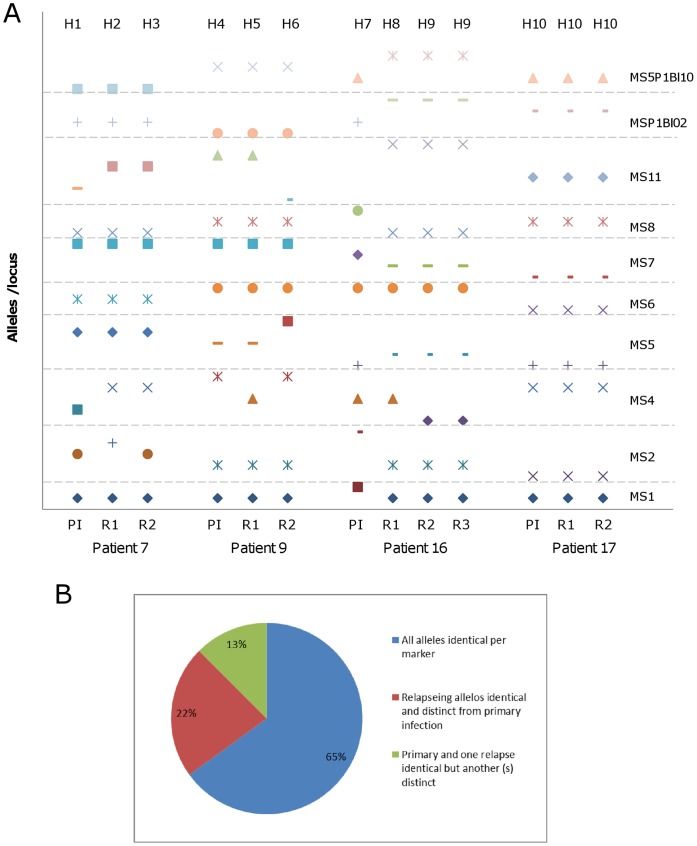
Few predominant alleles were detected by each marker, in general ranging from 5 to 12; one marker (MS11) identified 31 alleles (Table 1 and Table S2). The number of all alleles (predominant and rare) detected in the primary and relapse episodes was low. The average number of alleles was 9.2±2.3 and 10.8±2.7 for the predominant and all alleles, respectively, without including MS11 data, and 11.4±7.3 and 13.4±8.7 with MS11 data. A high frequency of predominant alleles of the same size was observed in the majority of markers. For all alleles (predominant and rare), this frequency increased from 40 to 77% (Fig. 1B). This finding reflects variation in how well the technique could detect low frequency alleles among the 10 markers used and suggested a significant temporal variability in predominant and rare alleles during the course of infection. In four patients with multiple recurrent infections, it was possible to confirm this temporal variation in predominant alleles (Fig. 2A). In these patients, each marker identified between two and 6 predominant alleles. Usually, those markers identified the same predominant allele in all consecutive samples from each patient in multiple malaria episodes (25 of 40, 65%) (Fig. 2B). Nine of the 15 remaining alleles were identical during relapses, but distinct from the primary infection. The combination of these alleles resulted in 10 haplotypes (H1 to H10). Only one patient harbored the same haplotype in all episodes (Patient 17; Fig. 2).
Table 1. Allele frequencies (%) and genetic variability of each molecular marker from Plasmodium vivax isolates.
| Percentage of episodes (%) | ||||||||||
| Alleles | MS1 | MS2 | MS4 | MS5 | MS6 | MS7 | MS8 | MS11 | MSP1bl2 | MSP1bl10 |
| 1 | 11 | 1 | 11 | 5 | 5 | 3 | 2 | 2 | 1 | 17 |
| 2 | 9 | 5 | 6 | 28 | 3 | 34 | 3 | 2 | 12 | 5 |
| 3 | 71 | 3 | 20 | 11 | 9 | 25 | 28 | 3 | 23 | 9 |
| 4 | 3 | 1 | 1 | 6 | 58 | 5 | 31 | 3 | 8 | 14 |
| 5 | 6 | 54 | 8 | 31 | 3 | 17 | 16 | 2 | 29 | 15 |
| 6 | 5 | 1 | 8 | 1 | 12 | 5 | 3 | 8 | 1 | |
| 7 | 8 | 5 | 2 | 1 | 3 | 16 | 8 | 18 | 14 | |
| 8 | 6 | 1 | 5 | 6 | 1 | 3 | 3 | |||
| 9 | 15 | 17 | 2 | 1 | 2 | 1 | ||||
| 10 | 1 | 12 | 3 | 3 | 2 | 15 | ||||
| 11 | 5 | 5 | 2 | 3 | ||||||
| 12 | 12 | 3 | 11 | 1 | ||||||
| 13 | 2 | |||||||||
| 14 | 6 | |||||||||
| 15 | 2 | |||||||||
| 16 | 2 | |||||||||
| 17 | 3 | |||||||||
| 18 | 2 | |||||||||
| 19 | 3 | |||||||||
| 20 | 3 | |||||||||
| 21 | 5 | |||||||||
| 22 | 2 | |||||||||
| 23 | 3 | |||||||||
| 24 | 3 | |||||||||
| 25 | 3 | |||||||||
| 26 | 3 | |||||||||
| 27 | 2 | |||||||||
| 28 | 8 | |||||||||
| 29 | 3 | |||||||||
| 30 | 2 | |||||||||
| 31 | 3 | |||||||||
| Total | 5 | 10 | 12 | 10 | 12 | 8 | 7 | 31 | 7 | 12 |
| H E | 0.482 | 0.681 | 0.888 | 0.808 | 0.647 | 0.789 | 0.783 | 0.969 | 0.813 | 0.886 |
MS: microsatellites [36]; MSP1bl2 and MSP1bl10: merozoite surface protein 1 block 2 and 10; H E: Expected heterozygosity; Total: number of distinct alleles for each marker. Alleles were numbered from smallest to the highest, each marker showed distinct sizes for the alleles, please see Supplementary Table S2 for the sizes of each allele.
Figure 2. Temporal variation of the predominant alleles.
(A) Comparison of predominant alleles among primary infection (PI), first relapse (R1), second relapse (R2), and third relapse (R3) from four P. vivax-infected patients genotyped using 10 molecular markers. Alleles are represented by different forms for each marker (indicated on the right side) and delimited by dotted lines. MS – microsatellite numbered according to Rezende et al. [36], MSP1bl2 and MSP1bl10– merozoite surface antigen 1 blocks 2 and 10, respectively. Haplotypes are indicated at the top of the Figure. (B) Frequencies of markers showing the same or distinct alleles at different times of blood collection for these four patients.
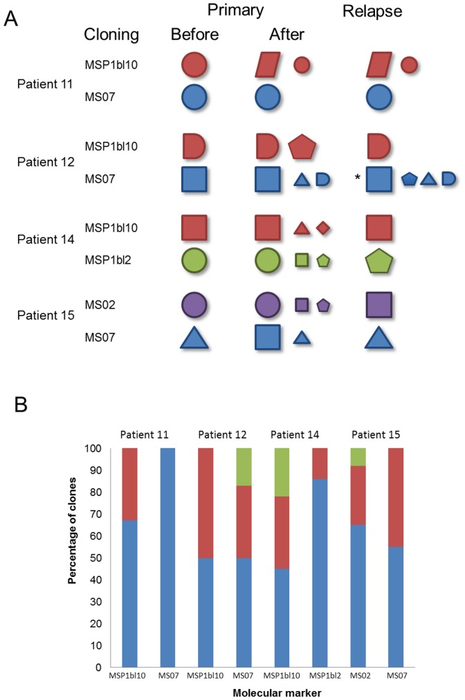
To clarify whether the detection of distinct alleles in relapses was due to their presence, although at low levels, in the preceding acute episodes, PCR products of two randomly selected markers from the primary infection of four patients were cloned, and up to 26 bacterial colonies (mean 11) were genotyped per product (Fig. 3). The majority of cloned samples were found to include more than one allele, representing a multiple-clone infection in both initial and recurrent infections. Up to 4 distinct alleles per marker were identified in a single patient (Fig. 3A). The frequency of colonies with the same allele ranged from 8 to 100% (Fig. 3B). These data strongly support the hypothesis that those distinct alleles detected in the recurrent infections correspond to undetected low frequency clones present in the primary infection.
Figure 3. Genotypic profile before and after PCR cloning.
PCR products from primary infection samples amplified using two randomly chosen molecular markers of four patients were cloned, and up to 26 colonies (mean of 11 colonies) were genotyped. (A) Each form represents an allele, size indicates predominant (larger) or rare alleles (smaller); color represents alleles of a distinct marker: MSP1bl10– red; MS07– blue; MSP1bl2– green; MS02– purple. The presence of two or more forms characterizes a multiple-clone infection. Before cloning the predominant allele was identified as the heighest peak in genotyping and the rare allele as the peak with one-third of the predominant peak height. After cloning the frequencies were inferred by the number of bacteria clones. The only relapse sample also cloned is indicated by an asterisk. (B) Frequency of each allele in primary infections after cloning measured by the percent of bacterial colonies genotyped with each allele (represented by different colors).
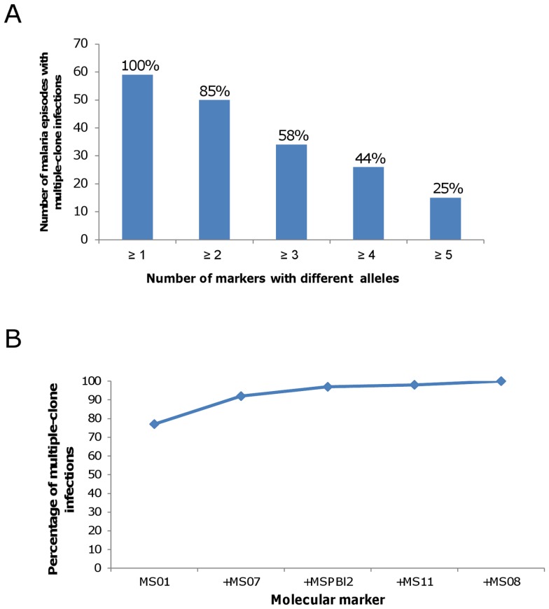
To further assess whether rare alleles were not identified in the primary infection because of low levels of fluorescence, we re-analyzed the original electropherogram peaks. Predominant peaks could have different heights, while the rare ones may have been of similar height (Figure S2). As an example of genotyping, in one patient the higher peak corresponding to the predominant allele had 455 rFU, and the rare allele had 153 rFU; that is, around 33% the predominant peak height. Genotyping the same marker for a second patient showed a higher peak of 4665 rFU and a lower peak with similar intensity of fluorescence as before (188 rFU); that is 4% of the predominant peak height. Reinforcing these data, the presence of rare alleles was further confirmed by cloning (data not shown). Thus, by properly adjusting the cut-off (≥150 rFU), the molecular markers detected a high frequency of multiple-clone infections. In 59 of 65 samples (91%) multiplicity of infection was detected by at least one marker (Fig. 4A). Based on a minimum of two markers, the percent of multiple infections detected decreased to 77%, and, with a minimum of three markers, it was 52% (Fig. 4A). We also observed a wide variation among the molecular markers in their ability to identify multiple infections, with two microsatellite markers (MS01 and MS07) accounting for most of the detection (Fig. 4B). Although five markers were able to detect all multiple-clone infections, the results suggested that few markers could be used to detect the majority of multiple-clone infections.
Figure 4. Detection of multiple-clone P. vivax infections using a panel of 10 markers.
(A) Number and percent of malaria episodes showing multiple-clone infections detected by different numbers of markers. (B) Minimum number of markers able to detect all multiple clone infections was five: MS01 (77%), MS01+ MS07 (92%), MS01+ MS07+ MSPBl2 (97%), MS01+ MS07+ MSPBl2+ MS11 (98%), MS01+ MS07+ MSPBl2+ MS11+ MS08 (100%).
Discussion
The activation of heterologous hypnozoites seems to be the most common cause of malaria recurrences [27], [29], [31], [39]. The results presented here reinforce previous studies, showing that the majority of relapse episodes were caused by a parasite population distinct from the primary infection. The novel finding of our study was the identification of high multiplicity not only in primary infections but also in relapses. These findings add complexity to the concept of heterologous activation, since they suggest that the allele present in relapses might also be present in the primary infection as a rare allele. Furthermore, the predominant parasite in the primary infection might not be predominant in the relapse, which shows that the frequency of circulating parasite clones alters considerably in P. vivax recurrences. Consequently, activation in relapse might be from homologous or heterologous hypnozoites or both (Table 1). Koepfli et al. [40] similarly reported temporal variation in the predominant allele during the course of P. vivax primary infection. In a study of P. vivax patients in the Peruvian Amazon, Van Den Eede et al. [39] reinforced this hypothesis of high turnover of parasite genotypes in recurrences. These findings point to the need for further studies to analyze multiple-clone infection during P. vivax recurrence, specifically with respect to primaquine resistance.
Numerous studies have addressed the mechanisms of hypnozoite activation. Recently, data from a study of infants in Thailand demonstrated that the first P. vivax relapse in life is usually caused by genetically homologous parasites. The authors suggest that this reflects the lack of previously acquired hypnozoites to be activated [41]. Accordingly, in the present study, five of seven patients showing a first malaria episode of their life exhibited a single-clone infection, with homologous parasites in recurrences. Notwithstanding, it is important to consider that malaria infection can be induced by the inoculation of more than one clone of sporozoites (multiple-clone infection) and, as hypothesized here, more than one clone of hypnozoites can remain dormant until some are activated. Consequently, it is not possible to determine if the heterologous hypnozoites are the first activated, which would explain their prevalence in relapses, or if the prevalence is related to the number of heterologous/homologous dormant clones that could be activated [27]. Since, in the current study, most of the initial infections did not correspond to the first sporozoite inoculation of life, previous infections could also be a source of heterologous hypnozoites.
Our aim was to improve sensitivity of detecting multiple-clone infections. The approach used to identify rare alleles was based on analysis of the electropherogram using a low cut-off level (≥150 rFU). By cloning PCR products, it was possible to confirm the specificity of this strategy and identify high levels of multiple-clone infections. By using a more common criterion to detect rare peaks, based on quantification of peak heights [42], [43], multiple-clone infection was confirmed in our primary/relapse samples (Fig. 5). However, this criterion has limitations, depending on the height of the predominant peak. Moreover, as multiplicity of infection has been demonstrated for different P. vivax populations [31], [32], [44]–[47], we believe that it is a common phenomenon of relapse parasites, that is not yet identified due to the low sensitivity of previous protocols. In order to reduce the artifact in genotyping, we used several strategies: repeat genotyping using different PCR products from the same patient to avoid dropout; confirming rare alleles using cloning before genotyping to detect null or silent alleles; and reduced stutter peaks (peaks closer to, or attached to, the main peak result from DNA slippage during PCR) in PCR standardization or discarding them from the analysis. In conclusion, our approach is useful to detect rare clones, but should be used with caution to avoid an overestimation of multiple-clone infections.
Figure 5. Percent of multiple-clone infections using different cut-off criteria.
The detection of rare alleles in the genotyping was based on three cut-off criteria: ≥150 rFU (here); peaks with more than one quarter [46]; or with more than one third of the predominant peak height [47].
Our results showed high haplotype variability and multiplicity of clones in parasites from relapsed patients. These findings complicate the concept of heterologous activation, suggesting the involvement of undetermined mechanisms based on host or environmental factors in the simultaneous activation of multiple clones of hypnozoites. This study provided new insights into molecular biology of malaria relapse that must to be considered in control strategies for the disease.
Supporting Information
Interval between primary infection and relapse infections. Time interval in months between primary and relapse per individual (A) and frequency of episodes at differing intervals (B). Repeated individual number represents a second relapse episode (red bar) and a third relapse episode (green bar) in the same patient. Interval of second and third relapses was measured in relation to previous acute malaria episode. Primary infections corresponding to the first malaria infection of the individual’s life are denoted by an asterisk (above the bars).
(TIFF)
Genotyping of two DNA samples with distinct profiles for the same marker (MSP1Bl02). (A) Multiple infection detected in which the minor peak showed around 33% of the fluorescence level (rFu) of the predominant peak (blue peaks). (B) Multiple infection detected in which the lower peak (rare allele) showed 4% of fluorescence level of the predominant peak. Fragment sizes are represented on the × axis of the graph. Red peaks represents the molecular marker used (MegaBACE™ ET550-R).
(TIFF)
Description of patient characteristics.
(DOCX)
Characteristics of predominant alleles from genotyping of 10 molecular markers of Plasmodium vivax infected patients.
(DOCX)
Acknowledgments
The authors thank Dr. Gehard Wunderlich for his critical review and suggestions for the manuscript. We are grateful to all patients who made this study possible. We also thank the Program for Technological Development in Tools for Health - PDTIS platform (FIOCRUZ) for DNA sequencing facilities.
Funding Statement
This work was supported by the Pronex malaria network: CNPq/Ministry of Health-DECIT; FAPEMAT; and FAPEMIG. CAB, LHC and CJF were supported by CNPq fellowships. FFA and AMR are supported by scholarships from CNPq and Fapemig, respectively. The funders had no role in study design, data collection and analysis, decision to publish, or preparation of the manuscript.
References
- 1. Hay SI, Guerra CA, Tatem AJ, Noor AM, Snow RW (2004) The global distribution and population at risk of malaria: past, present, and future. Lancet Infect Dis 4: 327–336. [DOI] [PMC free article] [PubMed] [Google Scholar]
- 2. Gething PW, Patil AP, Smith DL, Guerra CA, Elyazar IR, et al. (2011) A new world malaria map: Plasmodium falciparum endemicity in 2010. Malar J 10: 378. [DOI] [PMC free article] [PubMed] [Google Scholar]
- 3.World Health Organization (2010) World Malaria Report 2010. Geneva: World Health Organization.
- 4. Guerra CA, Howes RE, Patil AP, Gething PW, Van Boeckel TP, et al. (2010) The international limits and population at risk of Plasmodium vivax transmission in 2009. PLoS Negl Trop Dis 4: e774. [DOI] [PMC free article] [PubMed] [Google Scholar]
- 5.Ministério da Saude, Secretaria de Vigilancia em Saude, Departamento de Vigilancia Epidemiologic. (2010) Aspectos epidemiológicos da Malária. Brasilia: Ministerio da Saude.
- 6. Craige B, Alving AS, Jones R, Merrill Whorton C, Pullman TN, et al. (1947) The Chesson strain of Plasmodium vivax malaria. I. Relationship between prepatent period, latent period and relapse rate. J Infect Dis 80: 228–236. [DOI] [PubMed] [Google Scholar]
- 7. Adak T, Sharma V, Orlov V (1998) Studies on the Plasmodium vivax relapse pattern in Delhi, India. Am J Trop Med Hyg 59: 175–179. [DOI] [PubMed] [Google Scholar]
- 8. Contacos PG, Collins WE, Jeffery GM, Krotoski WA, Howard WA (1972) Studies on the characterization of Plasmodium vivax strains from Central America. Am J Trop Med Hyg 21: 707–712. [DOI] [PubMed] [Google Scholar]
- 9. Warren M, Garnham P (1970) Plasmodium cynomolgi: X-irradiation and development of exo-erythrocytic schizonts in Macaca mulatta. . Exp Parasitol 28: 551–556. [DOI] [PubMed] [Google Scholar]
- 10. Douglas NM, Nosten F, Ashley EA, Phaiphun L, van Vugt M, et al. (2011) Plasmodium vivax Recurrence Following Falciparum and Mixed Species Malaria: Risk Factors and Effect of Antimalarial Kinetics. Clin Infect Dis 52: 612–620. [DOI] [PMC free article] [PubMed] [Google Scholar]
- 11. Hulden L, Hulden L (2011) Activation of the hypnozoite: a part of Plasmodium vivax life cycle and survival. Malar J 10: 90. [DOI] [PMC free article] [PubMed] [Google Scholar]
- 12. James S, Shute P (1926) Report on the first results of laboratory work on malaria in England. League of Nations. Health Organization. Geneva. C. H. Malaria 57: 30. [Google Scholar]
- 13. Boyd M, Stratman T, Kitchen S (1936) On the duration of homologous immunity to Plasmodium vivax . Am J Trop Med 16: 311–315. [Google Scholar]
- 14. Boyd M (1947) A review of studies on immunity to vivax malaria. J Natl Malar Soc 6: 12–31. [PubMed] [Google Scholar]
- 15. Sinton J, Bird W (1928) Studies in malaria with special reference to treatment; plasmoquine in treatment of malaria. Indian J Med Research 16: 159. [Google Scholar]
- 16. Gogtay NJ, Desai S, Kamtekar KD, Kadam VS, Dalvi SS, et al. (1999) Efficacies of 5- and 14-day primaquine regimens in the prevention of relapses in Plasmodium vivax infections. Ann Trop Med Parasitol 93: 809–812. [DOI] [PubMed] [Google Scholar]
- 17.Gill C (1938) The seasonal periodicity of malaria and the mechanism of the epidemic wave. JA Churchill Ltd, London.
- 18. Goller J, Jolley D, Ringwald P, Biggs BA (2007) Regional differences in the response of Plasmodium vivax malaria to primaquine as anti-relapse therapy. Am J Trop Med Hyg 76: 203–207. [PubMed] [Google Scholar]
- 19. Howe C, Duff F (1946) Effect of altitude anoxia in provoking relapse in malaria Science. 103: 223. [DOI] [PubMed] [Google Scholar]
- 20. White N (2011) Determinants of relapse periodicity in Plasmodium vivax malaria. Malar J 10: 297. [DOI] [PMC free article] [PubMed] [Google Scholar]
- 21. Orjuela-Sánchez P, da Silva N, da Silva-Nunes M, Ferreira MU (2009) Recurrent parasitemias and population dynamics of Plasmodium vivax polymorphisms in rural Amazonia. Am J Trop Med Hyg 81: 961–968. [DOI] [PubMed] [Google Scholar]
- 22. Katsuragawa TH, Gil LH, Tada MS, de Almeida e Silva A, Costa JD, et al. (2010) The Dynamics of Transmission and Spatial Distribution of Malaria in Riverside Areas of Porto Velho, Rondônia, in the Amazon Region of Brazil. PLoS ONE 5: e9245. [DOI] [PMC free article] [PubMed] [Google Scholar]
- 23.Galappaththy G, Omari A, Tharyan P (2007) Primaquine for preventing relapses in people with Plasmodium vivax malaria. Cochrane Database of Systematic Reviews 1. Art. No.: CD004389. [DOI] [PubMed]
- 24. Craig A (1996) Kain (1996) Molecular analysis of strains of Plasmodium vivax from paired primary and relapse infections. J Infect Dis 174: 373–379. [DOI] [PubMed] [Google Scholar]
- 25. Kirchgatter K, Del Portillo H (1998) Molecular analysis of Plasmodium vivax relapses using the MSP1 molecule as a genetic marker. J Infect Dis 177: 511–515. [DOI] [PubMed] [Google Scholar]
- 26. Khusmith S (1998) Antigenic disparity of Plasmodium vivax causing initial symptoms and causing relapse. Southeast Asian J Trop Med Public Health 29: 519–524. [PubMed] [Google Scholar]
- 27. Imwong M, Snounou G, Pukrittayakamee S, Tanomsing N, Kim JR, et al. (2007) Relapses of Plasmodium vivax infection usually result from activation of heterologous hypnozoites. J Infect Dis 195: 927–933. [DOI] [PubMed] [Google Scholar]
- 28. Chen N, Auliff A, Rieckmann K, Gatton M, Cheng Q (2007) Relapses of Plasmodium vivax infection result from clonal hypnozoites activated at predetermined intervals. J Infect Dis 195: 934–941. [DOI] [PubMed] [Google Scholar]
- 29. Restrepo E, Imwong M, Rojas W, Carmona-Fonseca J, Maestre A (2011) High genetic polymorphism of relapsing P. vivax isolates in northwest Colombia. Acta Trop 119: 23–29. [DOI] [PMC free article] [PubMed] [Google Scholar]
- 30. Havryliuk T, Orjuela-Sánchez P, Ferreira MU (2008) Plasmodium vivax: microsatellite analysis of multiple-clone infections. Exp Parasitol 120 330–336. [DOI] [PubMed] [Google Scholar]
- 31. Van den Eede P, Erhart A, Van der Auwera G, Van Overmeir C, Thang ND, et al. (2010) High complexity of Plasmodium vivax infections in symptomatic patients from a rural community in central Vietnam detected by microsatellite genotyping. Am J Trop Med Hyg 82: 223–227. [DOI] [PMC free article] [PubMed] [Google Scholar]
- 32. Van den Eede P, Van der Auwera G, Delgado C, Huyse T, Soto-Calle VE, et al. (2010) Multilocus genotyping reveals high heterogeneity and strong local population structure of the Plasmodium vivax population in the Peruvian Amazon. Malar J 9: 151. [DOI] [PMC free article] [PubMed] [Google Scholar]
- 33.Ministério da Saude, Secretaria de Vigilancia em Saude, Diretoria Técnica de Gestão (2009) Guia prático de tratamento da malária no Brasil. Brasilia: Ministerio da Saude.
- 34. Rezende AM, Tarazona-Santos E, Fontes CJ, Souza JM, Couto AD, et al. (2010) Microsatellite loci: determining the genetic variability of Plasmodium vivax . Trop Med Int Health 15: 718–726. [DOI] [PubMed] [Google Scholar]
- 35. Koepfli C, Mueller I, Marfurt J, Goroti M, Sie A, et al. (2009) Evaluation of Plasmodium vivax genotyping markers for molecular monitoring in clinical trials. J Infect Dis 199: 1074–1080. [DOI] [PubMed] [Google Scholar]
- 36. Nishimura A, Morita M, Nishimura Y, Sugino Y (1990) A rapid and highly efficient method for preparation of competent Escherichia coli cells. Nucleic Acids Res 18: 6169. [DOI] [PMC free article] [PubMed] [Google Scholar]
- 37. Excoffier L, Laval G, Schneider S (2005) Arlequin (version 3.0): an integrated software package for population genetics data analysis. Evol. Bioinform Online 1: 47–50. [PMC free article] [PubMed] [Google Scholar]
- 38. Excoffier L, Smouse P, Quattro J (1992) Analysis of molecular variance inferred from metric distances among DNA haplotypes: application to human mitochondrial DNA restriction data. Genetics 131: 479–491. [DOI] [PMC free article] [PubMed] [Google Scholar]
- 39. Van den Eede P, Soto-Calle VE, Delgado C, Gamboa D, Grande T, et al. (2011) Plasmodium vivax sub-patent infections after radical treatment are common in Peruvian patients: results of a 1-year prospective cohort study. PLoS ONE 6: e16257. [DOI] [PMC free article] [PubMed] [Google Scholar]
- 40. Koepfli C, Schoepflin S, Bretscher M, Lin E, Kiniboro B, et al. (2011) How Much Remains Undetected? Probability of Molecular Detection of Human Plasmodia in the Field. PLoS ONE 6: e19010. [DOI] [PMC free article] [PubMed] [Google Scholar]
- 41. Imwong M, Boel ME, Pagornrat W, Pimanpanarak M, McGready R, et al. (2012) The first Plasmodium vivax relapses of life are usually genetically homologous. J Infect Dis 205: 680–683. [DOI] [PMC free article] [PubMed] [Google Scholar]
- 42. Anderson TJ, Su XZ, Bockarie M, Lagog M, Day KP (1999) Twelve microsatellite markers for characterization of Plasmodium falciparum from finger prick blood samples. Parasitology 119: 113–126. [DOI] [PubMed] [Google Scholar]
- 43. Anderson TJ, Haubold B, Williams JT, Estrada-Franco JG, Richardson L, et al. (2000) Microsatellite markers reveal a spectrum of population structures in the malaria parasite Plasmodium falciparum . Mol Biol Evol 17: 1467–1482. [DOI] [PubMed] [Google Scholar]
- 44. Karunaweera ND, Ferreira MU, Munasinghe A, Barnwell JW, Collins WE, et al. (2008) Extensive microsatellite diversity in the human malaria parasite Plasmodium vivax . Gene 410: 105–112. [DOI] [PubMed] [Google Scholar]
- 45. Ferreira MU, Karunaweera ND, da Silva-Nunes M, da Silva NS, Wirth DF, et al. (2007) Population structure and transmission dynamics of Plasmodium vivax in rural Amazonia. J Infect Dis 195: 1218–1226. [DOI] [PubMed] [Google Scholar]
- 46. Imwong M, Nair S, Pukrittayakamee S, Sudimack D, Williams JT, et al. (2007) Contrasting genetic structure in Plasmodium vivax populations from Asia and South America. Int J Parasitol 37: 1013–1022. [DOI] [PubMed] [Google Scholar]
- 47. Gunawardena S, Karunaweera ND, Ferreira MU, Phone-Kyaw M, Pollack RJ, et al. (2010) Geographic structure of Plasmodium vivax: microsatellite analysis of parasite populations from Sri Lanka, Myanmar, and Ethiopia. Am J Trop Med Hyg 82: 235–242. [DOI] [PMC free article] [PubMed] [Google Scholar]
Associated Data
This section collects any data citations, data availability statements, or supplementary materials included in this article.
Supplementary Materials
Interval between primary infection and relapse infections. Time interval in months between primary and relapse per individual (A) and frequency of episodes at differing intervals (B). Repeated individual number represents a second relapse episode (red bar) and a third relapse episode (green bar) in the same patient. Interval of second and third relapses was measured in relation to previous acute malaria episode. Primary infections corresponding to the first malaria infection of the individual’s life are denoted by an asterisk (above the bars).
(TIFF)
Genotyping of two DNA samples with distinct profiles for the same marker (MSP1Bl02). (A) Multiple infection detected in which the minor peak showed around 33% of the fluorescence level (rFu) of the predominant peak (blue peaks). (B) Multiple infection detected in which the lower peak (rare allele) showed 4% of fluorescence level of the predominant peak. Fragment sizes are represented on the × axis of the graph. Red peaks represents the molecular marker used (MegaBACE™ ET550-R).
(TIFF)
Description of patient characteristics.
(DOCX)
Characteristics of predominant alleles from genotyping of 10 molecular markers of Plasmodium vivax infected patients.
(DOCX)


