Abstract
Transcriptional regulations are involved in many aspects of plant development and are mainly achieved through the actions of transcription factors (TF). To investigate the mechanisms of plant development, we carried out genetic screens for mutants with abnormal shoot development. Taking an activation tagging approach, we isolated a gain-of-function mutant abs2-1D (abnormal shoot 2-1D). abs2-1D showed pleiotropic growth defects at both the vegetative and reproductive developmental stages. We cloned ABS2 and it encodes a RAV sub-family of plant B3 type of transcriptional factors. Phylogenetic analysis showed that ABS2 was closely related to NGATHA (NGA) genes that are involved in flower development and was previously named NGATHA-Like 1 (NGAL1). NGAL1 was expressed mainly in the root and the filament of the stamen in flower tissues and sub-cellular localization assay revealed that NGAL1 accumulated in the nucleus. Interestingly, over-expression of NGAL1 driven by the constitutive 35S promoter led to transgenic plants with conspicuous flower defects, particularly a loss-of-petal phenotype. A loss-of-function ngal1-1 mutant did not show obvious phenotype, suggesting the existence of redundant activities and also the utility of gain-of-function genetic screens. Our results show that the over-expression of NGAL1 is capable of altering flower petal development, as well as shoot development.
Introduction
In eukaryotic organisms, gene expression regulations can occur at multiple levels to ensure the proper elaboration of the information stored in the genetic materials. Among the numerous factors that are involved in these intricate regulatory networks, transcription factors (TFs) play pivotal roles at the transcription level and they are intimately involved in many aspects of development [1]. Considering the central roles they play, it is not surprising to see the presence of large numbers of TFs in eukaryotic genomes. The model plant Arabidopsis thaliana genome contains more than 1500 transcription factors, accounting for ∼6% of its estimated ∼27,000 genes genome [2]. Typically, TFs contain distinct types of DNA-binding domains and transcriptional regulation regions and are capable of activating or repressing the expressions of a large number of target genes [3]–[6].
One family of transcription factors that has been under extensive investigation in plants is the plant-specific B3 superfamily TFs, which contain a characteristic ∼110 amino acids B3 domain responsible for DNA binding [7]. The B3 domain was originally named because it is the third basic domain in the maize protein VIVIPAROUS1 (VP1) [8]. In Arabidopsis and rice, there are at least 118 and 91 B3 family genes, respectively [7]. Arabidopsis B3 family of TFs can be further grouped into four subfamilies: ARF (AUXIN RESPONSE FACTOR), LAV (LEAFY COTYLEDON2 -ABSCISIC ACID INSENSITIVE3–VAL), RAV (RELATED TO ABI3 and VP1) and REM (REPRODUCTIVE MERISTEM) [7].
In Arabidopsis, the RAV subfamily consists of at least 13 members, including RAV1, RAV2/TEMPRANILLO2 (TEM2), TEM1, NGATHA1-4 (NGA1-4) and NGATHA-like 1–3 (NGAL1-3), and members of this subfamily of TFs have been implicated in many developmental and physiological processes in plants [9]. RAV1 and RAV2 were initially identified based on the B3 domain that they share with maize VP1 [10]. However, RAV1 and RAV2, as well as four other RAV subfamily members, contain a second DNA binding domain, the AP2 domain, which is the hallmark domain in AP2 family of TFs, in addition to the B3 domain, and both DNA binding domains are capable of binding DNA [10]. RAV1 expression is down-regulated by the application of the phytohormone brassinosteroid and it may be a factor that negatively regulates leaf initiation, lateral root development and flowering transition [11]. RAV2/TEM2, as well as TEM1, may be regulators of flowering time and TEM1 can directly bind to Flowering Locus T (FT) promoter and negatively represses FT expression and flowering [12]. RAV1 and RAV2 expressions are also up-regulated by mechanical stimuli such as touch, wind, spray and transfer [13]. The NGATHAs and NGATHA-likes (NGA1-NGA4; NGAL1-NGAL3) are members of RAV subfamily that only contain the B3 DNA binding domain [9], [14]. The nga1 mutant was isolated in genetic modifier screens in pickle-15 kanadi1-2 (pkl-15 kan1-2) or kan1-2 kan2-1/+ backgrounds, while NGA3 was identified through the gain-of-function tower-of-pisa1 (top1) mutant [9], [14]–[17]. Although single nga1 mutants only exhibit subtle developmental phenotypes, they promote valve-like outgrowth, instead of the style-like outgrowth in gynoecium tissues in pkl–15 kan1–2 or kan1–2 kan2-1/+ backgrounds, suggesting that they are involved in the regulation of carpel polarity [9]. Quadruple nga1 nga2 nga3 nga4 mutant exhibits a conversion of style to valve-like structures and this coincides with a reduced expression of the style-specific gene SHATTERPROOF1 [9], [14]. Overall, one common theme seems to be that most, if not all, RAV subfamily members are involved in aspects of flower development.
Although much is now known about the functions of plant transcription factors and how they regulate plant growth and development, functions of many transcription factors remain poorly characterized. Novel investigation approaches, such as gain-of-function mutant screens, are offering new insights into TF functions [18]. We have identified numerous dominant gain-of-function mutants with altered shoot development and one of the semi-dominant mutants that we named abs2-1D (abnormal shoot 2-1D) was characterized here. abs2-1D displays a spectrum of phenotypes including a small plant stature, a faster leaf initiation rate, smaller leaves with abnormal shape and early flowering. We determined that the phenotypes of abs2-1D were the result of an elevated expression of ABS2, also called NGAL1, which is a RAV subfamily member of B3 transcription factor family. NGAL1 was highly expressed in roots, flowers and siliques and its protein product is located in the cell nucleus. Interestingly, we found that the Cauliflower Mosaic Virus (CaMV) 35S promoter-driven NGAL1 over-expression led to additional flower defects including a loss-of-petal phenotype. Our results suggest that NGAL1, when over-expressed, is capable of altering many facades of plant development.
Results
The Identification of abs2-1D Mutant
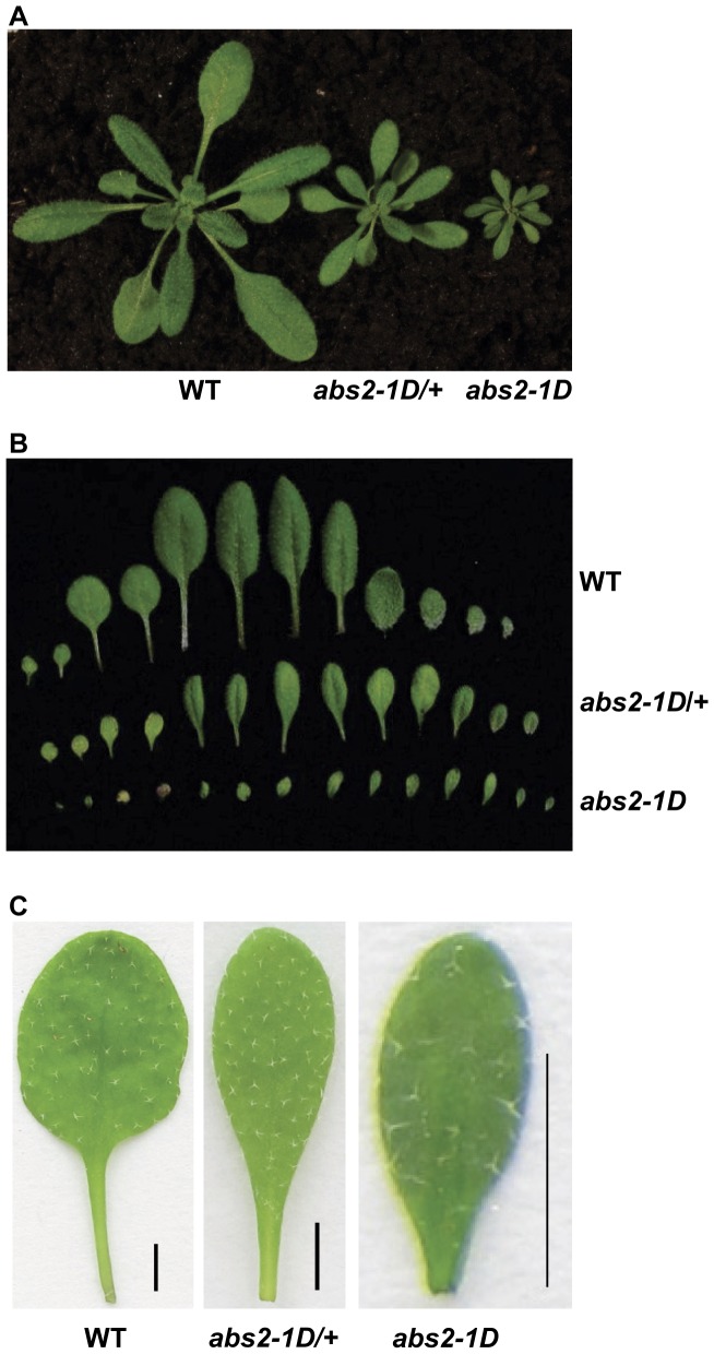
In our previous work looking for genetic suppressors of the Arabidopsis yellow variegated (var2) mutant, we carried out var2 activation tagging mutagenesis [19]. Activation tagging is a modified T-DNA insertional mutagenesis: on one hand it can generate loss-of-function insertional mutants like the traditional T-DNA mutagenesis; on the other hand this procedure is also capable of producing gain-of-function mutants with the inclusion of four copies of the CaMV 35S enhancers near the right border of the T-DNA [18]. The main mechanism underlying activation tagging is the activation of transcriptions of genes adjacent to the T-DNA insertion site. In our large-scale screens for var2 suppressors, we also identified a series of dominant mutants with altered shoot development that we named abs (abnormal shoot) mutants [20]. One semi-dominant mutant, abnormal shoot2-1D (abs2-1D; D for dominant), was characterized (Figure 1A). Both the heterozygous and the homozygous abs2-1D mutants were smaller in sizes than wild type (Figure 1A). One pronounced feature of the mutants was the abnormal leaf shape (Figure 1B–C). In wild type plants, there were clear distinctions between the leaf petiole and the leaf blade, while in abs2-1D the distinction between these two basic structures of a leaf was obscured and a gradual transition from petiole to blade was observed, and the leaves were also smaller (Figure 1B–C). In addition, abs2-1D mutant plants had a consistently higher leaf initiation rate at the vegetative stage (Figure 1A–B; Figure S1A). At the flowering stage, the heterozygous mutants developed shorter inflorescence than wild type and had reduced fertility, producing siliques that were much shorter than those of wild type (Figure S2A–B). The homozygous mutants were sterile under our growth conditions (Figure 1A).
Figure 1. Phenotypes of abs2-1D.
A. Three-week-old wild type, abs2-1D/+ heterozygous and abs2-1D homozygous plants. Plants were grown at 22°C under continuous illumination of ∼100 µmol·m−2·s−1. B. Cotyledons and rosette leaves of three-week-old wild-type, abs2-1D/+ heterozygous and abs2-1D homozygous plants. From left to right are two cotyledons and rosette leaves that were arranged in the order of their initiations. C. Comparison of the fifth rosette leaves of three-week-old wild type, abs2-1D/+ heterozygous and abs2-1D homozygous plants. Leaves were flattened between glass slides before photographing (Bars, 2 mm).
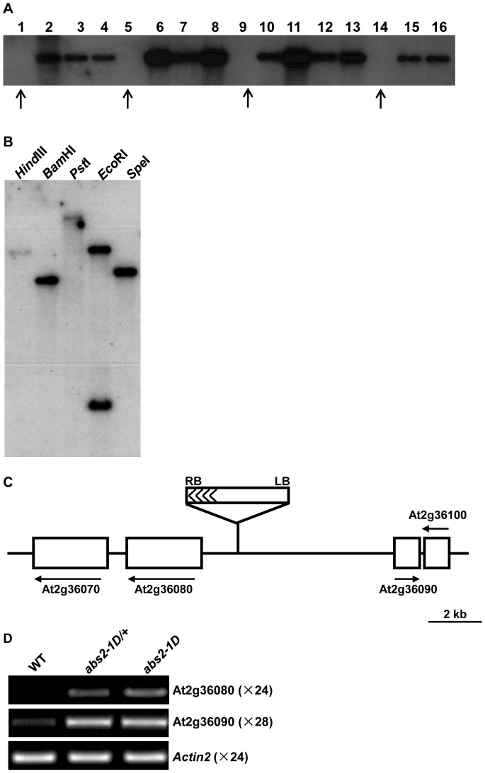
To investigate whether the phenotype of abs2-1D was linked with T-DNA insertion event(s), we carried out a co-segregation test. In a segregating population of abs2-1D, we performed southern blot analysis to see if T-DNA was associated with the abs2-1D phenotypes. Figure 2A shows that for all the plants that had the abs2-1D phenotypes, they also contained a T-DNA insertion, indicating that a T-DNA insertion is linked with abs2-1D phenotypes. Southern blot analysis with various restriction enzymes also indicated that the T-DNA insertion in abs2-1D was likely a single copy event (Figure 2B).
Figure 2. The cloning of ABS2.
A. abs2-1D was genetically linked with T-DNA. Total leaf DNAs were extracted from 16 progenies of an abs2-1D/+ heterozygous plant. The DNAs were digested with HindIII and restriction fragments were separated with electrophoresis followed by transfer to a nylon membrane. The blot was probed with 32P labeled BAR gene sequences. Plants that did not show abs2-1D phenotypes were indicated by arrows. B. Confirmation of a single T-DNA insertion in abs2-1D. Genomic DNAs from abs2-1D plants were digested with indicated restriction enzymes. After electrophoresis and transfer to a nylon membrane, the blot was hybridized with 32P labeled BAR gene sequences. There is one EcoRI site in the probe sequence so two hybridizing bands were observed. C. Cloning of abs2-1D. In the abs2-1D mutant, activation tagging T-DNA was inserted between At2g36080 and At2g36090. Solid lines represent intergenic regions, while white boxes represent genes in the vicinity of the T-DNA insertion. The right border of the T-DNA was facing At2g36080. D. Semi-quantitative RT-PCR analysis of the expression levels of At2g36080 and At2g36090 in wild-type, abs2-1D/+ heterozygous and abs2-1D homozygous mutants. Actin2 expression was shown as a control. Total cellular RNAs were extracted from the aerial parts of two-week-old seedlings. 1 µg DNase I treated RNA from each sample was used for cDNA synthesis. RT-PCRs were performed with indicated numbers of cycles.
The Cloning of ABS2
Since the abs2-1D phenotype was genetically linked with a single T-DNA insertion, we carried out plasmid rescue to identify the T-DNA insertion site in abs2-1D. We chose BamHI as the enzyme because of its relatively smaller southern signal size (Figure 2B). Sequencing of the rescued plasmid showed that plant genomic DNA sequence outside the left border of the T-DNA was recovered and sequence analysis showed that the T-DNA was inserted between genes At2g36080 and At2g36090 on chromosome 2, with the T-DNA right border facing At2g36080. The physical distance between the insertion site and the start codon of At2g36080 was 1367 base pairs (Figure 2C). Given the semi-dominant nature of abs2-1D and the feature of activation tagging, it is likely that the genes flanking the T-DNA, particularly the gene outside of the right border, could be the target gene whose expression is elevated in the mutant. We tested the expressions of At2g36080 and At2g36090 in abs2-1D and semi-quantitative RT-PCR showed increased levels of At2g36080 and At2g36090 transcripts in both the abs2-1D heterozygous and homozygous mutant plants (Figure 2D).
At2g36080 is ABS2
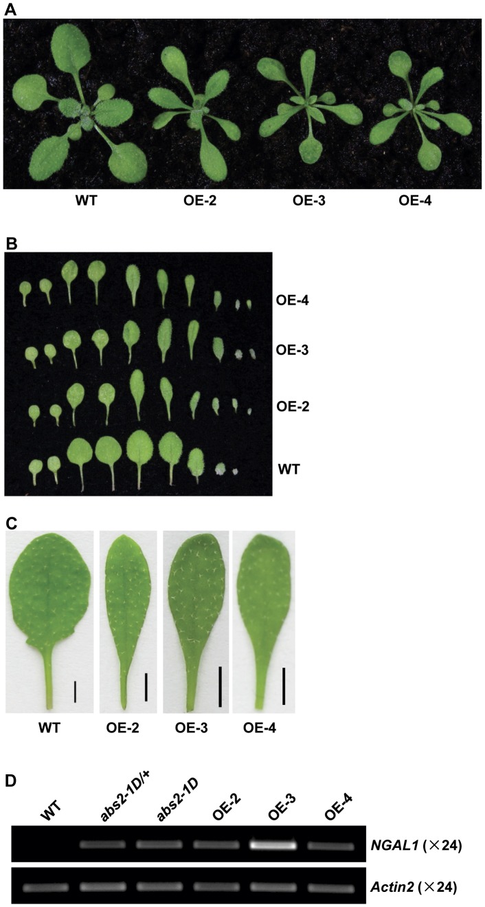
To confirm whether the over-expression of At2g36080 or At2g36090, was the cause behind the abs2-1D phenotypes, we carried out independent over-expression experiments. Full-length cDNA of At2g36080 was cloned into a binary vector and placed under the control of the CaMV 35S promoter. This vector was transformed into wild type Arabidopsis and transgenic At2g36080 over-expression (OE) plants were selected at T1 generation and allowed to self. At T2 generation, multiple At2g36080 OE transgenic lines showed leaf phenotypes similar to those of abs2-1D, in terms of the smaller plant size, the gradual leaf petiole to leaf blade transition and the faster initiation of leaves (Figure 3A–C; Figure S1B). In these lines, expressions of At2g36080 were increased to levels similar to or higher than those in abs2-1D (Figure 3D). These results indicate that the over-expression of At2g36080 is likely the cause for abs2-1D phenotypes and At2g36080 is ABS2.
Figure 3. Phenotypes of NGAL1 over-expression lines.
A. Phenotypes of two-week-old wild type and three independent NGAL1 over-expression (OE) lines, OE-2, OE-3 and OE-4. B. Cotyledons and rosette leaves detached from two-week-old wild-type, OE-2, OE-3 and OE-4 plants. From left to right were two cotyledons and rosette leaves that were arranged in the order of their initiations. C. Comparison of the fifth rosette leaf of two-week-old wild type, OE-2, OE-3 and OE-4 plants (Bars, 2 mm). D. Semi-quantitative RT-PCR analysis of the expression levels of NGAL1 in wild-type, abs2-1D/+ heterozygous, abs2-1D homozygous, OE-2, OE-3 and OE-4 plants. RT-PCRs were carried out as in Figure 2D.
Phylogenetic Analysis
The ORF Finder analysis showed that the coding region of ABS2/At2g36080 was 735 bp in length, encoding a protein product of 244 amino acids. Sequence analysis revealed that ABS2 is a member of the RAV subfamily of the plant B3 transcription factors and is closely related to the NGATHA (NGA) proteins [9]. In a previous study, ABS2/At2g36080 was named NGATHA-Like1 (NGAL1) and ABS2/At2g36080 will be referred to as NGAL1 hereafter [9]. To further determine the evolutionary distance among the RAV proteins, phylogenetic analysis was carried out and Figure S3A shows that NGAL1 and NGA proteins were in the same clade, among the seven genes in the RAV sub-family that only contain the B3 domain. To support the phylogeny reconstruction, we compared intron and exon structures of all the RAV subfamily genes from Arabidopsis (Figure S3B). RAV genes have simple gene structures with most members only include one exon. Genes grouped in the clade including NGAL1 have relatively complex gene structures of two or three exons (Figure S3B).
Expression Profiles of NGAL1
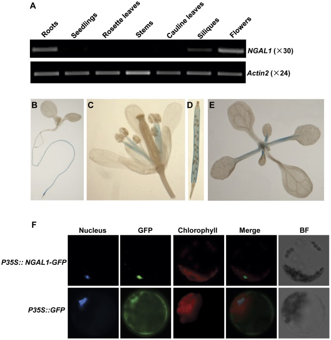
To examine the tissue expression pattern of NGAL1, we monitored its expressions in different Arabidopsis tissues including roots, two-week-old seedlings, rosette leaves, stems, cauline leaves, siliques, and flowers with semi-quantitative RT-PCR. Figure 4A shows that NGAL1 was most highly expressed in the flower and root tissues. Although at lower levels, NGAL1 transcripts were also present in siliques and two-week old seedlings (Figure 4A). Only very low levels of NGAL1 expression were detected from tissues of rosette leaves, stems and cauline leaves (Figure 4A). Our results indicate that NGAL1 expression shows some level of tissue specificity.
Figure 4. NGAL1 tissue expression profile and NGAL1 protein localization.
A. Expressions of NGAL1 in different tissues of wild type plants were determined by semi-quantitative RT-PCR. Total RNAs were extracted from roots, two-week-old seedlings, rosette leaves, stems, cauline leaves, siliques and flower tissues and semi-quantitative RT-PCRs were carried out as in Figure 2D. Actin2 expression was shown as a control. B–E. Tissue expression pattern of NGAL1 examined by histo-chemical GUS staining. Illustrated are one-week-old seedling (B), flower (C), silique (D) and two-week-old seedling (E) from transgenic plants expressing PNGAL1::GUS fusion construct. F. Nuclear localization of NGAL1-GFP fusion protein in Arabidopsis leaf protoplasts. Nuclei of protoplasts were stained by Hoechst 33342. GFP fluorescence and bright field (BF) images of Arabidopsis protoplasts were compared to show the sub-cellular localization of GFP (cytosol and nucleus) and NGAL1-GFP (nucleus).
To further investigate the NGAL1 expression profile, we constructed an NGAL1 promoter-GUS fusion vector using a ∼1.2 kb promoter region upstream of its start codon. Wild type plants were transformed with this PNGAL1-GUS construct and GUS activities were assayed at T2 generation in six independent transgenic lines and they showed similar GUS activities (Figure 4B–E). Consistent with our RT-PCR results, we found major GUS activities in roots and flowers (Figure 4B–C). In the flowers, NGAL1 expression was found primarily in the filament tissues of the stamen (Figure 4C). In the siliques, NGAL1 was expressed in tissues including pericarp, but not in developing seeds (Figure 4D). In the vegetative tissues, NGAL1 expression was found at leaf petioles (Figure 4E). Promoter-GUS assay further demonstrate the tissue specific expression profile of NGAL1.
NGAL1 is Targeted to the Nucleus
To study the sub-cellular localization of NGAL1, the full-length NGAL1 coding sequence was fused in-frame at its 3′ terminus with the GFP coding sequence to create a translational NGAL1-GFP fusion protein and the expression of this fusion protein was driven by the CaMV 35S promoter. The control vector contained GFP alone, which was also under the control of the 35S promoter. The two constructs were transformed and transiently expressed in Arabidopsis leaf protoplasts respectively. Fluorescent dye Hoechst 33342 was used to specifically indicate the nucleus [20], [21]. For the control vector, GFP green fluorescence signals were observed in both the cytosol and the nucleus (Figure 4F). In contrast, we observed NGAL1-GFP fluorescence signals that co-localized exclusively with the Hoechst 33342 fluorescence, suggesting that NGAL1 is a nuclear protein (Figure 4F).
The Over-expression of NGAL1 Leads to the Loss of Flower Petals
Upon closer examinations of the 35S promoter-driven NGAL1 OE lines, we observed that multiple NGAL1 OE lines showed pleiotropic development defects, in addition to the leaf phenotypes. Independent NGAL1 OE lines, including OE-2 and OE-3, showed clear formations of inflorescence after 4 weeks of growth, when wild type plants were just starting to bolt (Table 1; Figure S4A). NGAL1 OE lines also had more rosette leaves at bolting compared to wild type (Table 1). NGAL1 OE lines produced flowers with reduced sizes and flower organs, including sepals, stamens and carpels were smaller in NGAL1 OE lines compared with those of wild type (Figure 5A). Even more dramatic was the finding that strong NGAL1 OE lines showed the absence of flower petals, the second whorl of the typical four whorls of Arabidopsis wild type flower organs (Figure 5A–D; Table 2). There were also intermediate petal phenotypes as we observed abnormal filamentous structures instead of petals in some flowers (Figure 5E; Table 2). We determined that NGAL1 was indeed over-expressed in the petal-less flowers of independent OE lines, correlating the over-expression of NGAL1 with the loss-of-petal phenotype (Figure S4B). Our results indicate that the over-expression of NGAL1 is capable of altering flower development, particularly petal development in Arabidopsis.
Table 1. Comparison of the bolting times of wild type and NGAL1 over-expression lines.
| Number of leavesat bolting | Bolting time (DAG) | |
| wild type | 16.88±0.72 | 27.62±0.60 |
| OE-2 | 18.17±1.69, p<0.01 | 23.41±0.69, p<0.01 |
| OE-3 | 18.93±0.70, p<0.01 | 23.84±1.51, p<0.01 |
| OE-4 | 19.23±1.44, p<0.01 | 25.09±1.06, p<0.01 |
The average numbers of leaves of wild type and OE lines at bolting were calculated from randomly selected plants of each genotype (n≥28). Bolting times were calculated as days after germination (DAG). Data were presented in the form of mean±standard deviation (s.d.). Comparisons were made between wild type and each of the OE lines. Statistical significance was evaluated by p values generated by Student’s t-test.
Figure 5. Flower phenotypes of NGAL1 OE lines.
A. Floral tissues of wild type and NGAL1 OE plants. Note the conspicuous absence of the flower petals in NGAL1 OE lines. B–E. Individual flower phenotypes of wild type and NGAL1 OE plants. Individual flowers from wild type (B), two NGAL1 OE lines, OE-2 (C and E) and OE-3 (D), were shown. Note the filamentous structure found in some flowers from OE lines (pointed by the white arrow head).
Table 2. Quantification of the loss-of-petal phenotype in NGAL1 over-expression lines.
| OE Lines | Total1 | Flowers w/Four Normal Petals | Abnormal Flowers2 | Abnormality | |||||
| No Petal | One Abnormal Petal | Two Abnormal Petals | Three Abnormal Petals | Four Abnormal Petals | |||||
| OE-1 | 160 | 13 (8.1%) | 40 (25%) | 43 (26.9%) | 39 (24.4%) | 16 (10%) | 9 (5.6%) | 91.9% | |
| OE-2 | 206 | 7 (3.4%) | 56 (27.2%) | 61 (29.6%) | 51 (24.8%) | 13 (6.3%) | 18 (8.7%) | 96.6% | |
| OE-3 | 136 | 3 (2.2%) | 36 (26.5%) | 38 (27.9%) | 30 (22.1%) | 21 (15.4%) | 8 (5.9%) | 97.8% | |
| OE-4 | 182 | 8 (4.4%) | 32 (17.6%) | 58 (31.9%) | 62 (34.1%) | 18 (9.9%) | 4 (2.2%) | 95.6% | |
| OE-9 | 122 | 13 (10.7%) | 22 (18.0%) | 42 (34.4%) | 28 (23.0%) | 9 (7.4%) | 8 (6.6%) | 89.3% | |
Plants were randomly selected from each OE line (N≥28) to score the petal loss phenotype. Flowers produced by each plant were randomly picked and examined with a stereoscope.
Total numbers of flowers examined for each OE line.
Abnormal flowers are defined as flowers have at least one abnormal filament-like petal or petal less.
Identification of a NGAL1 Loss-of-function Mutant Allele
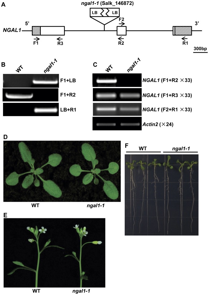
The phenotypes we observed with abs2-1D and NGAL1 OE lines were the consequences of gain-of-function genetic manipulations. To investigate potential loss-of-function phenotypes of NGAL1, we searched for its T-DNA insertional mutants in The Arabidopsis Information Resource (TAIR) and identified a putative insertion line SALK_146872 (Figure 6A). PCR analysis confirmed that the T-DNA was inserted in the first intron of NGAL1 (Figure 6B). We also found that the inserted T-DNA structure likely included two copies of T-DNAs because we identified both ends of the T-DNA insertion as the left border (Figure 6B). Homozygous line of SALK_146872 (named ngal1-1) was identified and RT-PCR results showed that full-length NGAL1 cDNAs cannot be detected in the floral tissues, where NGAL1 was normally expressed (Figure 6B–C). However, we did detect the presence of transcripts using primer sets that do not span the T-DNA, indicating that certain forms of abnormal transcripts of NGAL1 may exist in ngal1-1 (Figure 6C). Although NGAL1 over-expression led to the loss of flower petals, loss-of-function ngal1-1 plants showed no visible phenotypic changes at vegetative stages when compared with the wild type plants under our growth conditions (Figure 6D). We also did not observe abnormalities with ngal1-1 roots and flowers (Figure 6E–F). Our results suggest that the absence of NGAL1 does not dramatically alter plant development and its activity is likely buffered by redundant genes or activities.
Figure 6. Identification of a loss-of-function mutant allele of NGAL1.
A. T-DNA insertion site in Salk_146872 (ngal1-1). Lines represented introns and intergenic regions and boxes represented exons. 5′ and 3′ UTRs were indicated by shaded boxes. Approximate positions of the PCR primers used in B and C were marked with arrows. B. PCR-identification of ngal1-1 homozygous mutant. The T-DNA insertion was flanked by two LB sequences. C. Expressions of NGAL1 in wild type and ngal1-1 mutant. Total RNAs were extracted from flower tissues and RT-PCRs were carried out with indicated primers and cycle numbers. D. Phenotypes of two-week-old wild type and ngal1-1 homozygous seedlings. E. Floral tissues of five-week-old wild type and ngal1-1 homozygous plants. F. Comparison of root phenotypes of one-week-old wild type and ngal1-1 homozygous plants.
Discussion
The Over-expression of ABS2/NGAL1 is Capable of Altering Multiple Aspects of Plant Development
We are interested in exploring the genetic regulatory networks that govern the growth and development of higher plants. Taking a forward genetics approach, we screened for mutants with abnormal shoot development phenotypes in activation tagging mutant populations and investigated these abnormal shoot (abs) mutants further [20]. Here, we report the isolation and characterization of one semi-dominant abs mutant, abs2-1D, which has multiple developmental defects at both the vegetative and reproductive stages. The cloning of ABS2 revealed that it encodes a member of the RAV subfamily of the plant B3 family of transcription factors. Four major classes of transcription factors containing the B3 domain have been identified in plants, including the LAV, ARF, RAV, and REM subfamilies [7]. The 13 RAV subfamily members can be further placed into two groups based on the features of their DNA binding domains. ABS2, one of the seven RAV subfamily members that only have the B3 DNA binding domain, is closely related to NGA genes and is also named NGAL1 [9]. The other six members contain both the B3 domain and an additional AP2 DNA binding domain [22]. Transcription factors containing the B3 domain have been shown to be responsive to biotic and abiotic stresses, as well as to phytohormones such as abscisic acid, auxin and brassinosteroids [11], [23], [24]. Members of plant RAV subfamily are involved in multiple development programs including flower development [9], [14]–[17]. Two independent efforts have established that the NGA1–4 genes regulate style development [9], [14].
In both the abs2-1D mutant and the independent 35S-driven NGAL1 OE lines, shoot development programs were clearly altered. We also found that multiple 35S promoter-driven NGAL1 OE lines showed the loss-of-petal phenotype, suggesting that NGAL1 over-production can alter flower development. We did not observe the loss-of-petal phenotype in the abs2-1D mutant and one possibility is that the over-expression mechanisms were slightly different. In abs2-1D, over-expression was through the activation of NGAL1 expression by the presence of 35S enhancer sequences, whereas the intact 35S promoter was used in 35S promoter-driven over-expression. Phenotypes of our OE lines are also somewhat different from a previous OE of NGAL1, in which a different promoter was used [9]. However, we cannot rule out the possibility that the presence of 35S enhancers in abs2-1D, which can activate the expressions of many genes in their vicinity, might contribute to the difference.
Our RT-PCR results indicated that NGAL1 is normally expressed in many plant tissues including roots, flowers, siliques and young seedlings. Further promoter-GUS assay revealed that NGAL1 expressions also show clear tissue-specific patterns with the highest expressions in roots and the filament of the stamen. These data suggest NGAL1 functions are necessary in different tissues and it may be involved in many developmental processes in plants. Because NGAL1 is predicted to be a transcription factor and we have shown that it is targeted to the nucleus, it is likely NGAL1 regulates plant development in the nucleus, possibly through modulating the expressions of its target genes, directly or indirectly.
NGAL1 Over-expression Leads to the Loss of Flower Petals
In Arabidopsis, there are four whorls of floral organs: sepals, petals, stamens and carpels. The identities of floral organs are controlled by at least five classes of homeotic genes (ABCDE genes), and mutations in these floral regulators lead to homeotic conversions from one to another and work in this area has led to the establishment of the ABC model as the basis for the formation of floral organs [25].
During flower development, petal identity is determined by the combination of class A (AP1, AP2), class B (AP3, PI) and class E (SEP) genes [25], [26]. In weak alleles of homeotic mutants such as ap2 and ap3, if petal primordia are able to initiate, they are converted into stamens and sepals, respectively as primordia develop further [27], [28]. Another set of genes have been implicated in a different aspect of petal development, more specifically, petal initiation [29]–[34]. The Arabidopsis petal loss (ptl) mutant was isolated because of the loss-of-petal phenotype [29]. For the occasional petals that do develop, the orientations of petals are also disturbed in the ptl mutant [29]. PTL encodes a tri-helix transcription factor [30]. The rabbit ears mutant displays similar petal loss phenotypes to ptl and RABBIT EARS (RBE) encodes a zinc finger protein that is located in the nucleus and shares homology with SUPERMAN [31]. RBE may regulate second whorl initiation through the repression of AGAMOUS [32]. A third Arabidopsis gene, ROXY1, which encodes a glutaredoxin protein, is also necessary for petal initiation [33]. Interestingly, in all three cases, the petal phenotypes are position dependent, rather than identity dependent. Other factors, including UNUSUAL FLORAL ORGANS (UFO), have also been implicated in the petal development [34]. The exact mechanisms that these factors regulate petal initiation remain unclear.
Through the over-expression of NGAL1, we found that transgenic NGAL1 OE plants display flower defects, particularly an intriguing loss-of-petal phenotype, suggesting that the over-production of NGAL1 has the capacity to dramatically alter the normal development of flower petals. In contrast to the above-mentioned recessive mutations that lead to the loss-of-petal phenotype, our findings establish a case where a dominant gain-of-function mutation confers the loss-of-petal defect, similar to the over-expression of AG [35]. Based on the dominant nature, it can be argued that NGAL1, when over-expressed, may function as a negative regulator of petal initiation or petal development or both. Consistent with this notion of NGAL1 being a negative regulator, a recent report has shown that NGAL1 might function as a transcriptional repressor [36]. The impact of NGAL1 over-expression is not limited to flower development as its over-production also causes changes of leaf shape and the rate of leaf initiation. Our findings suggest that NGAL1, when over-expressed, is capable of impacting many aspects of plant development. It is important to point out that the phenotypes we observed with NGAL1 gain-of-function over-expression studies do not match completely with the tissue expression pattern of NGAL1. For example, the highest NGAL1 expression in the wild type flower is in the filament of the stamen but the most conspicuous flower defect in NGAL1 OE plants is the loss of petals. Although we cannot rule out that NGAL1 transcripts are present at low levels in other parts of the flower or in other stages of flower development, our findings suggests that NGAL1 may regulate plant development outside of its normal expression domains and this capacity offers a new gene resource for engineering plants with desirable traits such as flowers without the petals.
To further our understanding of NGAL1 functions, we also identified a loss-of-function NGAL1 mutant allele. However, we did not observe major developmental defects in this mutant, despite the reduction of NGAL1 expression. The lack of obvious phenotypes in the NGAL1 loss-of-function mutant is not entirely unexpected given the presence of multiple homologous genes in the RAV subfamily that might provide redundant functions [14]. However, this superficial lack-of-phenotype does not necessarily mean there are no subtle changes in the mutant that may only be manifest under certain circumstances, such as in another mutant’s background. For example, single mutants of NGA genes only show subtle mutant phenotypes but can cause dramatic floral defects in certain mutant backgrounds [9].
Materials and Methods
Plant Materials and Growth Conditions
The wild-type Arabidopsis thaliana and the abs2-1D mutant used in this study were of the Columbia-0 (Col) ecotype. The T-DNA insertion mutant (Salk_146872C) seeds were obtained from TAIR. Seeds were sown on commercial peat moss mix (Pindstrup, Denmark) and stratified at 4°C for 2 days. Plants were grown at 22°C under continuous illumination of ∼100 µmol·m−2·s−1.
Phylogenetic Analysis
To generate the phylogenetic tree of the RAV subfamily proteins, full-length amino acid sequences were retrieved from TAIR. Protein sequence alignment was carried out using the CLUSTALW software (http://www.ebi.ac.uk/Tools/clustalw2/). Phylogeny construction, bootstrap test of phylogeny by means of the neighbor-joining method and the Poisson correction model were performed using MEGA software version 4.0 [37]. Bootstrap analysis was performed using 1000 trials, and At2g30470 (VAL1) was used as an outgroup.
Genomic and coding sequences of RAV subfamily members were obtained from TAIR and used for the construction of gene structures.
Vector Constructions and Plant Transformation
To construct a NGAL1 over-expression vector, the full length At2g36080 cDNA was amplified with primers 36080F (5′-CATGGATCCTCTCTCATCACTATTTGCCATCTC-3′) and 36080R (5′-CATGGATCCCATCTATGACAACATAACAGGACC-3′) and ligated into pBluescript. After confirming the correct sequences, the cDNA was subcloned into a binary vector pBI111L and placed under the control of the CaMV 35S promoter. To generate ABS2 promoter-GUS fusion construct, a genomic fragment of ∼1.2 kb upstream of the start codon of At2g36080 was amplified with primers 36080PF (5′-CATTCTAGAAGAGGATTGAAACACGACTGTAGT-3′) and 36080PR (5′-CATGGATCCTTGGCTAGGTTACATGTATCTGC-3′) and cloned into a binary vector pCB308 [38]. Transgenic plants were generated by Agrobacterium tumefaciens-mediated floral dip method [39]. T1 seeds were screened on half-strength Murashige and Skoog (MS) solid medium containing 50 mg·L−1 kanamycin (for OE lines) or on soil for Basta resistance (for promoter-GUS lines). Histochemical GUS staining was carried out as described in [20]. GUS activities were examined in multiple independent transgenic lines in T1 and T2 generations.
Morphological Observations
Phenotypes of Arabidopsis flowers were observed and photographed using an Olympus SZ61 stereomicroscope equipped with a Canon G12 camera and greenhouse-grown Arabidopsis plants were photographed directly with a Canon G12 camera.
To quantify the leaf initiation rates of wild type, abs2-1D mutants and NGAL1 OE lines, numbers of rosette leaves were counted from randomly selected plants (n≥28) of each genotype on a daily basis starting from one-week-old plants until bolting. Mean and standard deviation of leaf numbers were calculated.
To quantify the loss-of-petal phenotype of NGAL1 OE lines, randomly picked flowers from each OE line were sorted into two categories first: normal flowers (defined as flowers with four normal shaped petals) and abnormal flowers (defined as flowers with filament-like abnormal petals or without petals). Percentage of each type of flowers and the total percentage of abnormality of each OE line were calculated.
DNA and RNA Manipulations
Genomic DNAs were isolated and Southern-blot analyses were performed as described in Yu et al [19].
Total cellular RNAs were extracted with Trizol Reagents (Invitrogen, USA) according to the manufacturer’s instructions and stored at −80°C. First-strand cDNA was synthesized with 1 µg of total RNA using PrimeScript™ II 1st strand cDNA synthesis Kit (Takara, Japan). Primers 36080F1 (5′-TCATCACTATTTGCCATCTC-3′), 36080R1 (5′-CTATGACAACATAACAGGAC-3′), 36080F2 (5′-AACCAATCACGACCAGTTTC-3′), 36080R2 (5′-AAACTGGTCGTGATTGGTTG-3′) and 36080R3 (5′-TGAACATGGCGATAAGAGTC-3′) were used for detecting At2g36080 transcripts. Actin2 gene expression was monitored using the primers ACT2F (5′-TCAAAGACCAGCTCTTCCATCGAGA-3′) and ACT2R (5′-ACACACAAGTGCATCATAGAAACGA-3′). At2g36090 transcripts were amplified with primers 36090F1 (5′-ACGACATCATAGAGTCTCAC-3′) and 36090R1 (5′-TCTACCTTGAGACTCACTTC-3′).
Transient Expression of NGAL1-GFP in Arabidopsis Protoplasts
To construct the NGAL1-GFP fusion gene, the coding region of NGAL1 was amplified by PCR, using primers ABS2-F1 (5′-CATGGGATCCTCATCACTATTTGCCATCTC-3′) and ABS2-GFPR (5′-CATGGGATCCACCACCACCACCACCACCGCTCGTCCGGTTCATATCTCC-3′). The resulting fragment was digested with BamHI and ligated in frame with the GFP coding sequence in the pTF486 vector and sequenced [19]. Arabidopsis leaf protoplast isolation and Hoechst 33342 nucleus staining were performed as described in [20], [21]. Fluorescence and bright field images were captured and analyzed with fluorescence microscopy (DM5000B, Leica, Germany).
Isolation of ngal1-1
To isolate NGAL1 T-DNA mutant (ngal1-1), we obtained a putative T-DNA insertion mutant line (Salk_146872C) from TAIR. Genomic DNAs were prepared and gene-specific primers, 36080F1, 36080R2, and T-DNA specific primers LB (5′-GAACAACACTCAACCCTATCTC-3′) were used to identify heterozygous and homozygous plants. PCR products were gel purified and sequenced.
Supporting Information
Comparison of leaf initiation rates of wild type, abs2-1D mutants and NGAL1 overexpression lines.
(PDF)
Phenotypes of abs2-1D mutants at flowering stages.
(PDF)
The Arabidopsis RAV sub-family of transcription factors.
(PDF)
Phenotypes of NGAL1 OE lines at flowering stage.
(PDF)
Funding Statement
This work was supported by funding to FY from National Natural Science Foundation of China (31071073) and Chinese Ministry of Education Program for New Century Excellent Talents in University (NCET-09-0657), and by funding to XL from National Natural Science Foundation of China (31100864). The funders had no role in study design, data collection and analysis, decision to publish, or preparation of the manuscript.
References
- 1. Ariel AD, Manavella PA, Dezar CA, Chan RC (2007) The true story of the HD-Zip family. Trends Plant Sci 12: 419–426. [DOI] [PubMed] [Google Scholar]
- 2. Riechmann JL, Heard J, Martin G, Reuber L, Jiang CZ, et al. (2000) Arabidopsis transcription factors: genome-wide comparative analysis among eukaryotes. Science 290: 2105–2110. [DOI] [PubMed] [Google Scholar]
- 3. Tran LS, Nakashima K, Shinozaki K, Yamaguchi-Shinozaki K (2007) Plant gene networks in osmotic stress response: from genes to regulatory networks. Method Enzymol 428: 109–128. [DOI] [PubMed] [Google Scholar]
- 4. Yamaguchi-Shinozaki K, Shinozaki K (2005) Organization of cis-acting regulatory elements in osmotic- and cold-stress-responsive promoters. Trends Plant Sci 10: 88–94. [DOI] [PubMed] [Google Scholar]
- 5. Nakashima K, Ito Y, Yamaguchi-Shinozaki K (2009) Transcriptional regulatory networks in response to abiotic stresses in Arabidopsis and grasses. Plant Physiol 149: 88–95. [DOI] [PMC free article] [PubMed] [Google Scholar]
- 6. Zhao L, Luo QL, Yang CHL, Han YP, Li WB (2008) A RAV-like transcription factor controls photosynthesis and senescence in soybean. Planta 227: 1389–1399. [DOI] [PubMed] [Google Scholar]
- 7. Swaminathan K, Peterson K, Jack T (2008) The plant B3 superfamily. Trends Plant Sci 13: 647–655. [DOI] [PubMed] [Google Scholar]
- 8. McCarty DR, Hattori T, Carson CB, Vasil V, Lazar M, et al. (1991) The viviparous-1 developmental gene of maize encodes a novel transcriptional activator. Cell 66: 895–905. [DOI] [PubMed] [Google Scholar]
- 9. Alvarez JP, Goldshmidt A, Efroni I, Bowman JL, Eshed Y (2009) The NGATHA distal organ development genes are essential for style specification in Arabidopsis . Plant Cell 21: 1373–1393. [DOI] [PMC free article] [PubMed] [Google Scholar]
- 10. Kagaya Y, Ohmiya K, Hattori T (1999) RAV1, a novel DNA-binding protein, binds to bipartite recognition sequence through two distinct DNA-binding domains uniquely found in higher plant. Nucleic Acids Res 27: 470–478. [DOI] [PMC free article] [PubMed] [Google Scholar]
- 11. Hu YX, Wang YH, Liu XF, Li JY (2004) Arabidopsis RAV1 is down-regulated by brassinosteroid and may act as a negative regulator during plant development. Cell Res 14: 8–15. [DOI] [PubMed] [Google Scholar]
- 12. Castillejo C, Pelaz S (2008) The Balance between CONSTANS and TEMPRANILLO activities determines FT expression to trigger flowering. Curr Biol 18: 1338–1343. [DOI] [PubMed] [Google Scholar]
- 13. Kagaya Y, Hattori T (2009) Arabidopsis transcription factors, RAV1 and RAV2, are regulated by touch-related stimuli in a dose-dependent and biphasic manner. Genes Genet Syst 84: 95–99. [DOI] [PubMed] [Google Scholar]
- 14. Trigueros M, Navarrete-Gómez M, Sato S, Christensen SK, Pelaz S, et al. (2009) The NGATHA genes direct style development in the Arabidopsis gynoecium. Plant Cell 21: 1394–1409. [DOI] [PMC free article] [PubMed] [Google Scholar]
- 15. Eshed Y, Baum SF, Bowman JL (1999) Distinct mechanisms promote polarity establishment in carpels of Arabidopsis. Cell 99: 199–209. [DOI] [PubMed] [Google Scholar]
- 16. Bowman JL, Eshed Y, Baum SF (2002) Establishment of polarity in angiosperm lateral organs. Trends Genet 18: 134–141. [DOI] [PubMed] [Google Scholar]
- 17. Alvarez JP, Pekker I, Goldshmidt A, Blum E, Amsellem Z, et al. (2006) Endogenous and synthetic microRNAs stimulate simultaneous, efficient, and localized regulation of multiple targets in diverse species. Plant Cell 18: 1134–1151. [DOI] [PMC free article] [PubMed] [Google Scholar]
- 18. Weigel D, Ahn JH, Blázquez MA, Borevitz JO, Christensen SK, et al. (2000) Activation tagging in Arabidopsis. Plant Physiol 122: 1003–1013. [DOI] [PMC free article] [PubMed] [Google Scholar]
- 19. Yu F, Liu X, Alsheikh M, Park S, Rodermel S (2008) Mutations in SUPPRESSOR OF VARIEGATION1, a factor required for normal chloroplast translation, suppress var2-mediated leaf variegation in_Arabidopsis. Plant Cell 20: 1786–1804. [DOI] [PMC free article] [PubMed] [Google Scholar]
- 20. Wang M, Liu X, Wang R, Li W, Rodermel S, et al. (2012) The overexpression of a putative Arabidopsis BAHD acyl-transferase causes dwarfism that can be rescued by brassinosteroid. J Exp Bot 63: 5787–5801. [DOI] [PMC free article] [PubMed] [Google Scholar]
- 21. Meadows MG, Potrykus I (1981) Hoechst 33258 as a Vital Stain for Plant Cell Protoplasts. Plant Cell Rep 1: 77–79. [DOI] [PubMed] [Google Scholar]
- 22. Kim S, Soltis PS, Wall K, Soltis DE (2006) Phylogeny and domain evolution in the APETALA2-like gene family. Mol Biol Evol 23: 107–120. [DOI] [PubMed] [Google Scholar]
- 23. McCarty DR, Carson CB, Stinard PS, Robertson DS (1989) Molecular analysis of viviparous-1: An abscisic acid-insensitive mutant of maize. Plant Cell 1: 523–532. [DOI] [PMC free article] [PubMed] [Google Scholar]
- 24. Okushima Y, Mitina I, Quach HL, Theologis A (2005) AUXIN RESPONSE FACTOR 2 (ARF2): a pleiotropic developmental regulator. Plant J 43: 29–46. [DOI] [PubMed] [Google Scholar]
- 25. Alvarez-Buylla ER, Benítez M, Corvera-Poiré A, Chaos Cador A, de Folter S, et al. (2010) Flower development. Arabidopsis Book 8: e0127. [DOI] [PMC free article] [PubMed] [Google Scholar]
- 26. Krizek BA, Fletcher JC (2005) Molecular mechanisms of flower development: an armchair guide. Nature 6: 688–698. [DOI] [PubMed] [Google Scholar]
- 27. Theissen G, Saedler H (2001) Plant biology: Floral quartets. Nature 409: 469–471. [DOI] [PubMed] [Google Scholar]
- 28. Lohmann JU, Weigel D (2002) Building beauty: The genetic control of floral patterning. Dev Cell 2: 135–142. [DOI] [PubMed] [Google Scholar]
- 29. Griffith ME, Conceição AS, Smyth DR (1999) PETAL LOSS gene regulates initiation and orientation of second whorl organs in the Arabidopsis flower. Development 126: 5635–5644. [DOI] [PubMed] [Google Scholar]
- 30. Brewer PB, Howles PA, Dorian K, Griffith ME, Ishida T, et al. (2004) PETAL LOSS, a trihelix transcription factor gene, regulates perianth architecture in the Arabidopsis flower. Development 131: 4035–4045. [DOI] [PubMed] [Google Scholar]
- 31. Takeda S, Matsumoto N, Okada K (2004) RABBIT EARS, encoding a SUPERMAN-like zinc finger protein, regulates petal development in Arabidopsis thaliana . Development 13: 425–434. [DOI] [PubMed] [Google Scholar]
- 32. Krizek BA, Lewis MW, Fletcher JC (2006) RABBIT EARS is a second-whorl repressor of AGAMOUS that maintains spatial boundaries in Arabidopsis flowers. Plant J 45: 369–383. [DOI] [PubMed] [Google Scholar]
- 33. Xing S, Rosso MG, Zachgo S (2005) ROXY1, a member of the plant glutaredoxin family, is required for petal development in Arabidopsis thaliana. . Development 132: 1555–1565. [DOI] [PubMed] [Google Scholar]
- 34. Durfee T, Roe JL, Sessions RA, Inouye C, Serikawa K, et al. (2003) The F-box-containing protein UFO and AGAMOUS participate in antagonistic pathways governing early petal development in Arabidopsis. Proc Natl Acad Sci USA 100: 8571–8576. [DOI] [PMC free article] [PubMed] [Google Scholar]
- 35. Mizukami Y, Ma H (1992) Ectopic expression of the floral homeotic gene AGAMOUS in transgenic Arabidopsis plants alters floral organ identity. Cell 71: 119–131. [DOI] [PubMed] [Google Scholar]
- 36. Ikeda M, Ohme-Takagi M (2009) A novel group of transcriptional repressors in Arabidopsis. Plant Cell Physiol 50: 970–975. [DOI] [PubMed] [Google Scholar]
- 37. Tamura K, Dudley J, Nei M, Kumar S (2007) MEGA4: Molecular Evolutionary Genetics Analysis (MEGA) software version 4.0. Mol Biol Evol 24: 1596–1599. [DOI] [PubMed] [Google Scholar]
- 38. Xiang C, Han P, Lutziger I, Wang K, Oliver DJ (1999) A mini binary vector series for plant transformation. Plant Mol Biol 40: 711–717. [DOI] [PubMed] [Google Scholar]
- 39. Clough SJ, Bent AF (1998) Floral dip: a simplified method for Agrobacterium mediated transformation of Arabidopsis thaliana . Plant J 16: 735–743. [DOI] [PubMed] [Google Scholar]
Associated Data
This section collects any data citations, data availability statements, or supplementary materials included in this article.
Supplementary Materials
Comparison of leaf initiation rates of wild type, abs2-1D mutants and NGAL1 overexpression lines.
(PDF)
Phenotypes of abs2-1D mutants at flowering stages.
(PDF)
The Arabidopsis RAV sub-family of transcription factors.
(PDF)
Phenotypes of NGAL1 OE lines at flowering stage.
(PDF)


