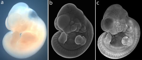
Figure 1.
Comparison of E11.5 whole mount mouse embryo imaged by brightfield, and by nuclear fluorescent staining combined with widefield or confocal microscopy. (a) Unstained embryo imaged by conventional bright field microscopy, (b) Embryo stained with Hoechst 33342 imaged by conventional fluorescence microscopy using a fluorescent stereomicroscope. (c) DAPI-stained embryo imaged by confocal microscopy wherein a z-stack of images spanning the depth of the embryo is collapsed to form a single projection image file. This image is a composite generated by splicing multiple flattened z-stack images taken with 5× objective.

