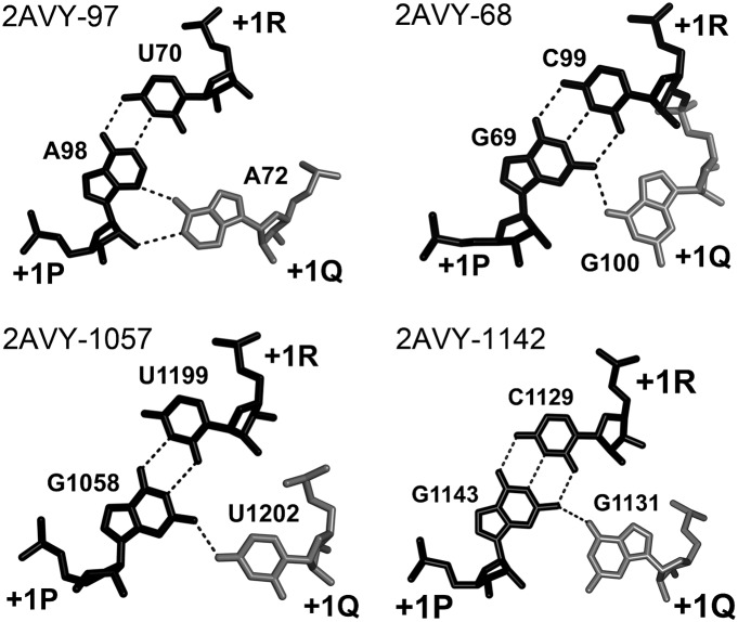
FIGURE 5.
A-DTJ: structure of layer +1. In all A-DTJs, nucleotide +1Q (white) interacts with the minor groove of base pair [+1P;+1R] (black), making either the cis WC/SE (2AVY-97, 2AVY-1142) or cis HG/SE (2AVY-68 and 2AVY-1057) juxtaposition with +1P. Although in all motifs, the base of nucleotide +1Q is positioned in vicinity of the ribose +1P, only in motif 2AVY-97 are the two entities close enough to form an H-bond.

