

Abstract
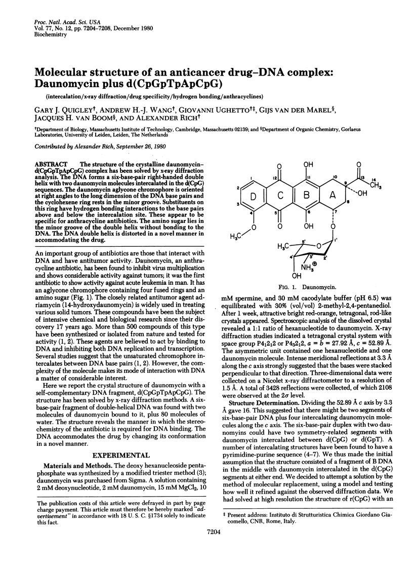
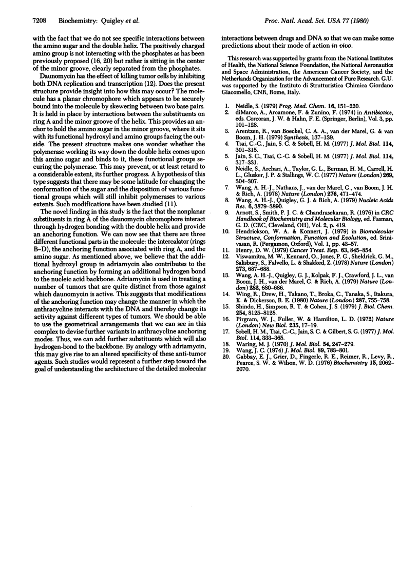
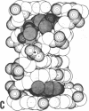
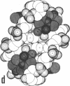
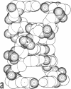
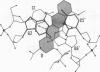
The structure of the crystalline daunomycin-d(CpGpTpApCpG) complex has been solved by x-ray diffraction analysis. The DNA forms a six-base-pair right-handed double helix with two daunomycin molecules intercalated in the d(CpG) sequences. The daunomycin aglycone chromophore is oriented at right angles to the long dimension of the DNA base pairs and the cyclohexene ring rests in the minor groove. Substituents on this ring have hydrogen bonding interactions to the base pairs above and below the intercalation site. These appear to be specific for anthracycline antibiotics. The amino sugar lies in the minor groove of the double helix without bonding to the DNA. The DNA double helix is distorted in a novel manner in accommodating the drug.
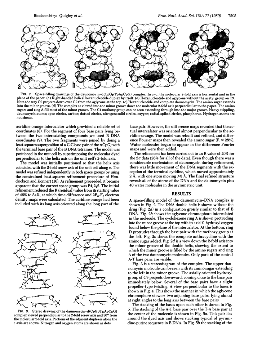
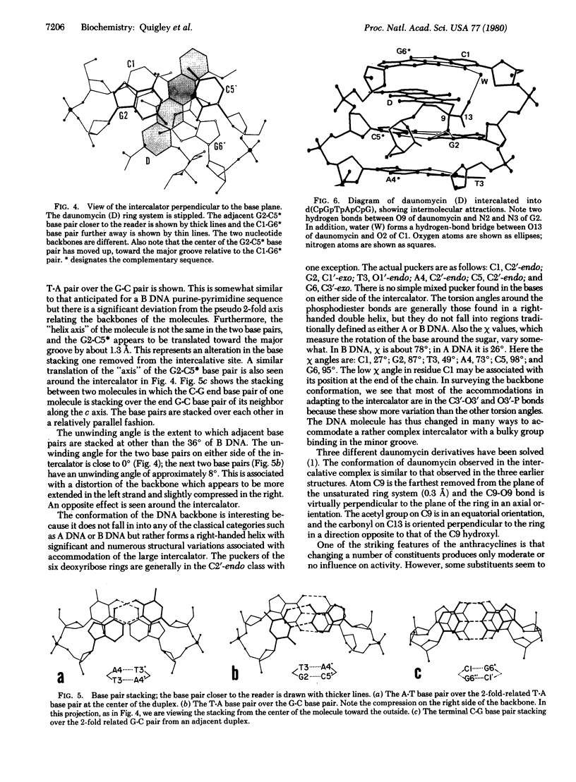
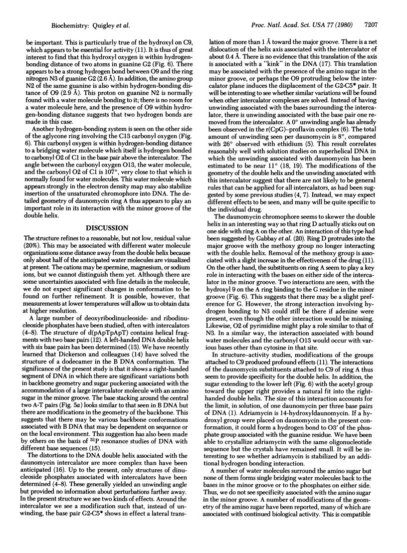
Full text
PDF
Images in this article
Selected References
These references are in PubMed. This may not be the complete list of references from this article.
- Gabbay E. J., Grier D., Fingerle R. E., Reimer R., Levy R., Pearce S. W., Wilson W. D. Interaction specificity of the anthracyclines with deoxyribonucleic acid. Biochemistry. 1976 May 18;15(10):2062–2070. doi: 10.1021/bi00655a006. [DOI] [PubMed] [Google Scholar]
- Henry D. W. Structure-activity relationships among daunorubicin and adriamycin analogs. Cancer Treat Rep. 1979 May;63(5):845–854. [PubMed] [Google Scholar]
- Jain S. C., Tsai C. C., Sobell H. M. Visualization of drug-nucleic acid interactions at atomic resolution. II. Structure of an ethidium/dinucleoside monophosphate crystalline complex, ethidium:5-iodocytidylyl (3'-5') guanosine. J Mol Biol. 1977 Aug 15;114(3):317–331. doi: 10.1016/0022-2836(77)90253-4. [DOI] [PubMed] [Google Scholar]
- Neidle S., Achari A., Taylor G. L., Berman H. M., Carrell H. L., Glusker J. P., Stallings W. C. Structure of a dinucleoside phosphate--drug complex as model for nucleic acid--drug interaction. Nature. 1977 Sep 22;269(5626):304–307. doi: 10.1038/269304a0. [DOI] [PubMed] [Google Scholar]
- Neidle S. The molecular basis for the action of some DNA-binding drugs. Prog Med Chem. 1979;16:151–221. doi: 10.1016/s0079-6468(08)70188-7. [DOI] [PubMed] [Google Scholar]
- Pigram W. J., Fuller W., Hamilton L. D. Stereochemistry of intercalation: interaction of daunomycin with DNA. Nat New Biol. 1972 Jan 5;235(53):17–19. doi: 10.1038/newbio235017a0. [DOI] [PubMed] [Google Scholar]
- Shindo H., Simpson R. T., Cohen J. S. An alternating conformation characterizes the phosphodiester backbone of poly(dA-dT) in solution. J Biol Chem. 1979 Sep 10;254(17):8125–8128. [PubMed] [Google Scholar]
- Sobell H. M., Tsai C. C., Jain S. C., Gilbert S. G. Visualization of drug-nucleic acid interactions at atomic resolution. III. Unifying structural concepts in understanding drug-DNA interactions and their broader implications in understanding protein-DNA interactions. J Mol Biol. 1977 Aug 15;114(3):333–365. doi: 10.1016/0022-2836(77)90254-6. [DOI] [PubMed] [Google Scholar]
- Tsai C. C., Jain S. C., Sobell H. M. Visualization of drug-nucleic acid interactions at atomic resolution. I. Structure of an ethidium/dinucleoside monophosphate crystalline complex, ethidium:5-iodouridylyl (3'-5') adenosine. J Mol Biol. 1977 Aug 15;114(3):301–315. doi: 10.1016/0022-2836(77)90252-2. [DOI] [PubMed] [Google Scholar]
- Viswamitra M. A., Kennard O., Jones P. G., Sheldrick G. M., Salisbury S., Favello L., Shakked Z. DNA double helical fragment at atomic resolution. Nature. 1978 Jun 22;273(5664):687–688. doi: 10.1038/273687a0. [DOI] [PubMed] [Google Scholar]
- Wang A. H., Nathans J., van der Marel G., van Boom J. H., Rich A. Molecular structure of a double helical DNA fragment intercalator complex between deoxy CpG and a terpyridine platinum compound. Nature. 1978 Nov 30;276(5687):471–474. doi: 10.1038/276471a0. [DOI] [PubMed] [Google Scholar]
- Wang A. H., Quigley G. J., Kolpak F. J., Crawford J. L., van Boom J. H., van der Marel G., Rich A. Molecular structure of a left-handed double helical DNA fragment at atomic resolution. Nature. 1979 Dec 13;282(5740):680–686. doi: 10.1038/282680a0. [DOI] [PubMed] [Google Scholar]
- Wang A. H., Quigley G. J., Rich A. Atomic resolution analysis of a 2:1 complex of CpG and acridine orange. Nucleic Acids Res. 1979 Aug 24;6(12):3879–3890. doi: 10.1093/nar/6.12.3879. [DOI] [PMC free article] [PubMed] [Google Scholar]
- Wang J. C. The degree of unwinding of the DNA helix by ethidium. I. Titration of twisted PM2 DNA molecules in alkaline cesium chloride density gradients. J Mol Biol. 1974 Nov 15;89(4):783–801. doi: 10.1016/0022-2836(74)90053-9. [DOI] [PubMed] [Google Scholar]
- Waring M. Variation of the supercoils in closed circular DNA by binding of antibiotics and drugs: evidence for molecular models involving intercalation. J Mol Biol. 1970 Dec 14;54(2):247–279. doi: 10.1016/0022-2836(70)90429-8. [DOI] [PubMed] [Google Scholar]
- Wing R., Drew H., Takano T., Broka C., Tanaka S., Itakura K., Dickerson R. E. Crystal structure analysis of a complete turn of B-DNA. Nature. 1980 Oct 23;287(5784):755–758. doi: 10.1038/287755a0. [DOI] [PubMed] [Google Scholar]