Abstract
The GABAA receptors are the major inhibitory neurotransmitter receptors in mammalian brain. Each isoform consists of five homologous or identical subunits surrounding a central chloride ion-selective channel gated by GABA. How many isoforms of the receptor exist is far from clear. GABAA receptors located in the postsynaptic membrane mediate neuronal inhibition that occurs in the millisecond time range; those located in the extrasynaptic membrane respond to ambient GABA and confer long-term inhibition. GABAA receptors are responsive to a wide variety of drugs, e.g. benzodiazepines, which are often used for their sedative/hypnotic and anxiolytic effects.
Keywords: GABA Receptors, Ion Channels, Membrane Proteins, Neurobiology, Protein Purification, Benzodiazepines, Cys Loop Receptors
Introduction
Fast synaptic transmission is effected by neurotransmitters that bind to and thereby induce channel opening in postsynaptic receptors. The two major inhibitory ligand-gated ion channels, the GABAA receptor and the glycine receptor, are anion-selective. While the cation-selective nicotinic acetylcholine receptor, which belongs to the same protein family, is already strongly enriched in nature in the form of Torpedo marmorata electroplax membranes, no rich source exists for anion-selective receptors and their isolation has been hampered by poor abundance (<1 pmol/mg of brain membrane protein). From this value, a purification factor of >4000-fold may be calculated for a 250-kDa protein complex. This formidable problem was solved with the help of affinity chromatography. Within 2 months in 1982, two reports appeared, one on the purification of a glycine receptor using a strychnine affinity column (1) and a preliminary report on the purification of a GABA receptor characterized by [3H]muscimol binding using a benzodiazepine affinity column (2). A detailed account showing that both of the binding sites, the one for the agonist GABA and that for benzodiazepines, reside on the same protein complex was published shortly after in the Journal of Biological Chemistry (3). In this article, the two documented subunits were given the names α and β. An improved purification procedure using alternative detergents was published 1 year later in the same journal (4). Protein sequencing led to cloning of α and β subunits (5), and the homology of these two subunits with those of nicotinic acetylcholine receptors and glycine receptors became evident. Erroneously, the cloning paper claimed stimulation by benzodiazepines of the GABAA receptor composed of α and β subunits, but it is clear now that this modulation requires an additional subunit (see below). In the following years, many subunit isoforms were cloned. Isoform diversity was anticipated early on the basis of photolabeling experiments (6). What differentiates GABAA receptors from the other members of the Cys loop family of receptors (7) is their rich pharmacology, which may be due partially to the large number of cavities observed in the intramembrane space (8). In contrast, natural toxins targeting GABAA receptors are very rare in comparison with other channel proteins.
In the following, we attempt to give a brief overview of our knowledge of the biochemistry of GABAA receptors, including structure, function, and modulation. A more detailed review encompassing all Cys loop receptors has been published recently (9). GABAB receptors are G-protein-coupled receptors and differ strongly in structure, function, and sequence from GABAA receptors and will not be discussed here. GABAC receptors are now generally assumed to be one of the many isoforms of GABAA receptors, and the International Union of Basic and Clinical Pharmacology discourages further use of this term (10).
The structural features of GABAA receptors are shared by the entire superfamily of Cys loop-type ligand-gated ion channels. The unique property that keeps GABAA receptors apart from other members of the superfamily is the activating ligand GABA. From the amino acid sequence of a single subunit, it is impossible to identify the type of ligand and ion selectivity of the channel, except maybe whether it contributes to a cation- or anion-selective channel. This makes a discussion on the origins of GABAA receptors speculative.
GABAA receptors are generally pentameric proteins composed of different subunits. Individual subunits may be well characterized concerning sequence, expression level, and localization in a neuron, but in many cases, it is far from clear which subunits collaborate together to form a pentameric receptor. Even if this is known, the subunit arrangement of the pentamer is not evident.
The agonist of GABAA receptors is generally being called after Gamma-AminoButyric Acid. This is despite the fact that, under physiological conditions, the acid form of the neurotransmitter hardly exists, and a more appropriate name for this neurotransmitter would be γ-aminobutyrate.
Gene Organization
As mentioned above, the GABAA receptors are members of the Cys loop ligand-gated ion channel superfamily (for review, see Refs. 7 and 9), which also includes the nicotinic acetylcholine receptors (11), the glycine receptors (12), the ionotropic 5-HT3 receptors (13), the Zn2+-activated ion channels (14), and three recently crystallized receptors: the Caenorhabditis elegans glutamate-gated chloride channel (15) and the two bacterial receptors GLIC (Gloeobacter violaceus ligand-gated ion channel) (16) and ELIC (Erwinia chrysanthemi ligand-gated ion channel) (17). All eukaryotic members of the Cys loop family share a motif composed of two cysteine residues separated by 13 amino acid residues. Simon et al. (18) tried to find a motif identifying GABAA receptors. A genome search made with a consensus sequence could identify all known GABAA receptor sequences but also many subunits of the other Cys loop receptor family members.
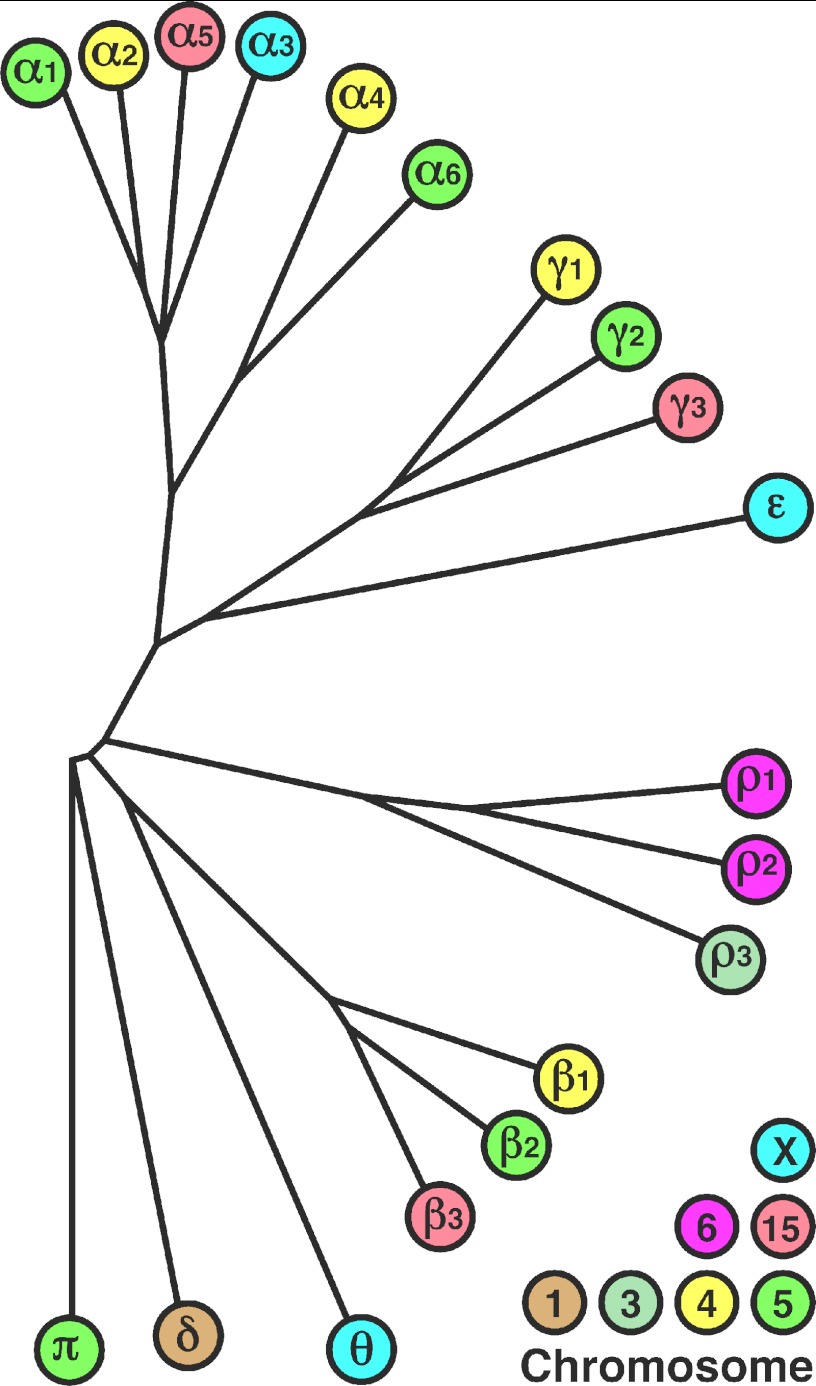
In this minireview, we concentrate on human GABAA receptors. The complexity of the GABAA receptors rests on the fact that numerous subunits exist. Ignoring splice variants and point mutations, we know that, in human, there are six α subunits, three β subunits, three γ subunits, three ρ subunits (building blocks of the formerly called GABAC receptors), and one each of the ϵ, δ, θ, and π subunits. Within a family of subunits, there is ∼70% sequence identity, and between members of different families, 20% sequence identity or 50% sequence similarity (19). The GABAA receptor subunit genes exhibit a basic pattern of nine coding exons with eight introns with two exceptions: δ has 8 exons, and γ3 has 10 exons. For some of the subunits, there exist splice variants (for details, see Ref. 19 and references cited therein), but the function/physiological role of these variants is not yet fully determined. Fig. 1 shows a phylogenetic tree of the human GABAA receptor subunits and their chromosomal localization. The majority of genes coding for the GABAA receptor subunits are organized into four clusters on chromosomes 4, 5, 15, and X in the human genome (20). This gene clustering is assumed to contribute to coordinated gene expression. Two clusters each group four genes, α2, α4, β1, and γ1 on chromosome 4 and α1, α6, β2, and γ2 on chromosome 5; and two clusters have three genes, α5, β3, and γ3 on chromosome 15 and α3, ϵ, and θ on chromosome X. Common to all clusters is that the β subunit exhibits an opposite transcriptional orientation. By phylogenetic tree analysis, it becomes apparent that the ϵ subunit is “γ-like” and the θ subunit is “β-like” (21). Based on these facts, it has been assumed that all four clusters have evolved from an ancestral cluster containing an “α-like”, a β-like, and a γ-like subunit. Two rounds of tetraploidization (whole genome duplication) during the early chordate evolution and a tandem duplication of an α subunit after the first tetraploidization event can account for the current structural organization within the genome. This is supported by the fact that α1 and α2, as well as α6 and α4, which are in the same position within the cluster on chromosomes 5 and 4, respectively, have a higher sequence similarity to each other than to any other pair of α subunits (reviewed in Ref. 20).
FIGURE 1.
Phylogenetic tree analysis of the 19 known genes coding for human GABAA receptor subunits. The immature amino acid sequences were obtained from the UniProt database (94). The alignment was done with ClustalX (95), and Dendroscope (64) was used for depiction of the dendrogram.
Structure of GABAA Receptors
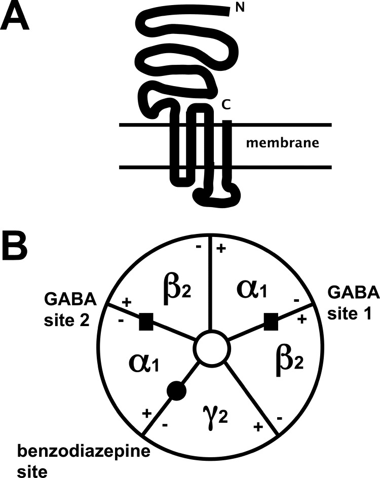
The GABAA receptors are multisubunit proteins (for review, see Refs. 22–24). Before we discuss their structure, we will examine the properties of a single subunit. Mature subunits are ∼450 amino acid residues in length and share a common topological organization, as illustrated in Fig. 2A. About half of the subunit consists of a hydrophilic extracellular N-terminal domain containing the Cys loop, followed by four transmembrane sequences (M1–M4). M2 lines the ion channel. Between M3 and M4 there is a large intracellular loop involved in modulation by phosphorylation (see below). A number of proteins have been described that interact with the intracellular loop between M3 and M4 of specific GABAA receptor subunits (25, 26). These proteins have been shown to play important roles in receptor trafficking and anchoring of receptors in the cytoskeleton and in the postsynaptic membrane. The N and C termini of the subunits are both located extracellularly.
FIGURE 2.
Schematic representation of the major isoform of GABAA receptors, α1β2γ2. The GABAA receptors are integral membrane proteins. Five subunits are grouped around the central ion pore. A, topology of a single subunit. All subunits share this topology. B, top view of the pentamer. The sidedness of the subunits is symbolized + and −.
In theory, a huge number of GABAA receptors may be assembled even in a single cell, as in some cases, more than eight subunit isoforms have been found to be expressed in a single cell. Furthermore, alternative splicing and RNA editing (27) contribute to receptor diversity. The major adult isoform is generally accepted to be composed of α1, β2, and γ2 subunits. There is considerable uncertainty as to the number of different GABAA receptor isoforms existing in nature (28). For a discussion of the isoforms that may be really expressed in nature, the reader may consult Ref. 10.
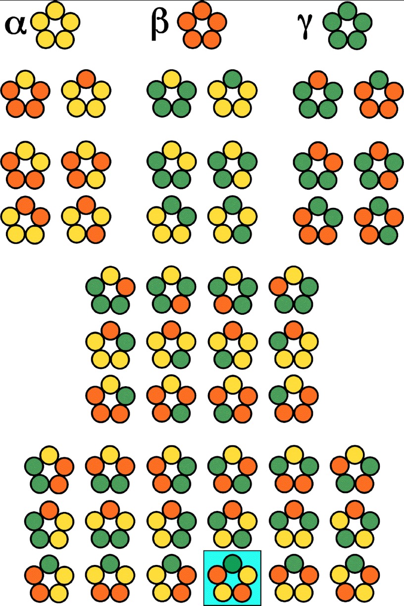
As mentioned above, five subunits selected from 19 isoforms form a complex and surround a central ion channel, with all five M2 segments contributing to the channel (Fig. 2B). Even if the same subunits contribute to channel formation, they may be differently arranged. Whereas the α, β, and γ subunits seem to combine into defined subunit arrangements (29–31), the ϵ and δ subunits seem to have promiscuous assembly properties (32–34). The functional properties of the receptors depend on both subunit composition (35) and arrangement (36). Fig. 3 illustrates the 30 possibilities to build a pentameric receptor from three different subunits. Approaches characterizing assembly and studies using concatenated subunits have concluded that, in the major subunit isoform, the α1, β2, and γ2 subunits are arranged γ2β2α1β2α1 counterclockwise around the central pore as viewed from the cell exterior (29–31).
FIGURE 3.
Possible arrangements in a pentamer of subunits taken from three different types, α (yellow), β (red), and γ (green). There are three homomeric receptors, 18 receptors composed of two subunits, and 30 receptors composed of three different subunits. The receptor on the blue square represents the current consensus of the subunit arrangement in α1β2γ2 GABAA receptors as seen from the cell exterior.
Unfortunately, a high resolution structure of the GABAA receptor is missing. Two-dimensional crystals of the nicotinic acetylcholine receptor with 4 Å resolution in the membrane part (37) have given us a good idea of the general architecture of Cys loop receptors. Some bacterial Cys loop receptors have been crystallized, but the degree of homology with GABAA receptors is low (15–17). Also, an acetylcholine-binding protein homologous to the N-terminal extracellular portion of the nicotinic acetylcholine receptor has been crystallized (38). The GABAA receptor shares structural and, to a small extent, sequence homology with all of these crystallized proteins. The crystal structure of the acetylcholine-binding protein has triggered an early homology modeling attempt (39). Additionally, the modulatory compound diazepam (see below) has been docked into this model. It should be noted, however, that the sequence identity of these two proteins amounts to only ∼18% and that the binding pocket is, to a large extent, made up of loop structures, which are difficult to model. The caveats in interpretation of homology models based on such a low degree of homology have been discussed in detail (40). The recent crystallization of a hybrid of the extracellular domain of the α7 subunit of the nicotinic acetylcholine receptor with acetylcholine-binding protein (41) may improve the situation.
Cellular Localization
The subunits of rat GABAA receptors have been localized in the central nervous system at the protein level using subunit-specific antibodies (42). Some subunits have a broad expression throughout the central nervous system. Other subunits show a restricted expression. An extreme example is the α6 subunit, which is expressed in only a single cell type, the cerebellar granule cells (42). Another example of a restricted distribution is the ρ subunit, which is expressed mainly, but not exclusively, in the retina (42). Outside the central nervous system, GABAA receptors have been found in the liver (43), in smooth airway muscles of the lung (44), and in several types of immune cells (45, 46).
Subcellular Localization
For a long time, GABAA receptors have been assumed to localize to postsynaptic sites in the brain. Synaptic transmission leads here to the release of GABA, which, in turn, can open GABAA receptor chloride channels, thereby creating a short (millisecond) increase in the anion conductance, leading to hyperpolarization of a depolarized membrane. These short events have been termed phasic inhibition. It is now clear that GABAA receptors can also localize to extrasynaptic sites and confer here the so-called tonic inhibition. Low ambient GABA concentrations open these receptors for a longer time period (for review, see Ref. 47). Some subunits only assume an extrasynaptic location, as it has been postulated for the δ subunit (for review, see Ref. 47). In many cells, the long-term charge translocation through extrasynaptic receptors exceeds that through synaptic receptors. Modulation of this tonic inhibition has been implicated in disease states (48).
Ion Conductance
The GABAA receptors are generally GABA-gated anion channels selective for Cl− ions, with some permeability for bicarbonate anions (49). Exceptionally, in C. elegans, a cation-selective GABA-gated channel has been discovered (50). Excitatory neurotransmitters increase the cation conductance to depolarize the membrane, whereas inhibitory neurotransmitters increase the anion conductance to tendentially hyperpolarize the membrane. However, if the gradient for Cl− ions decreases due to down-regulation of KCC2 chloride ion transporters, opening of GABAA receptors may cause an outward flux of these anions, leading to depolarization of the membrane and thereby to excitation. This phenomenon has been implicated in neuropathic pain (51). During early development (52) and in neuronal subcompartments (53), GABA similarly confers excitation.
Mechanism
For a discussion of functional details, we concentrate on the major subunit isoforms αβγ, which are best characterized. In a first stage, the agonist GABA binds to its two binding sites located at the α/β subunit interfaces, which have been shown to display slightly different apparent affinities for channel opening (54). Agonist site occupancy is then followed by a conformational change that locks the agonist in the binding pocket (55). The protein alters conformation and enters one or several closed states that have been termed “flipped states” (56). Further conformational changes lead to an opening of the ion pore. The open channel can then make short visits to a ligand-bound closed state (57).
How binding site occupancy leads to channel opening is a matter of debate. A stretch of amino acid residues termed the F-loop projects from the agonist site toward the ion channel. In the α subunit, this loop has been implicated in this process, and analogously, in the γ subunit, the F-loop has been implicated in channel modulation by benzodiazepines (see below). Recent experiments rather argue against this model (for review, see Ref. 58). A second region that has been implicated in channel gating is the short loop between M2 and M3. A lysine residue located in this region has been suggested to undergo formation of a salt bridge during the process of gating (59) on the basis of S-S bridge formation after mutation of the two corresponding residues. A third region seems to be located within the β4-β5 linker of the β subunit (60). Insertion of glycine residues leads to a strongly reduced apparent potency for channel gating by GABA. Conformational changes during gating have also been probed by site-specific introduction of fluorescent reporters pioneered in the conceptually more simple homomeric ρ receptor (61) or by ligand-induced alteration of chemical reaction rates between cysteine-reactive reagents and cysteine-mutated receptors (55). Most probably, large portions of the receptor protein undergo conformational changes during channel gating.
The channel has been investigated using the water accessibility method, in which individual amino acid residues are mutated to cysteine, and the mutant receptors are exposed to a water-soluble cysteine-reactive compound. Altered channel activity was then taken as evidence for exposure of the corresponding residue to the channel lumen (62).
Modulation of GABAA Receptors
As many other proteins, GABAA receptors are modulated by post-translational modification, e.g. several different protein kinases have been shown to phosphorylate specific amino acid resides on specific receptor subunits and thereby modulate channel activity. This modification has also been shown to affect surface stability or trafficking (63). Besides covalent modification, a number of receptor-associated proteins have been described (reviewed in Ref. 25).
Apart from these two mechanisms, GABAA receptors are modulated by two endogenous molecules (65, 66) and a wide range exogenous small molecules (67). It has been hypothesized that GABAA receptors are modulated by such a large number of compounds because the latter can occupy numerous cavities located within the part of the receptor embedded in the membrane (8). Selected examples of modulators are benzodiazepines and other drugs acting at the same site, barbiturates, etomidate, the competitive antagonist bicuculline, and the channel blocker picrotoxin. Subunit composition and arrangement determine drug selectivity. It is beyond the scope of this minireview to discuss all modulators.
The first class of endogenous modulators to be recognized were the neurosteroids (65). These substances allosterically modulate channel opening and also work as channel agonists at high concentrations. Both functions have been molecularly localized in the transmembrane regions of α and β subunits (68). Interestingly, some δ subunit-containing isoforms of the GABAA receptor depend on the presence of neurosteroids to be gated by GABA (34, 69). For some time, it was assumed that specifically δ subunit-containing GABAA receptors were susceptible to modulation by neurosteroids, but it is clear now that γ subunit-containing receptors are similarly affected (70). Varying steroid levels during the female cycle have been hypothesized to reduce the state of anxiety and seizure susceptibility via the neurosteroid-binding site (48). Recently, it was recognized that the endocannabinoid 2-arachidonoylglycerol specifically positively modulates β2 subunit-containing GABAA receptors (66) through a binding site located in M4. So far, little is known about the physiological role of this modulation. Despite an intensive search, no endogenous modulator that acts via the benzodiazepine-binding site has been identified so far.
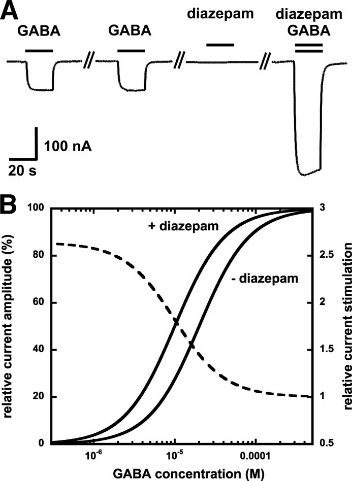
As a first example of exogenous modulators, we will have a closer look at a group of popular drugs, the benzodiazepines. Benzodiazepines bind to a high affinity binding site located at the α/γ subunit interface in a homologous position to the agonist site (for review, see Refs. 71 and 72). Classical benzodiazepines like valium are positive allosteric modulators of the response to GABA. Benzodiazepines do not open the channel by themselves (Fig. 4A). High affinity binding of these substances to their recognition site leads to a conformational change in the receptor such as to increase the apparent affinity for channel gating by GABA at both agonist sites. As a result, maximal currents elicited by GABA remain unaffected, and the GABA concentration channel opening curve is shifted to lower GABA concentrations (Fig. 4B). At the single channel level, benzodiazepines increase mean open times (73). Classical benzodiazepines have also been termed “benzodiazepine agonists” despite the fact that they induce channel opening with only exceedingly low efficacy. Compounds exist that compete at the binding site for benzodiazepines but do not affect the GABA concentration dependence, the benzodiazepine antagonists. Furthermore, there are such compounds that compete and shift the GABA concentration dependence to the right, the negative allosteric modulators, which have also been termed “benzodiazepine inverse agonists.”
FIGURE 4.
A, current trace of an electrophysiological recording showing an application of GABA to a cell expressing GABAA receptors. Positive allosteric modulators of the benzodiazepine type (in this case, 1 μm diazepam) do not induce any current by themselves but increase the current amplitude upon co-application with low concentrations of the agonist GABA. B, GABA concentration response curves in the absence (EC50 = 20 μm, n = 1.5) and presence (EC50 = 10 μm, n = 1.5) of diazepam. The maximal current amplitude (100%) is not affected by diazepam. The dashed line shows GABA concentration dependence of the calculated relative current stimulation. Please note that there is no stimulation at high concentrations of GABA.
The relative positioning of benzodiazepines in their binding site has been approached using the proximity-accelerated chemical reaction (74). GABAA receptor residues thought to reside in the site were individually mutated to cysteine, and the mutant receptors were exposed to a modified benzodiazepine molecule carrying a cysteine-reactive substituent at a defined atom. Direct apposition of the reactive group and cysteine leads to a covalent reaction. From this work, we know that the C1 atom of diazepam is located close to α1His-101 (75) and that the 3′-atom is located close to α1Ser-205 and α1Thr-206 (76). Very recently, diazepam has been docked into a multitude of conformational variants of a large number of homology models of the GABAA receptor, and the docking poses were rated according to all available experimental observations (77). Future work will show if homology modeling approaches are adequate to predict relative positioning of benzodiazepines in their binding pocket. In addition to the classical benzodiazepine-binding site at the α/γ subunit interface, an unusual site has recently been described at the α/β subunit interface (78, 79). The implications of this additional site still remain to be elucidated.
The second example of an exogenous modulator discussed here is ethanol. There is general agreement that high lethal concentrations (>50 mm) of ethanol modulate the function of many membrane proteins, among them the GABAA receptor. However, it is intensely disputed whether ethanol concentrations influencing human behavior (<20 mm) affect GABAA receptors. Although one group reported positive allosteric modulation on α6β3δ receptors (80), several groups were unable to reproduce the findings (34, 81).
Role of Individual Receptors
Although it is relatively simple to address questions at the level of individual receptor subunit isoforms, we can only speculate how many GABAA receptors are expressed in our brain and what their subunit composition is, not to mention subunit arrangement. Experiments have therefore been limited to the role of defined receptor subunit isoforms. Knock-out mice in which a functional gene for a certain GABAA receptor subunit is lacking have been prepared. Elimination of a particular subunit isoform would be expected to suppress synthesis of all those GABAA receptor isoforms containing the subunit in question and cause alteration in the behavior of the mutant mice. This is expected to give information on the function of the corresponding subunit isoform. Such studies have the inherent drawback that hundreds of genes show adaptive changes as a consequence of the lack of a subunit. A more elegant approach to this question is knock-in mice carrying the point mutation α1H101R (and homologous mutations in α2, α3, and α5), rendering the site for benzodiazepines at the α/γ subunit interface insensitive to these drugs. In behavioral experiments, it was tested which benzodiazepine effects were missing in the mutant animals. Such experiments associated the α1 subunit with sedation (82, 83), the α2/3 subunits with anxiety (84, 85), and the α5 subunit with temporal and spatial memory (86, 87). In attempts for simplification, it was stated that “α1 receptors” or “α1 subunit-containing receptors” confer sedation, “α2/3 receptors” or “α2/3 subunit-containing receptors” confer anxiolysis, and inhibition of “α5 receptors” or inhibition of “α5 subunit-containing receptors” confers cognitive enhancement. Both types of simplification are problematic. As many receptors contain two different α subunits, such statements should be avoided. For example, for α6-containing GABAA receptors in the cerebellum, α1α6βxγ2 and α1α6βxδ receptors dominate over α6βxγ2 and α1βxδ receptors, respectively (88). As only the α subunit neighboring the γ subunit contributes to the benzodiazepine action (36), a receptor with mixed α subunits may contain an α1 subunit exclusively at the α/β interface, which would not affect the response to classical benzodiazepines.
As a further complication, several experimental drugs exist that contradict the above assignment. For example, the experimental compound ocinaplon (DOV-273547), which stimulates αx (x = 1, 2, 3, or 5) receptors without subunit preference, is a non-sedative anxiolytic (89). Another example is pyrazolopyrimidine (DOV-51892), which is specific for α1-containing receptors. Therefore, DOV-51892 is predicted to have sedative properties, but behavioral studies demonstrate that DOV-51892 is a non-sedative anxiolytic (90). At the moment, we should be careful to make any simplified statements. Behavior is a complex phenomenon, and most probably, there are several types of GABAA receptors involved even in simple behavioral traits.
A more precise approach to the role of defined GABAA receptor subunits in defined cells involves a two-step procedure (91, 92). In the first step, knock-in mice were prepared in which all γ subunits were rendered insensitive to zolpidem, a drug acting at the benzodiazepine-binding site. In the second step, these subunits were replaced in a defined cell type with zolpidem-responsive wild-type γ2 subunits. Zopidem administration instantaneously activated exclusively receptors containing the responsive γ2 subunits. Zolpidem-induced alterations in behavior could then be attributed to these receptors.
Conclusions
Many scientists and companies are put off by the complexity of the field of GABAA receptors, but it is exactly this complexity that offers numerous possibilities of fine-tuned pharmacological interventions. Further work is needed in diverse areas. Most importantly, protein crystallization of the closed, open, and desensitized conformations of GABAA receptors is needed. Crystallization of different benzodiazepine-sensitive isoforms of the receptor in the absence and presence of benzodiazepines will contribute to our understanding of the action of these drugs and help in the design of receptor isoform-specific drugs. Information on the structure of the different receptor isoforms, their subunit composition and subunit arrangement, and specifically how the cell controls which subunits are assembled in what sequence is one focus of interest. Specifically, approaches to determine the subunit composition and arrangement in situ at synapses and at extrasynaptic sites are needed, even if this seems difficult with the presently available techniques. Modulation of these receptor isoforms by endogenous and exogenous ligands and how conformational changes induced by ligand binding induce channel opening have to be better understood. A further focus of interest concerns positioning of the receptors to their synaptic and extrasynaptic place of action, as well as synapse-specific clustering. It may be anticipated that genetic alterations of subunits of the GABAA receptor affect any of the abovementioned processes and thereby contribute to inherited human diseases. A start has been made with the analysis of point mutations that cause epilepsy (93).
Acknowledgment
We thank Dr. V. Niggli for carefully reading the manuscript.
Footnotes
This work was supported by Swiss National Foundation Grant 31003A_132806/1. This is the third article in the Thematic Minireview Series on Celebrating the Discovery of the Cysteine Loop Ligand-gated Ion Channel Superfamily.
REFERENCES
- 1. Pfeiffer F., Graham D., Betz H. (1982) Purification by affinity chromatography of the glycine receptor of rat spinal cord. J. Biol. Chem. 257, 9389–9393 [PubMed] [Google Scholar]
- 2. Sigel E., Mamalaki C., Barnard E. A. (1982) Isolation of a GABA receptor from bovine brain using a benzodiazepine affinity column. FEBS Lett. 147, 45–48 [DOI] [PubMed] [Google Scholar]
- 3. Sigel E., Stephenson F. A., Mamalaki C., Barnard E. A. (1983) A γ-aminobutyric acid/benzodiazepine receptor complex of bovine cerebral cortex. J. Biol. Chem. 258, 6965–6971 [PubMed] [Google Scholar]
- 4. Sigel E., Barnard E. A. (1984) A γ-aminobutyric acid/benzodiazepine receptor complex from bovine cerebral cortex. Improved purification with preservation of regulatory sites and their interactions. J. Biol. Chem. 259, 7219–7223 [PubMed] [Google Scholar]
- 5. Schofield P. R., Darlison M. G., Fujita N., Burt D. R., Stephenson F. A., Rodriguez H., Rhee L. M., Ramachandran J., Reale V., Glencorse T. A., Seeburg P. H., Barnard E. A. (1987) Sequence and functional expression of the GABAA receptor shows a ligand-gated receptor superfamily. Nature 328, 221–227 [DOI] [PubMed] [Google Scholar]
- 6. Sieghart W., Karobath M. (1980) Molecular heterogeneity of benzodiazepine receptors. Nature 286, 285–287 [DOI] [PubMed] [Google Scholar]
- 7. Betz H. (1990) Ligand-gated ion channels in the brain: the amino acid receptor superfamily. Neuron 5, 383–392 [DOI] [PubMed] [Google Scholar]
- 8. Ernst M., Bruckner S., Boresch S., Sieghart W. (2005) Comparative models of GABAA receptor extracellular and transmembrane domains: important insights in pharmacology and function. Mol. Pharmacol. 68, 1291–1300 [DOI] [PubMed] [Google Scholar]
- 9. Miller P. S., Smart T. G. (2010) Binding, activation, and modulation of Cys loop receptors. Trends Pharmacol. Sci. 31, 161–174 [DOI] [PubMed] [Google Scholar]
- 10. Olsen R. W., Sieghart W. (2008) International Union of Pharmacology. LXX. Subtypes of γ-aminobutyric acid A receptors: classification on the basis of subunit composition, pharmacology, and function. Update. Pharmacol. Rev. 60, 243–260 [DOI] [PMC free article] [PubMed] [Google Scholar]
- 11. Corringer P. J., Le Novère N., Changeux J. P. (2000) Nicotinic receptors at the amino acid level. Annu. Rev. Pharmacol. Toxicol. 40, 431–458 [DOI] [PubMed] [Google Scholar]
- 12. Breitinger H. G., Becker C. M. (2002) The inhibitory glycine receptor–simple views of a complicated channel. ChemBioChem 3, 1042–1052 [DOI] [PubMed] [Google Scholar]
- 13. Thompson A. J., Lummis S. C. (2006) 5-HT3 receptors. Curr. Pharm. Des. 12, 3615–3630 [DOI] [PMC free article] [PubMed] [Google Scholar]
- 14. Davies P. A., Wang W., Hales T. G., Kirkness E. F. (2003) A novel class of ligand-gated ion channel is activated by Zn2+. J. Biol. Chem. 278, 712–717 [DOI] [PubMed] [Google Scholar]
- 15. Hibbs R. E., Gouaux E. (2011) Principles of activation and permeation in an anion-selective Cys loop receptor. Nature 474, 54–60 [DOI] [PMC free article] [PubMed] [Google Scholar]
- 16. Bocquet N., Prado de Carvalho L., Cartaud J., Neyton J., Le Poupon C., Taly A., Grutter T., Changeux J. P., Corringer P. J. (2007) A prokaryotic proton-gated ion channel from the nicotinic acetylcholine receptor family. Nature 445, 116–119 [DOI] [PubMed] [Google Scholar]
- 17. Hilf R. J., Dutzler R. (2008) X-ray structure of a prokaryotic pentameric ligand-gated ion channel. Nature 452, 375–379 [DOI] [PubMed] [Google Scholar]
- 18. Simon J., Wakimoto H., Fujita N., Lalande M., Barnard E. A. (2004) Analysis of the set of GABAA receptor genes in the human genome. J. Biol. Chem. 279, 41422–41435 [DOI] [PubMed] [Google Scholar]
- 19. Olsen R. W., Tobin A. J. (1990) Molecular biology of GABAA receptors. FASEB J. 4, 1469–1480 [DOI] [PubMed] [Google Scholar]
- 20. Darlison M. G., Pahal I., Thode C. (2005) Consequences of the evolution of the GABAA receptor gene family. Cell. Mol. Neurobiol. 25, 607–624 [DOI] [PMC free article] [PubMed] [Google Scholar]
- 21. Sinkkonen S. T., Hanna M. C., Kirkness E. F., Korpi E. R. (2000) GABAA receptor ϵ and θ subunits display unusual structural variation between species and are enriched in the rat locus ceruleus. J. Neurosci. 20, 3588–3595 [DOI] [PMC free article] [PubMed] [Google Scholar]
- 22. Barnard E. A., Skolnick P., Olsen R. W., Mohler H., Sieghart W., Biggio G., Braestrup C., Bateson A. N., Langer S. Z. (1998) International Union of Pharmacology. XV. Subtypes of γ-aminobutyric acid A receptors: classification on the basis of subunit structure and receptor function. Pharmacol. Rev. 50, 291–313 [PubMed] [Google Scholar]
- 23. Macdonald R. L., Olsen R. W. (1994) GABAA receptor channels. Annu. Rev. Neurosci. 17, 569–602 [DOI] [PubMed] [Google Scholar]
- 24. Rabow L. E., Russek S. J., Farb D. H. (1995) From ion currents to genomic analysis: recent advances in GABAA receptor research. Synapse 21, 189–274 [DOI] [PubMed] [Google Scholar]
- 25. Chen Z. W., Olsen R. W. (2007) GABAA receptor-associated proteins: a key factor regulating GABAA receptor function. J. Neurochem. 100, 279–294 [DOI] [PubMed] [Google Scholar]
- 26. Jacob T. C., Moss S. J., Jurd R. (2008) GABAA receptor trafficking and its role in the dynamic modulation of neuronal inhibition. Nat. Rev. Neurosci. 9, 331–343 [DOI] [PMC free article] [PubMed] [Google Scholar]
- 27. Daniel C., Ohman M. (2009) RNA editing and its impact on GABAA receptor function. Biochem. Soc. Trans. 37, 1399–1403 [DOI] [PubMed] [Google Scholar]
- 28. Whiting P. J., McKernan R. M., Wafford K. A. (1995) Structure and pharmacology of vertebrate GABAA receptor subtypes. Int. Rev. Neurobiol. 38, 95–138 [DOI] [PubMed] [Google Scholar]
- 29. Tretter V., Ehya N., Fuchs K., Sieghart W. (1997) Stoichiometry and assembly of a recombinant GABAA receptor subtype. J. Neurosci. 17, 2728–2737 [DOI] [PMC free article] [PubMed] [Google Scholar]
- 30. Baumann S. W., Baur R., Sigel E. (2002) Forced subunit assembly in α1β2γ2 GABAA receptors. Insight into the absolute arrangement. J. Biol. Chem. 277, 46020–46025 [DOI] [PubMed] [Google Scholar]
- 31. Baur R., Minier F., Sigel E. (2006) A GABAA receptor of defined subunit composition and positioning: concatenation of five subunits. FEBS Lett. 580, 1616–1620 [DOI] [PubMed] [Google Scholar]
- 32. Bollan K. A., Baur R., Hales T. G., Sigel E., Connolly C. N. (2008) The promiscuous role of the ϵ subunit in GABAA receptor biogenesis. Mol. Cell. Neurosci. 37, 610–621 [DOI] [PubMed] [Google Scholar]
- 33. Kaur K. H., Baur R., Sigel E. (2009) Unanticipated structural and functional properties of δ subunit-containing GABAA receptors. J. Biol. Chem. 284, 7889–7896 [DOI] [PMC free article] [PubMed] [Google Scholar]
- 34. Baur R., Kaur K. H., Sigel E. (2009) Structure of α6β3δ GABAA receptors and their lack of ethanol sensitivity. J. Neurochem. 111, 1172–1181 [DOI] [PubMed] [Google Scholar]
- 35. Sigel E., Baur R., Trube G., Möhler H., Malherbe P. (1990) The effect of subunit composition of rat brain GABAA receptors on channel function. Neuron 5, 703–711 [DOI] [PubMed] [Google Scholar]
- 36. Minier F., Sigel E. (2004) Positioning of the α subunit isoforms confers a functional signature to γ-aminobutyric acid type A receptors. Proc. Natl. Acad. Sci. U.S.A. 101, 7769–7774 [DOI] [PMC free article] [PubMed] [Google Scholar]
- 37. Unwin N. (2005) Refined structure of the nicotinic acetylcholine receptor at 4 Å resolution. J. Mol. Biol. 346, 967–989 [DOI] [PubMed] [Google Scholar]
- 38. Brejc K., van Dijck W. J., Klaassen R. V., Schuurmans M., van der Oost J., Smit A. B., Sixma T. K. (2001) Crystal structure of an ACh-binding protein reveals the ligand-binding domain of nicotinic receptors. Nature 411, 269–276 [DOI] [PubMed] [Google Scholar]
- 39. Berezhnoy D., Gibbs T. T., Farb D. H. (2009) Docking of 1,4-benzodiazepines in the α1/γ2 GABAA receptor modulator site. Mol. Pharmacol. 76, 440–450 [DOI] [PMC free article] [PubMed] [Google Scholar]
- 40. Ernst M., Brauchart D., Boresch S., Sieghart W. (2003) Comparative modeling of GABAA receptors: limits, insights, future developments. Neuroscience 119, 933–943 [DOI] [PubMed] [Google Scholar]
- 41. Li S. X., Huang S., Bren N., Noridomi K., Dellisanti C. D., Sine S. M., Chen L. (2011) Ligand-binding domain of an α7-nicotinic receptor chimera and its complex with agonist. Nat. Neurosci. 14, 1253–1259 [DOI] [PMC free article] [PubMed] [Google Scholar]
- 42. Sieghart W., Sperk G. (2002) Subunit composition, distribution, and function of GABAA receptor subtypes. Curr. Top. Med. Chem. 2, 795–816 [DOI] [PubMed] [Google Scholar]
- 43. Minuk G. Y., Zhang M., Gong Y., Minuk L., Dienes H., Pettigrew N., Kew M., Lipschitz J., Sun D. (2007) Decreased hepatocyte membrane potential differences and GABAA-β3 expression in human hepatocellular carcinoma. Hepatology 45, 735–745 [DOI] [PubMed] [Google Scholar]
- 44. Mizuta K., Xu D., Pan Y., Comas G., Sonett J. R., Zhang Y., Panettieri R. A., Jr., Yang J., Emala C. W., Sr. (2008) GABAA receptors are expressed and facilitate relaxation in airway smooth muscle. Am. J. Physiol. Lung Cell. Mol. Physiol. 294, L1206–L1216 [DOI] [PMC free article] [PubMed] [Google Scholar]
- 45. Alam S., Laughton D. L., Walding A., Wolstenholme A. J. (2006) Human peripheral blood mononuclear cells express GABAA receptor subunits. Mol. Immunol. 43, 1432–1442 [DOI] [PubMed] [Google Scholar]
- 46. Bjurstöm H., Wang J., Ericsson I., Bengtsson M., Liu Y., Kumar-Mendu S., Issazadeh-Navikas S., Birnir B. (2008) GABA, a natural immunomodulator of T lymphocytes. J. Neuroimmunol. 205, 44–50 [DOI] [PubMed] [Google Scholar]
- 47. Farrant M., Nusser Z. (2005) Variations on an inhibitory theme: phasic and tonic activation of GABAA receptors. Nat. Rev. Neurosci. 6, 215–229 [DOI] [PubMed] [Google Scholar]
- 48. Maguire J. L., Stell B. M., Rafizadeh M., Mody I. (2005) Ovarian cycle-linked changes in GABAA receptors mediating tonic inhibition alter seizure susceptibility and anxiety. Nat. Neurosci. 8, 797–804 [DOI] [PubMed] [Google Scholar]
- 49. Kaila K., Pasternack M., Saarikoski J., Voipio J. (1989) Influence of GABA-gated bicarbonate conductance on potential, current, and intracellular chloride in crayfish muscle fibers. J. Physiol. 416, 161–181 [DOI] [PMC free article] [PubMed] [Google Scholar]
- 50. Beg A. A., Jorgensen E. M. (2003) EXP-1 is an excitatory GABA-gated cation channel. Nat. Neurosci. 6, 1145–1152 [DOI] [PubMed] [Google Scholar]
- 51. Coull J. A., Beggs S., Boudreau D., Boivin D., Tsuda M., Inoue K., Gravel C., Salter M. W., De Koninck Y. (2005) BDNF from microglia causes the shift in neuronal anion gradient underlying neuropathic pain. Nature 438, 1017–1021 [DOI] [PubMed] [Google Scholar]
- 52. Ben-Ari Y., Gaiarsa J. L., Tyzio R., Khazipov R. (2007) GABA: a pioneer transmitter that excites immature neurons and generates primitive oscillations. Physiol. Rev. 87, 1215–1284 [DOI] [PubMed] [Google Scholar]
- 53. Wright R., Raimondo J. V., Akerman C. J. (2011) Spatial and temporal dynamics in the ionic driving force for GABAA receptors. Neural Plast. 2011, 728395. [DOI] [PMC free article] [PubMed] [Google Scholar]
- 54. Baumann S. W., Baur R., Sigel E. (2003) Individual properties of the two functional agonist sites in GABAA receptors. J. Neurosci. 23, 11158–11166 [DOI] [PMC free article] [PubMed] [Google Scholar]
- 55. Wagner D. A., Czajkowski C. (2001) Structure and dynamics of the GABA binding pocket: a narrowing cleft that constricts during activation. J. Neurosci. 21, 67–74 [DOI] [PMC free article] [PubMed] [Google Scholar]
- 56. Lape R., Colquhoun D., Sivilotti L. G. (2008) On the nature of partial agonism in the nicotinic receptor superfamily. Nature 454, 722–727 [DOI] [PMC free article] [PubMed] [Google Scholar]
- 57. Bormann J., Clapham D. E. (1985) γ-Aminobutyric acid receptor channels in adrenal chromaffin cells: a patch-clamp study. Proc. Natl. Acad. Sci. U.S.A. 82, 2168–2172 [DOI] [PMC free article] [PubMed] [Google Scholar]
- 58. Khatri A., Weiss D. S. (2010) The role of loop F in the activation of the GABA receptor. J. Physiol. 588, 59–66 [DOI] [PMC free article] [PubMed] [Google Scholar]
- 59. Kash T. L., Jenkins A., Kelley J. C., Trudell J. R., Harrison N. L. (2003) Coupling of agonist binding to channel gating in the GABAA receptor. Nature 421, 272–275 [DOI] [PubMed] [Google Scholar]
- 60. Venkatachalan S. P., Czajkowski C. (2012) Structural link between γ-aminobutyric acid type A (GABAA) receptor agonist-binding site and inner β-sheet governs channel activation and allosteric drug modulation. J. Biol. Chem. 287, 6714–6724 [DOI] [PMC free article] [PubMed] [Google Scholar]
- 61. Chang Y., Weiss D. S. (2002) Site-specific fluorescence reveals distinct structural changes with GABA receptor activation and antagonism. Nat. Neurosci. 5, 1163–1168 [DOI] [PubMed] [Google Scholar]
- 62. Xu M., Akabas M. H. (1993) Amino acids lining the channel of the γ-aminobutyric acid type A receptor identified by cysteine substitution. J. Biol. Chem. 268, 21505–21508 [PubMed] [Google Scholar]
- 63. Kittler J. T., Moss S. J. (2003) Modulation of GABAA receptor activity by phosphorylation and receptor trafficking: implications for the efficacy of synaptic inhibition. Curr. Opin. Neurobiol. 13, 341–347 [DOI] [PubMed] [Google Scholar]
- 64. Huson D. H., Richter D. C., Rausch C., Dezulian T., Franz M., Rupp R. (2007) Dendroscope: An interactive viewer for large phylogenetic trees. BMC Bioinformatics 8, 460. [DOI] [PMC free article] [PubMed] [Google Scholar]
- 65. Belelli D., Lambert J. J. (2005) Neurosteroids: endogenous regulators of the GABAA receptor. Nat. Rev. Neurosci. 6, 565–575 [DOI] [PubMed] [Google Scholar]
- 66. Sigel E., Baur R., Rácz I., Marazzi J., Smart T. G., Zimmer A., Gertsch J. (2011) The major central endocannabinoid directly acts at GABAA receptors. Proc. Natl. Acad. Sci. U.S.A. 108, 18150–18155 [DOI] [PMC free article] [PubMed] [Google Scholar]
- 67. Sieghart W. (1995) Structure and pharmacology of γ-aminobutyric acid A receptor subtypes. Pharm. Rev. 47, 181–234 [PubMed] [Google Scholar]
- 68. Hosie A. M., Wilkins M. E., da Silva H. M., Smart T. G. (2006) Endogenous neurosteroids regulate GABAA receptors through two discrete transmembrane sites. Nature 444, 486–489 [DOI] [PubMed] [Google Scholar]
- 69. Zheleznova N., Sedelnikova A., Weiss D. S. (2008) α1β2δ, a silent GABAA receptor: recruitment by tracazolate and neurosteroids. Br. J. Pharmacol. 53, 1062–1071 [DOI] [PMC free article] [PubMed] [Google Scholar]
- 70. Shu H. J., Bracamontes J., Taylor A., Wu K., Eaton M. M., Akk G., Manion B., Evers A. S., Krishnan K., Covey D. F., Zorumski C. F., Steinbach J. H., Mennerick S. (2012) Characteristics of concatemeric GABAA receptors containing α4/δ subunits expressed in Xenopus oocytes. Br. J. Pharmacol. 165, 2228–2243 [DOI] [PMC free article] [PubMed] [Google Scholar]
- 71. Sigel E., Buhr A. (1997) The benzodiazepine-binding site of GABAA receptors. Trends Pharmacol. Sci. 18, 425–429 [DOI] [PubMed] [Google Scholar]
- 72. Sigel E., Lüscher B. P. (2011) A closer look at the high affinity benzodiazepine-binding site on GABAA receptors. Curr. Top. Med. Chem. 11, 241–246 [DOI] [PubMed] [Google Scholar]
- 73. Twyman R. E., Rogers C. J., Macdonald R. L. (1989) Differential regulation of γ-aminobutyric acid receptor channels by diazepam and phenobarbital. Ann. Neurol. 25, 213–220 [DOI] [PubMed] [Google Scholar]
- 74. Foucaud B., Perret P., Grutter T., Goeldner M. (2001) Cysteine mutants as chemical sensors for ligand-receptor interactions. Trends Pharmacol. Sci. 22, 170–173 [DOI] [PubMed] [Google Scholar]
- 75. Berezhnoy D., Nyfeler Y., Gonthier A., Schwob H., Goeldner M., Sigel E. (2004) On the benzodiazepine-binding pocket in GABAA receptors. J. Biol. Chem. 279, 3160–3168 [DOI] [PubMed] [Google Scholar]
- 76. Tan K. R., Baur R., Charon S., Goeldner M., Sigel E. (2009) Relative positioning of diazepam in the benzodiazepine-binding pocket of GABA receptors. J. Neurochem. 111, 1264–1273 [DOI] [PubMed] [Google Scholar]
- 77. Richter L., de Graaf C., Sieghart W., Varagic Z., Mörzinger M., de Esch I. J., Ecker G. F., Ernst M. (2012) Diazepam-bound GABAA receptor models identify new benzodiazepine-binding site ligands. Nat. Chem. Biol. 8, 455–464 [DOI] [PMC free article] [PubMed] [Google Scholar]
- 78. Baur R., Tan K. R., Lüscher B. P., Gonthier A., Goeldner M., Sigel E. (2008) Covalent modification of GABAA receptor isoforms by a diazepam analogue provides evidence for a novel benzodiazepine-binding site that prevents modulation by these drugs. J. Neurochem. 106, 2353–2363 [DOI] [PubMed] [Google Scholar]
- 79. Ramerstorfer J., Furtmüller R., Sarto-Jackson I., Varagic Z., Sieghart W., Ernst M. (2011) The GABAA receptor α+β interface: a novel target for subtype selective drugs. J. Neurosci. 31, 870–877 [DOI] [PMC free article] [PubMed] [Google Scholar]
- 80. Wallner M., Hanchar H. J., Olsen R. W. (2003) Ethanol enhances α4β3δ and α6β3δ γ-aminobutyric acid type A receptors at low concentrations known to affect humans. Proc. Natl. Acad. Sci. U.S.A. 100, 15218–15223 [DOI] [PMC free article] [PubMed] [Google Scholar]
- 81. Borghese C. M., Stórustovu S., Ebert B., Herd M. B., Belelli D., Lambert J. J., Marshall G., Wafford K. A., Harris R. A. (2006) The δ subunit of γ-aminobutyric acid type A receptors does not confer sensitivity to low concentrations of ethanol. J. Pharmacol. Exp. Ther. 316, 1360–1368 [DOI] [PubMed] [Google Scholar]
- 82. Rudolph U., Crestani F., Benke D., Brünig I., Benson J. A., Fritschy J. M., Martin J. R., Bluethmann H., Möhler H. (1999) Benzodiazepine actions mediated by specific γ-aminobutyric acid A receptor subtypes. Nature 401, 796–800 [DOI] [PubMed] [Google Scholar]
- 83. McKernan R. M., Rosahl T. W., Reynolds D. S., Sur C., Wafford K. A., Atack J. R., Farrar S., Myers J., Cook G., Ferris P., Garrett L., Bristow L., Marshall G., Macaulay A., Brown N., Howell O., Moore K. W., Carling R. W., Street L. J., Castro J. L., Ragan C. I., Dawson G. R., Whiting P. J. (2000) Sedative but not anxiolytic properties of benzodiazepines are mediated by the GABAA receptor α1 subtype. Nat. Neurosci. 3, 587–592 [DOI] [PubMed] [Google Scholar]
- 84. Löw K., Crestani F., Keist R., Benke D., Brünig I., Benson J. A., Fritschy J. M., Rülicke T., Bluethmann H., Möhler H., Rudolph U. (2000) Molecular and neuronal substrate for the selective attenuation of anxiety. Science 290, 131–134 [DOI] [PubMed] [Google Scholar]
- 85. Crestani F., Löw K., Keist R., Mandelli M., Möhler H., Rudolph U. (2001) Molecular targets for the myorelaxant action of diazepam. Mol. Pharmacol. 59, 442–445 [DOI] [PubMed] [Google Scholar]
- 86. Crestani F., Keist R., Fritschy J. M., Benke D., Vogt K., Prut L., Blüthmann H., Möhler H., Rudolph U. (2002) Trace fear conditioning involves hippocampal α5 GABAA receptors. Proc. Natl. Acad. Sci. U.S.A. 99, 8980–8985 [DOI] [PMC free article] [PubMed] [Google Scholar]
- 87. Collinson N., Kuenzi F. M., Jarolimek W., Maubach K. A., Cothliff R., Sur C., Smith A., Otu F. M., Howell O., Atack J. R., McKernan R. M., Seabrook G. R., Dawson G. R., Whiting P. J., Rosahl T. W. (2002) Enhanced learning and memory and altered GABAergic synaptic transmission in mice lacking the α5 subunit of the GABAA receptor. J. Neurosci. 22, 5572–5580 [DOI] [PMC free article] [PubMed] [Google Scholar]
- 88. Jechlinger M., Pelz R., Tretter V., Klausberger T., Sieghart W. (1998) Subunit composition and quantitative importance of hetero-oligomeric receptors: GABAA receptors containing α6 subunits. J. Neurosci. 18, 2449–2457 [DOI] [PMC free article] [PubMed] [Google Scholar]
- 89. Lippa A., Czobor P., Stark J., Beer B., Kostakis E., Gravielle M., Bandyopadhyay S., Russek S. J., Gibbs T. T., Farb D. H., Skolnick P. (2005) Selective anxiolysis produced by ocinaplon, a GABAA receptor modulator. Proc. Natl. Acad. Sci. U.S.A. 102, 7380–7385 [DOI] [PMC free article] [PubMed] [Google Scholar]
- 90. Popik P., Kostakis E., Krawczyk M., Nowak G., Szewczyk B., Krieter P., Chen Z., Russek S. J., Gibbs T. T., Farb D. H., Skolnick P., Lippa A. S., Basile A. S. (2006) The anxioselective agent 7-(2-chloropyridin-4-yl)pyrazolo-[1,5-a]-pyrimidin-3-yl(pyridin-2-yl)methanone (DOV 51892) is more efficacious than diazepam at enhancing GABA-gated currents at α1 subunit-containing GABAA receptors. J. Pharmacol. Exp. Ther. 319, 1244–1252 [DOI] [PubMed] [Google Scholar]
- 91. Wulff P., Goetz T., Leppä E., Linden A. M., Renzi M., Swinny J. D., Vekovischeva O. Y., Sieghart W., Somogyi P., Korpi E. R., Farrant M., Wisden W. (2007) From synapse to behavior: rapid modulation of defined neuronal types with engineered GABAA receptors. Nat. Neurosci. 10, 923–929 [DOI] [PMC free article] [PubMed] [Google Scholar]
- 92. Sieghart W. (2012) Unravelling the role of GABAA receptor subtypes in distinct neurons and behaviour. J. Physiol. 590, 2181–2182 [DOI] [PMC free article] [PubMed] [Google Scholar]
- 93. Macdonald R. L., Kang J. Q., Gallagher M. J. (2010) Mutations in GABAA receptor subunits associated with genetic epilepsies. J. Physiol. 588, 1861–1869 [DOI] [PMC free article] [PubMed] [Google Scholar]
- 94. Wu C., Nebert D. W. (2004) Update on genome completion and annotations: Protein Information Resource. Hum. Genomics 1, 229–233 [DOI] [PMC free article] [PubMed] [Google Scholar]
- 95. Thompson J. D., Gibson T. J., Plewniak F., Jeanmougin F., Higgins D. G. (1997) The CLUSTAL_X windows interface: flexible strategies for multiple sequence alignment aided by quality analysis tools. Nucleic Acids Res. 25, 4876–4882 [DOI] [PMC free article] [PubMed] [Google Scholar]


