

Abstract
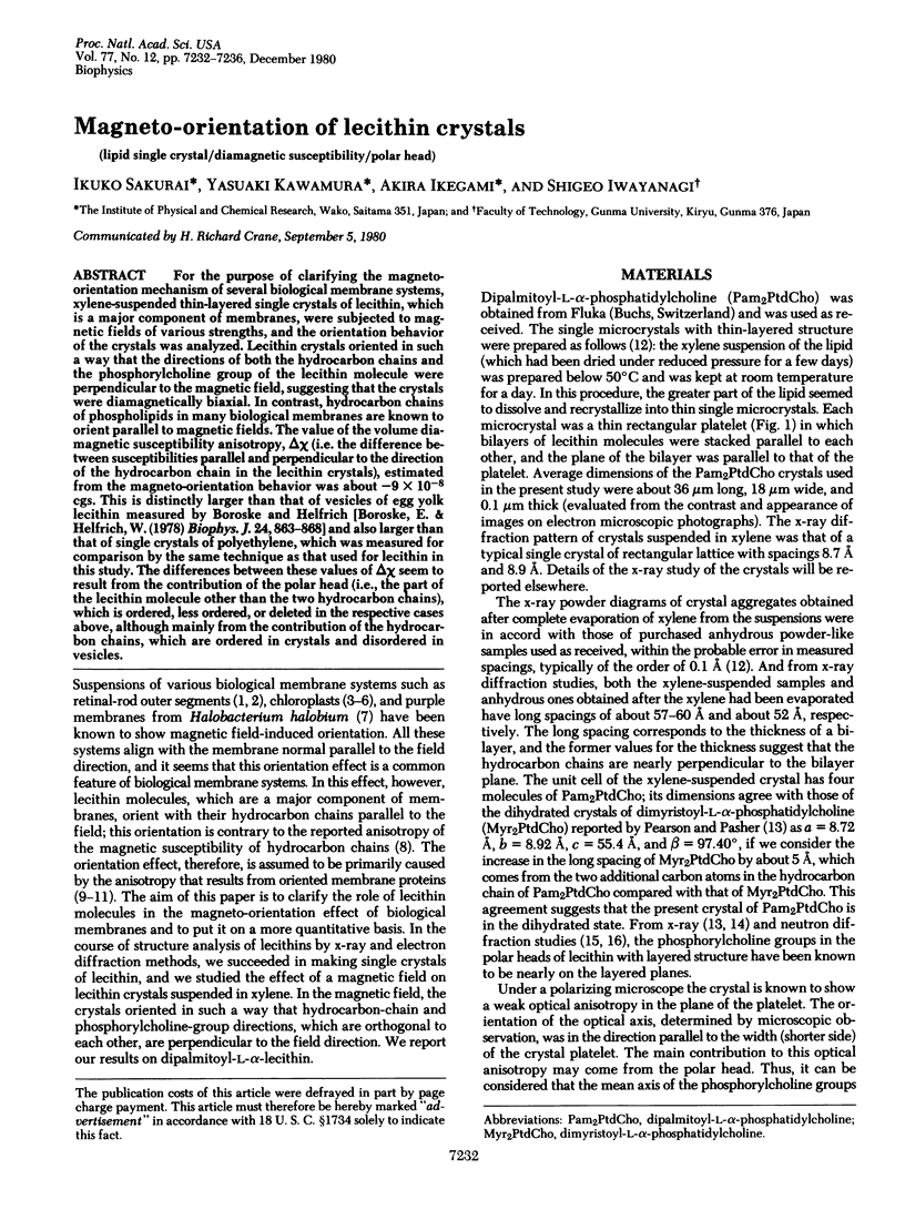
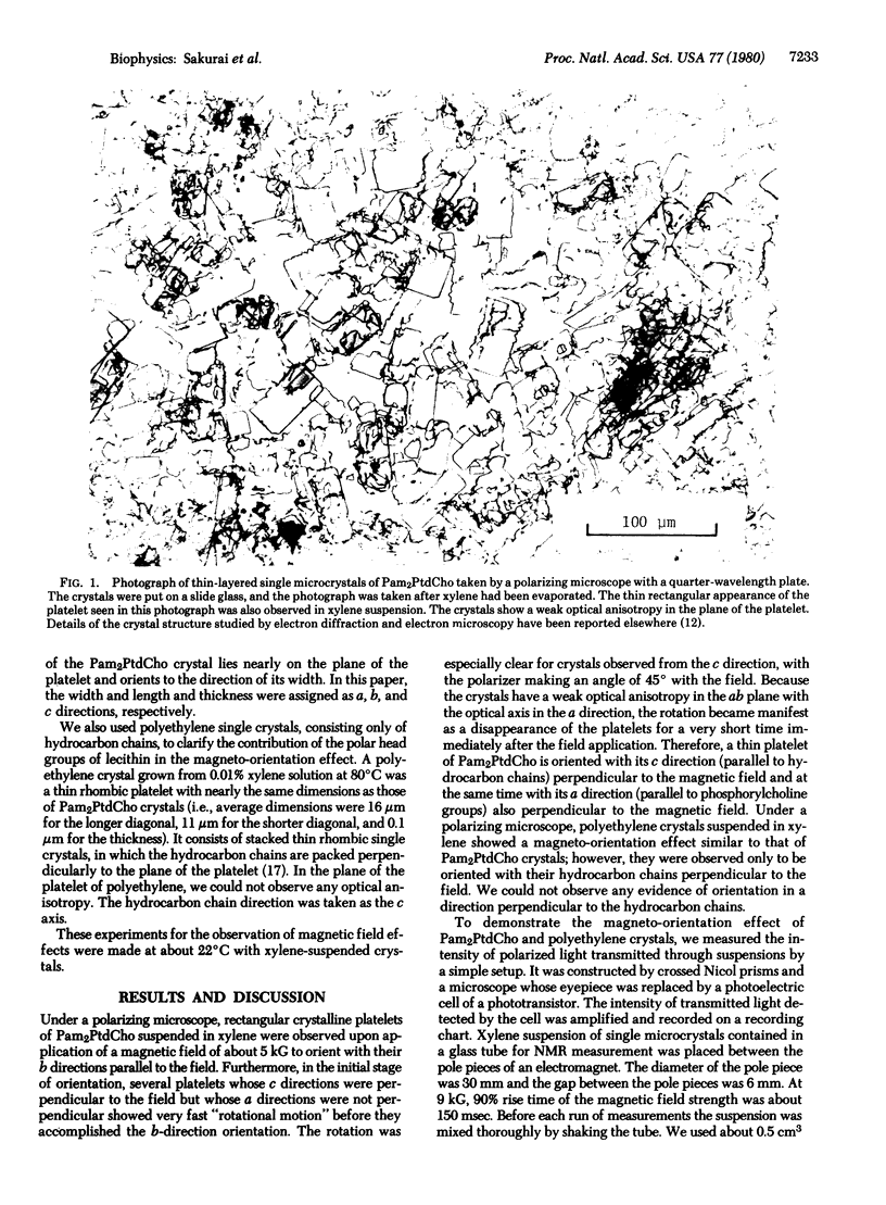
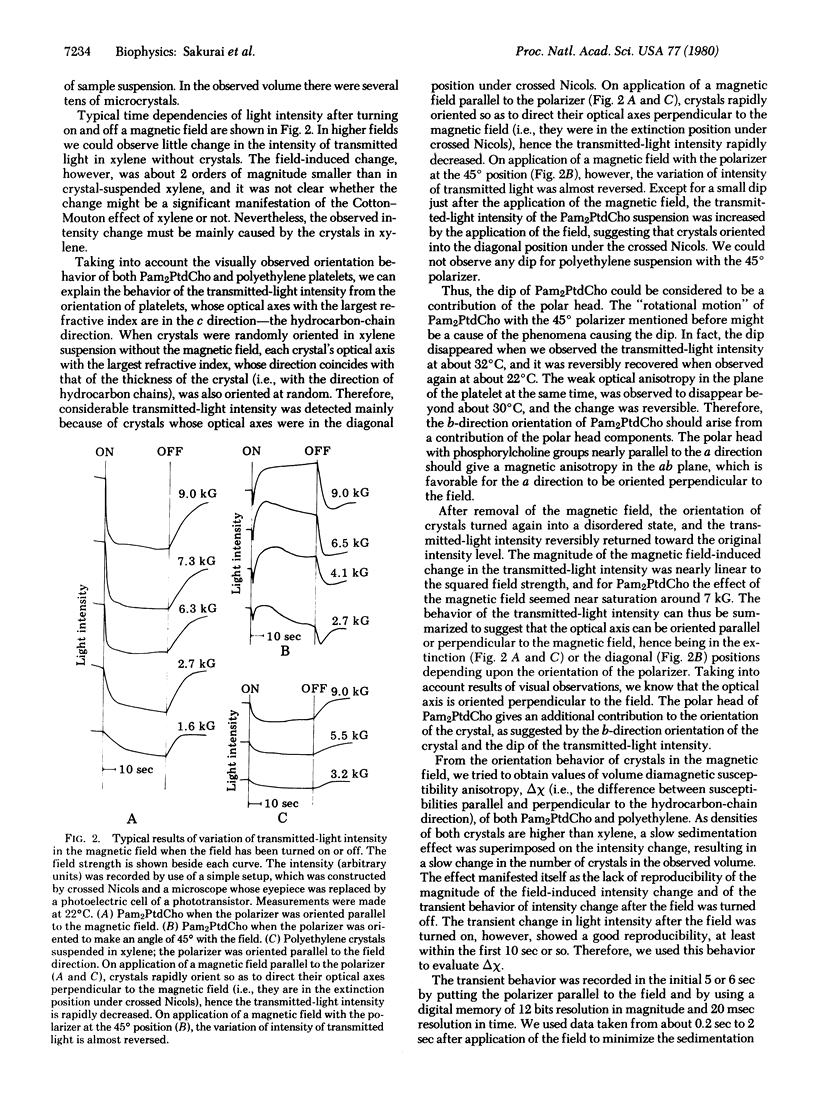
For the purpose of clarifying the magneto-orientation mechanism of several biological membrane systems, xylene-suspended thin-layered single crystals of lecithin, which is a major component of membranes, were subjected to magnetic fields of various strengths, and the orientation behavior of the crystals was analyzed. Lecithin crystals oriented in such a way that the directions of both the hydrocarbon chains and the phosphorylcholine group of the lecithin molecule were perpendicular to the magnetic field, suggesting that the crystals were diamagnetically biaxial. In contrast, hydrocarbon chains of phospholipids in many biological membranes are known to orient parallel to magnetic fields. The value of the volume diamagnetic susceptibility anisotropy, delta chi (i.e. the difference between susceptibilities parallel and perpendicular to the direction of the hydrocarbon chain in the lecithin crystals), estimated from the magneto-orientation behavior was about -9 X 10(-8) cgs. This is distinctly larger than that of vesicles of egg yolk lecithin measured by Boroske and Helfrich [Boroske, E. & Helfrich, W. (1978) Biophys. J. 24, 863-868] and also larger than that of single crystals of polyethylene, which was measured for comparison by the same technique as that used for lecithin in this study. The differences between these values of delta chi seem to result from the contribution of the polar head (i.e., the part of the lecithin molecule other than the two hydrocarbon chains), which is ordered, less ordered, or deleted in the respective cases above, although mainly from the contribution of the hydrocarbon chains, which are ordered in crystals and disordered in vesicles.
Full text
PDF
Images in this article
Selected References
These references are in PubMed. This may not be the complete list of references from this article.
- Becker J. F., Geacintov N. E., Van Nostrand F., Van Metter R. Orientation of chlorophyll in vivo. Studies with magnetic field oriented chlorella. Biochem Biophys Res Commun. 1973 Apr 2;51(3):597–602. doi: 10.1016/0006-291x(73)91356-9. [DOI] [PubMed] [Google Scholar]
- Boroske E., Helfrich W. Magnetic anisotropy of egg lecithin membranes. Biophys J. 1978 Dec;24(3):863–868. doi: 10.1016/S0006-3495(78)85425-3. [DOI] [PMC free article] [PubMed] [Google Scholar]
- Breton J., Michel-Villaz M., Paillotin G. Orientation of pigments and structural proteins in the photosynthetic membrane of spinach chloroplasts: a linear dichroism study. Biochim Biophys Acta. 1973 Jul 26;314(1):42–56. doi: 10.1016/0005-2728(73)90062-5. [DOI] [PubMed] [Google Scholar]
- Chagneux R., Chalazonitis N. Evaluation de l'anisotropie magnétique des cellules multimembranaires dans un champ magnétique constant (segments externes des bâtonnets de la rétine de grenouille. C R Acad Sci Hebd Seances Acad Sci D. 1972 Jan 10;274(2):317–320. [PubMed] [Google Scholar]
- Chalazonitis N., Chagneux R., Arvanitaki A. Rotation des segments externes des photorécepeurs dans le champ magnétique constant. C R Acad Sci Hebd Seances Acad Sci D. 1970 Jul 6;271(1):130–133. [PubMed] [Google Scholar]
- Franks N. P. Structural analysis of hydrated egg lecithin and cholesterol bilayers. I. X-ray diffraction. J Mol Biol. 1976 Jan 25;100(3):345–358. doi: 10.1016/s0022-2836(76)80067-8. [DOI] [PubMed] [Google Scholar]
- Geacintov N. E., Van Norstrand F., Becker J. F., Tinkel J. B. Magnetic field induced orientation of photosynthetic systems. Biochim Biophys Acta. 1972 Apr 20;267(1):65–79. doi: 10.1016/0005-2728(72)90138-7. [DOI] [PubMed] [Google Scholar]
- Geacintov N. E., Van Nostrand F., Pope M., Tinkel J. B. Magnetic field effect on the chlorophyll fluorescence in Chlorella. Biochim Biophys Acta. 1971 Mar 2;226(2):486–491. doi: 10.1016/0005-2728(71)90118-6. [DOI] [PubMed] [Google Scholar]
- Hitchcock P. B., Mason R., Thomas K. M., Shipley G. G. Structural chemistry of 1,2 dilauroyl-DL-phosphatidylethanolamine: molecular conformation and intermolecular packing of phospholipids. Proc Natl Acad Sci U S A. 1974 Aug;71(8):3036–3040. doi: 10.1073/pnas.71.8.3036. [DOI] [PMC free article] [PubMed] [Google Scholar]
- Hong F. T. Magnetic anisotropy of the visual pigment rhodopsin. Biophys J. 1980 Feb;29(2):343–346. doi: 10.1016/S0006-3495(80)85138-1. [DOI] [PMC free article] [PubMed] [Google Scholar]
- Neugebauer D. C., Blaurock A. E. Magnetic orientation of purple membranes demonstrated by optical measurements and neutron scattering. FEBS Lett. 1977;78(1):31–35. doi: 10.1016/0014-5793(77)80266-4. [DOI] [PubMed] [Google Scholar]
- Pauling L. Diamagnetic anisotropy of the peptide group. Proc Natl Acad Sci U S A. 1979 May;76(5):2293–2294. doi: 10.1073/pnas.76.5.2293. [DOI] [PMC free article] [PubMed] [Google Scholar]
- Pearson R. H., Pascher I. The molecular structure of lecithin dihydrate. Nature. 1979 Oct 11;281(5731):499–501. doi: 10.1038/281499a0. [DOI] [PubMed] [Google Scholar]
- Worcester D. L., Franks N. P. Structural analysis of hydrated egg lecithin and cholesterol bilayers. II. Neutrol diffraction. J Mol Biol. 1976 Jan 25;100(3):359–378. doi: 10.1016/s0022-2836(76)80068-x. [DOI] [PubMed] [Google Scholar]
- Worcester D. L. Structural origins of diamagnetic anisotropy in proteins. Proc Natl Acad Sci U S A. 1978 Nov;75(11):5475–5477. doi: 10.1073/pnas.75.11.5475. [DOI] [PMC free article] [PubMed] [Google Scholar]