Abstract
Background:
Pancreaticoduodenectomy remains a major undertaking. A preoperative blood test, which could confidently predict the benefits of surgery would improve the selection of pancreatic cancer patients for surgery. This study aimed to identify protein biomarkers prognostic for long-term survival and to validate them with clinico-pathological information.
Methods:
Serum from 40 preoperative patients was used to train for predictive biomarkers using surface-enhanced laser desorption/ionisation time-of-flight mass spectrometry (SELDI), and the results were verified on 21 independent samples. Two predictive proteins were identified by tryptic peptide mass fingerprinting and sequencing, and validated on serum from another 57 patients by enzyme-linked immunosorbent assay (ELISA). The influence of these proteins on growth and invasion of two cancer cell lines was tested in-vitro.
Results:
The SELDI panel of m/z 3700, 8222 and 11 522 peaks predicted <12 months’ survival (ROC AUC: 0.79, 0.64–0.90; P<0.039). When CA19-9 was added, the ROC AUC increased to 0.95 (0.84–0.99; P<0.0001). The six subjects in the verification group who died within 12 months were correctly classified. The m/z 8222 and 11 522 proteins were identified as Serum ApoC-II and SAA-1, respectively. In the validation samples, ELISA results confirmed that ApoC-II was predictive of survival (Kaplan–Meier P<0.009), but not SAA-I. ApoC-II, CA19-9 and major-vessel involvement independently predicted survival. ApoC-II and SAA-1 increased cell growth and invasion of both cancer cell lines.
Conclusion:
Serum ApoC-II, CA19-9 and major-vessel invasion independently predict survival and improves selection of patients for pancreaticoduodenectomy.
Keywords: pancreatic neoplasms, pancreaticoduodenectomy, prognosis, apolipoproteins, survival, biomarkers
Surgical resection and adjuvant chemotherapy for pancreatic cancer provide the best chance of long-term survival (Castellanos et al, 2011). The outcome appears to be independent of pylorus preservation (Akizuki et al, 2008), but may be improved by more extensive resection of mesopancreatic tissue (Samra et al, 2006) and vascular resection (Tang et al, 2011). Despite the introduction of these improvements over the last decade, one-fifth of patients do not survive 12 months; an unsatisfactory result, given that the survival time includes prolonged recovery from major surgery and that palliative treatments may have achieved the same outcome (Distler et al, 2010). When faced with the decision to undergo surgery, it is important for patients to be given an accurate prognosis, particularly regarding the chance of long-term survival.
Prognosis generally relies on established pathological classification such as the TNM staging of malignant tumours (TNM) (American Joint Committee on Cancer, 2009). To further improve the prognosis, the expression of a range of tissue proteins has been reported to have prognostic value in cohorts of patients undergoing pancreatic resection, but none has been studied in a prospective randomised manner (Jamieson et al, 2011), and their usefulness in the clinic remains unproven. Plasma biomarkers are more available in the preoperative setting, with Carbohydrate Antigen 19-9 (CA 19-9) being the most commonly used for diagnosis and for prognosis in clinical practice. However, although reviews have supported the prognostic use of CA 19-9 (Mehta et al, 2010), others have reported it to be unreliable (Kondo et al, 2010). An elevated preoperative value of CA 19-9 was found not to be prognostic of survival, while its continual elevation in the postoperative setting was a significant indicator of poor outcome (Hata et al, 2012). Carbohydrate Antigen 19-9 is frequently elevated in benign conditions and more so in the jaundiced patient (Molina et al, 2012). Other serum markers of the host’s response to cancer such as human C-reactive protein (CRP) (Pine et al, 2009), serum amyloid A-1 (SAA-1) (Firpo et al, 2009) and the neutrophil to lymphocyte ratio (Bhatti et al, 2010) are also reported to predict survival.
Such studies justify an exploratory examination of serum in an attempt to improve prediction of outcome, particularly in the era of extensive surgery and neoadjuvant therapy. One potential source of such biomarkers is the pool of low-abundance small proteins in serum. Surface-Enhanced Laser Desorption/Ionisation Time-of-Flight Mass Spectrometry (SELDI) was used to discover diagnostic protein biomarkers, including ApoA-II and ApoC-I (Xue et al, 2010), which were not previously recognised. Further, the SELDI technique has also revealed biomarkers of pancreatic cancer-related outcomes such as invasiveness (Valkovskaya et al, 2007), responsiveness to gemcitabine (Cao et al, 2010) and cachexia (Felix et al, 2011). It is therefore possible that SELDI could identify prognostic markers of long-term survival in the serum.
This study uses the SELDI platform to discover markers of prognosis in a training set of serum samples, and verifies the markers by SELDI in an independent set of serum samples. The prognostic capability of these proteins is then assessed using ELISA, along with other predictors of survival, in a further independent validation set of serum samples.
Materials and methods
Patients and sera
The study was approved by the Northern Sydney Health Human Research Ethics Committee, Sydney, Australia (HREC 909 228M) and the University of Verona, Ethics Committee, Verona, Italy.
The training set
Sera from 40 consecutive patients about to undergo pancreaticoduodenectomy for pancreatic ductal carcinoma (PDCa) at the Uniersity of Verona (UniVer) from 2006–2007 were prospectively collected and stored at −80 °C as the ‘training set’ until analysed by SELDI.
The verification set
Sera from 21 consecutive patients (the ‘verification set’), collected before pancreatic resection at Royal North Shore Hospital (RNSH) in 2007 were analysed by SELDI.
The validation set
Further 82 serum samples from subjects at RNSH from 2009 to 2010, consisting of sera from 57 PDCa patients before pancreatic resection, 13 with unresectable (advanced) pancreatic cancer (APDCa) and 12 healthy volunteers (HV), which were controls for the analysis technique were collected prospectively as a ‘validation set’. Clinical and pathological data for the validation set were prospectively recorded in a Microsoft Access database. The analysis of the data was delayed until 12 months after surgery. Two ‘validation set’ patients were lost to follow-up at 4 and 10 months, and their data were censored at the last review day, when they were alive. Tumours were synoptically classified according to defined pathological prognostic criteria by one of the authors, AG, as previously described (Gill et al, 2009). Carbohydrate Antigen 19-9 and CRP were measured by enzyme-linked immunosorbent assay (ELISA) and the neutrophil–lymphocyte ratio was recorded for the validation-set patients.
Preparation of serum and proteinchip arrays for SELDI-TOF MS
Serum was separated into aliquots 30 min after collection and stored at −80 °C. From each sample, 20 μl was diluted 1 : 1 with denaturing buffer (8 ℳ urea per 1% CHAPS), then centrifuged at 12 000 rpm for 5 min. Protein levels were measured by Micro BCH protein assay kits (Thermo Scientific, Chicago, IL, USA), and the amount of buffer added to the samples adjusted to provide 2 μg per spot. The supernatant was diluted to 1 : 25 with 10% acetonitrile (ACN)/0.1% trifluoroacetic acid (TFA). Treated samples were applied to hydrophobic (H50) proteinchip arrays and processed, as previously described (Xue et al, 2010). The proteinchip arrays were analysed using the Bio-Rad Enterprise SELDI System (Bio-Rad, Hercules, CA, USA).
SELDI-TOF MS Analysis
Duplicate spectra were obtained in the mass/charge (m/z) range 2500–75 000 with a laser intensity set at 220 (arbitrary units), detector sensitivity at 8 and a signal to noise ratio of 5%. The m/z value for each peak was determined using external calibration with standards (Sigma–Aldrich, St Louis, MO, USA) which are the following: bovine insulin (5734.51+1H), equine cytochrome c (12 361.96+1H), equine apomyoglobin (16 952.27+1H) and rabbit muscle aldolase (39 212.28 +1H). Spectra were analysed using ProteinChip Software, Version 3.1 (Bio-Rad).
Protein peak identification
Serum samples were separated into six fractions eluted at pH 9, 7, 5, 4, 3 and the organic phase using the ProteinChip Serum Fractionation Kit (Bio-Rad). The fractions of interest were immediately applied to a C18 reverse-phase HPLC column eluted with a gradient of 15–60% ACN in 0.1% TFA. Eluted fractions containing the protein peaks of interest were lyophilised and then subjected to 1D SDS–PAGE, staining with Coomassie Blue to visualise the proteins. The bands containing the putative proteins of interest were cut and sent to the Australian Proteome Analysis Facility (APAF, Macquarie University, Sydney, Australia) who used nano-LC-ESI MS/MS (nano-liquid chromatography electro-spray ionisation tandem MS) for protein identification by MS sequencing. Peak lists were generated using MASCOT script in Analyst 2.0 (AB Sciex, Framingham, MA, USA). The MS/MS spectra with >10 peaks were identified using a centroid identification protocol, de-isotoped, and the peak areas >1% maximum reported. The search parameters used by MASCOT were: database: SwissProt 55.2 (362782 sequences; 130497792 residues); taxonomy: homo sapiens (human) (19117 sequences); type of search: MS/MS ion search; enzyme: trypsin; mass values: monoisotopic; protein mass: unrestricted; peptide mass tolerance: ±300 ppm; fragment mass tolerance: ±0.6 Da; maximum missed cleavages: 1; criteria for acceptance: MudPIT scoring, significant threshold P<0.05, peptide ion score >25 and bold red only.
Protein identities were confirmed by a SELDI immuno-adsorption approach. An ApoC-II antibody (AF-4497, R&D System, McKinley, Minneapolis, MN, USA) and a SAA-1 antibody (sc-59679, Santa Cruz Biotechnology, Santa Cruz, CA, USA), both diluted 1 : 2000, were separately conjugated to RS100 proteinchips and immunoreactive proteins were allowed to bind, then the chips were washed and analysed by SELDI.
Measurement of serum levels of CA19-9, SAA-1 and ApoC-II by ELISA
The levels of CA 19-9, ApoC-II and SAA-1 in serum were measured using a CA 19-9 ELISA Kit (Alpha Diagnostic International, San Antonio, TX, USA), AssayMax ApoC-II ELISA kit (AssayPro, St Charles, MO, USA) and Human SAA Assay Kit (Invitrogen, Carlsbad, CA, USA), respectively. Sample dilution was fivefold for CA 19-9 and 200-fold for ApoC-II and SAA-1. The absorbance values for the three analytes were measured at 450 nm using a Microplate Reader (Tecan, Salzburg, Austria) within 10 min. Duplicate samples were measured blind in a random order with controls and the average coefficient of variation was 3.7%.
Pancreatic cancer cell lines and cell proliferation assay
Two human pancreatic cancer cell lines, CFPAC-1 (ATCC CRL-1918; ATCC, Manassas, VA, USA) and PANC-1 (ATCC CRL-1469) were kindly provided by Professor Barry Allen (St George Hospital, Sydney, Australia). CFPAC-1 cells were cultured in RPMI 1640 containing 10% foetal bovine serum (FBS; Gibco BRL, Gaithersburg, MD, USA). PANC-1 cells were cultured in Dubecco’s Modified Eagle’s Medium (DMEM) with 10% FBS. Cells were grown in a humidified (37 °C, 5% CO2) incubator and passaged upon reaching 80% confluence at a ratio of 1 : 3. The effect of the candidate prognostic markers on cell growth was determined in CFPAC-1 and PANC-1 cells by the colorimetric 3-(4,5-dimethylthiazol-2-yl)-2,5-diphenyl tetrazolium bromide (MTT, Sigma–Aldrich) assay method described previously (Xue et al, 2009).
Invasion assay
Cell invasion was studied using a modification of the Cytoselect 24-well Cell Invasion Assay (Cell Biolabs, San Diego, CA, USA). Briefly, 0.5–1.0 × 10⁶ cells ml−1 were seeded onto the insert chamber of the plate in medium containing 1% FBS, while medium containing 10% FBS was added to the lower chamber of the invasion plate. The plate was incubated at room temperature for 1 h. Purified human plasma-derived ApoC-II (Sigma–Aldrich) or recombinant human SAA-1 (PeproTech, Rocky Hill, NJ, USA) were added to the inner chamber. The plate was incubated for 24-48 h at 37 °C in 5% CO2. The non-invading cells were removed and invading cells stained using the cell-stain solution and counted with a light microscope under × 10 magnification, with at least three individual fields counted per insert.
Statistics and bioinformatics
For SELDI analysis, spectra were normalised to the total ion current between 2500 and 75 000 m/z and peaks were detected using the biomarker wizard utility (Bio-Rad). In the training set, univariate analysis by Mann–Whitney test at P<0.05 was used to distinguish patients who survived more or <12 m. Six-fold crossover validation and logistic regression were used to predict >12-m survival after surgery. The discriminatory power for each marker was characterised by receiver operating characteristics (ROC) area under the curve (AUC) analysis, and the AUCs were compared using the DeLong test (MedCalc Version 11.6.1.0, Miriakerke, Belgium).
ELISA measurements of protein concentrations were normalised by logarithmic transformation for analysis by analysis of variance (ANOVA) and survival analysis and the geometric means presented. Post hoc analysis was by the Fishers least-significance difference (LSD) method. To determine the influence of ApoC-II and SAA-1 on survival in Kaplan–Meier analysis of the validation-set samples, the cutoff values, which demonstrated the most significant difference between groups were selected. Co-correlation of protein indices with clinical and pathological indices was assessed by Spearman’s test. Univariate and multivariate effects on survival were tested using univariate Cox proportional hazard regression analysis and the five most significant independent variables were analysed by backward multivariate regression.
Results
Characteristics of patients in the three sets
The three patient sets were similar with respect to age, sex, serum and liver enzyme levels with the exception of serum bilirubin, which was lower in the validation set, probably because of the wider use of stenting in these patients (Table 1). In the 40 training-set patients, the surgical procedures undertaken were the following: pyloric preservation resection in 28, total pancreatectomy in eight and standard pancreaticoduodenectomy in four. Twenty-nine received adjuvant chemotherapy, six received chemo-radiotherapy and five had no adjuvant therapy. Eight patients died from cancer progression within 12 months of surgery. Median survival was 15 months (IQR 11–22 months).
Table 1. Demographic and pathological details of the study groups.
|
Patient groups
|
||||||
|---|---|---|---|---|---|---|
| Training set | Verification set | Validation set | ||||
|
23 : 17
|
12 : 9
|
33 : 24
|
||||
| Sex (M:F) | Mean | Range | Mean | Range | Mean | Range |
| Age (years) | 60.5 | 57.7–70.2 | 70.9 | 28–80 | 67 | 61–75 |
| Bilirubin (μmol l−1) | 66 | 16–243 | 60 | 9–189 | 24** | 9–41 |
| ALP (IU l−1) | 194 | 93–361.5 | 193 | 69–433 | 170 | 66–524 |
| AST (IU l−1) | 82 | 30–135 | 66 | 43–133 | 63 | 12–400 |
| ALT (IU L−1) | 110 | 25–248 | 87 | 35–218 | 76 | 8–622 |
| Albumin (g l−1) | 36.5 | 33–41 | 31 | 23–38 | ||
| CA 19-9 (U ml−1) | 128 | 28–703 | 130 | 147–229 | 96 | 31–266 |
| Number of Whipples procedures | 32 | 21 | 57 | |||
| Total pancreatectomy | 8 | |||||
| Portal vein resection | 3 | 22 | ||||
| Added arterial resection | 4 | |||||
| Adjuvant chemotherapy | 29 | 4 | 42 | |||
| R0 | 24 | 7 | 25 | |||
| R1 | 11 | 10 | 31 | |||
| R2 | 5 | 4 | 1 | |||
| Stage I–II:III | 25 : 15 | 13 : 8 | 41 : 16 | |||
| Grade I–II:III | 25 : 15 | 16 : 5 | 39 : 18 | |||
| T 1, 2, 3, 4 | 4, 21, 11, 4 | 5, 10, 6, 1 | 5, 6, 40, 6 | |||
| N 1, 2, 3 | 24, 12, 4 | 10, 11 | 19, 10, 28 | |||
| Median survival (m) | 15 | 11–22 | 17.3 | 11–23 | 14 | 10–18 |
Abbreviations: ALP=serum alkaline phosphatase; ALT=alanine amino transferase; AST=aspartate amino transferase; CA 19-9=Carbohydrate Antigen 19-9.
Geometric mean values with the inner quartile range; Bilirubin (serum bilirubin), ALP, AST, ALT, CA 19-9. Resection clearance R0, R1, R2 indicate clear margins >1 mm, cancer within 1 mm of the margin and macroscopic tumour at to the margins. Otherwise values are number of cases.
Stage I and II indicate the tumour is confined to the pancreas with or without early nodal spread. Stage III indicates the tumour has reached the margin of resection or that there is more extensive nodal spread.
Grade I, II and III indicates well, moderate and poor differentiation.
** indicates geometric mean was different from both training and verification sets values by analysis of variance with LSD post hoc analysis, P<0.005.
Of the verification-set patients, twenty-one underwent a pancreaticoduodenectomy. Six died from cancer progression within 12 months of surgery, whereas the median survival was 17.3 months (IQR 11–23 months).
Of the validation-set patients, fifty-seven underwent a pancreaticoduodenectomy with 22 requiring vascular resection, indicating that they had more advanced tumours. Twelve died within 12 months of surgery because of recurrence, with a median of 14.3 months (IQR 10.3–17.6 months).
SELDI-TOF MS derivation of a prognostic panel
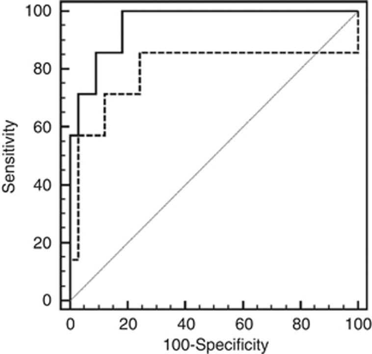
Analysis of the training set of serum samples by SELDI-TOF identified 59 protein peaks with intensities at least 10-fold greater than baseline. Logistic regression and sixfold crossover validation analysis demonstrated that 18 peaks (m/z=3208, 3432, 3476, 3700, 3879, 4398, 4461, 5051, 5367, 5785, 7333, 8222, 9422, 11522, 12862, 13889, 16963, 17367) along with CA 19-9 were possibly predictive of survival of <1 year. Seven of these peaks and CA19-9 were chosen on two or more occasions in the sixfold cross-validation analysis. Three of these peaks, m/z 3700, 8222, 11 522, were independent of each other by Spearman’s correlation, and AUC for the combination of these peaks to predict 1-year survival was 0.79 (CI, 0.64–0.90). The mean value was greater in those surviving <1 year, but this was only significant for the m/z 8222 peak (P=0.034). When CA 19-9 was included in the panel (m/z 3700, 8222, 11 522+CA 19-9), survival of <1 year was predicted by ROC analysis with AUC=0.957 (CI, 0.84–0.996; Figure 1). Two patients who were predicted to live >12 months died at 336 and 364 days after surgery. One patient predicted to live <12 months lived for 972 days. The sensitivity of this panel was 0.86 and specificity was 0.82 at the strongest criterion value. When this equation was tested on the SELDI results from the verification set, the six patients predicted to have a poor prognosis died within the first 12 months after surgery, whereas 10 of 15 patients were correctly predicted to live >12 months. The other five died at 21, 26, 45, 50 and 51 weeks after surgery. Thus, although the sensitivity was only 0.55, the specificity was 1.0 and the post-test probability of outcome, given a positive test, was also 1.0.
Figure 1.
ROC curves for training samples. Dashed line indicates the three-member panel of m/z 3700, 8222 and 11 522 with an AUC of 0.79 (s.e., 0.13). Solid line indicates the panel, including CA 19-9 with AUC of 0.97 (s.e., 0.03). These lines were not significantly different (P=0.24, DeLong test).
Identification of the m/z 8222 protein as ApoC-II and the m/z 11 522 protein as SAA-1
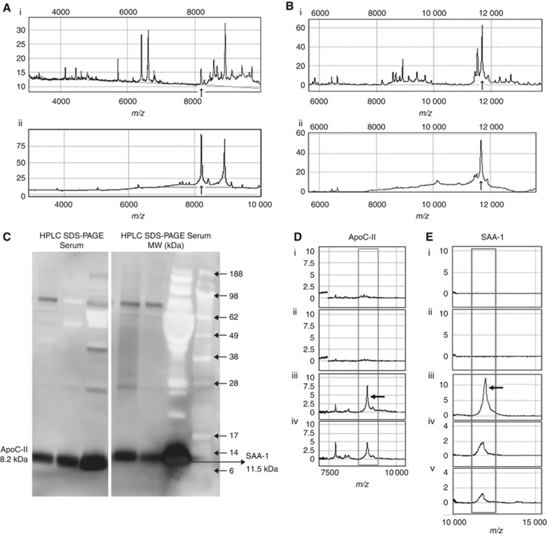
Figure 2 illustrates the steps involved in identification of the two proteins m/z 8222 and 11 522 after the ion-exchange fractionation step. The data obtained by LC-ESI/MS/MS showed five major tryptic fragments from the protein peak at m/z 8222, two of them overlapping. MASCOT analysis revealed that the biomarker at 8222 m/z corresponded to ApoC-II with sequence coverage of 82.6%. Two major tryptic fragments were obtained from the protein peak at m/z 11 522, which was identified as SAA-1 with sequence coverage of 47.2%. The intensity of the protein peak at m/z 3700 was too low for identification.
Figure 2.
Purification and identification of the proteins corresponding to m/z 8222 and 11 522 peaks. (Ai) Initial SELDI profile from serum; (Aii) semi-purified peak after HPLC fractionation showing the m/z 8222 peak (arrowed) selected for sequencing. (Bi) Initial SELDI for serum; (Bii) semi-purified sample for the m/z 11 522 peak (arrowed). (C) Western-blot lanes are labelled: serum (crude serum), HPLC (the HPLC fraction containing the peaks of interest), SDS–PAGE (the protein extracted from 1D-SDS–PAGE gel bands, which were subjected to protein sequencing and molecular weight (MW) (the protein standards, which are presented in kDa) for ApoC-II in the panel on the left and SAA-1 in the panel on the right. (D) Confirmation of m/z 8222 peak identity using SELDI immuno-adsorption on RS100 chips containing ApoC-II antibody. (Di) Affinity-purified IgG negative control; (Dii) unprocessed serum without antibody; (Diii) unprocessed serum with antibody; (Div) antibody with semi-purified ApoC-II from patient serum. (E) Confirmation of m/z 11 522 peak identity using SAA-1 antibody. (Ei) Affinity-purified IgG (negative control); (Eii) unprocessed serum without antibody; (Eiii) recombinant human SAA-1 (positive control); (Eiv) unprocessed serum with antibody; and (Ev) semi-purified SAA-1 from patient serum after HPLC purification with antibody.
SELDI immuno-adsorption and western-blotting validation
Western-blotting analysis was performed for ApoC-II (m/z 8222 peak) comparing crude serum samples, HPLC-purified serum and the excised SDS–PAGE band. A similar comparison was performed for SAA-1 (m/z 11 522 peak). The results showed that the anti-ApoC-II and anti-SAA-1 antibodies recognised the respective target proteins in purification fractions and SDS–PAGE gel-bands excised for MS/MS analysis. The peaks migrated in a similar manner to the originally targeted proteins (Figure 2C). Other less intense peaks were also seen in the 26 and 90 kDa region, but these were considered to be owing to the polyclonal nature of the antibodies used, although they were purified to be >90% specific for the protein of interest.
Further immunological confirmation that the protein peaks were ApoC-II and SAA-1 was undertaken with SELDI immuno-adsorption using commercial ApoC-II and SAA-1 antibodies covalently bound to RS100 chips (Figure 2D and E). Here the m/z 8222 and 11 522 proteins bound to ApoC-II and SAA-1 antibodies, respectively, with minimal non-specific binding to IgG control. Further, ApoC-II antibody binds to the m/z 8222 peak in unprocessed serum (Figure 2D(iii)) and HPLC-purified serum (Figure 2D(iv)). The SAA-1 antibody bound to the m/z 11 522 peak with a recombinant human SAA-1 sample (Figure 2E(iii)) (positive control), with unprocessed serum (Figure 2E(iv)) and with semi-purified SAA-1 (Figure 2E(v)).
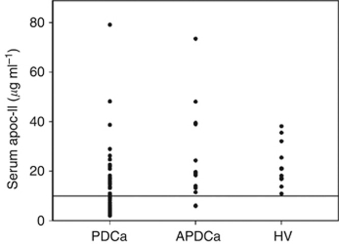
ELISA study on validation-set samples
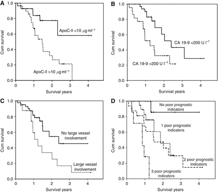
ELISA serum measurements (Table 2) demonstrated that ApoC-II (analysed as geometric means) were lower in resectable cases than in advanced cases (APDCa) (P<0.001) with about half of the values being <10 μg ml−1, the lowest value for HV cases (Figure 3). Serum SAA-1 levels were higher in PDCa serum than APDCa (P=0.028) or HV samples (P=0.003) but, CRP was not different among the groups. Kaplan–Meier survival analysis demonstrated that ApoC-II at the cut-off values of 10 μg ml−1 significantly influenced survival (Figure 4, A, P=0.009), whereas SAA-1 and CRP were not predictive of survival (data not shown). CA 19-9 at a cut-off of 200 IU l−1 (Figure 4B, P=0.009) and the pathological finding of large-vessel invasion (Figure 4C, P=0.025) also demonstrated influence on survival. The combination of these three measures indicated that survival was excellent for the 15 subjects (26%) with all three values in the low-risk range but patients were unlikely to survive 12 months when the three measures were in high-risk range (8 subjects, 14%). The remaining patients with one or two high-risk measures had a median survival of 2 years (Figure 4D).
Table 2. Measurement of SAA, ApoC-II, CRP and CA19.9 by ELISA in validation set.
|
SAA (ng ml−1) |
ApoC-II (μg ml−1) |
CRP (ng ml−1) |
CA19.9 U l
−1
|
||||||
|---|---|---|---|---|---|---|---|---|---|
| N | Mean | s.d. | Mean | s.d. | Mean | s.d. | Mean | s.d. | |
| PDCA | 57 | 104.0 | 3.9 | 6.2 | 2.5 | 3.7 | 4.1 | 105 | 4.1 |
| APCA | 13 | 32.9 | 3.8 | 15.3 | 3.7 | 3.4 | 4.3 | 267 | 2.1 |
| HV | 12 | 9.9 | 2.0 | 13.7 | 1.6 | 2.4 | 4.9 | — | — |
| P by ANOVA | 0.000 | 0.001 | n.s. | n.s. | |||||
| P for PDC vs APCA | 0.013 | 0.007 | |||||||
| P for PDC vs HV | 0.000 | 0.027 | |||||||
Abbreviations: ANOVA=analysis of variance; APCA=advanced pancreatic carcinoma; HV=healthy volunteers; PDCA=pancreatic ductal adenocarcinoma. Values are geometric means±s.d.
Figure 3.
Plot of serum concentrations of ApoC-II for the different groups in the Validation study. The horizontal line indicates the cut-off value used for prognosis (10 μg ml−1).
Figure 4.
Kaplan–Meier survival curves for factors influencing survival after resection for PDCa in validation-2 samples. (A) The survival was significantly reduced if ApoC-II was >10 μg ml−1 (solid line, n=28) compared with <10 μg ml−1 (dotted line, n=29), log rank P=0.009. (B) The survival curves for CA 19-9 at the cutoff of 200 U l−1 where dotted line represents patients with CA 19-9>200 U l−1 (n=24) and solid line represents patients with values were <200 U l−1 (n=33), P=0.009. (C) The influence of pathologically determined large-vessel invasion (dotted line, n=16) vs rest of the cases (solid line, n=41) P=0.025. (D) Combination of the three measures with the solid line representing 15 subjects with three low-risk measures, dotted line indicates survival of 8 subjects with 3 abnormal values, the dashed line indicates survival of 25 subjects with 1 abnormal measure and the dashed-dotted line indicated survival of 9 subjects with 2 abnormal values. Note that three patients with large-vessel involvement had low values of ApoC-II and CA19-9. The curves were different, χ2=20.4, υ=3, P<0.001.
Cox proportional hazard assessment of prognostic factors
Multiple potential prognostic factors were assessed by univariate Cox proportional hazard analysis (Supplementary Table S1). This allowed comparison of the proteins of interest, SAA-1 and ApoC-II, with CRP, CA 19-9, serum bilirubin and the ratio of total neutrophil to lymphocytes in the preoperative blood film (Neu/LCC), along with pathological indices. Spearman’s correlation indicated that ApoC-II was independent of other measures, whereas SAA-1 had a weak but non-significant correlation with the Neu/LCC ratio (P=0.07), and CA19-9 only correlated with the need for vascular reconstruction. Large-vessel invasion correlated with T-stage (P=0.03), retroperitoneal nerve invasion (P=0.02), perineural invasion (P=0.006) and small-vessel invasion (P=0.008). Lymph node involvement correlated with T-stage and retroperitoneal extension. Therefore to avoid interference by co-correlation, the following indices were entered into the Cox proportional hazards model using backward regression: log10ApoC-II, log10SAA-1, log10CA19-9, lymph node involvement and large-vessel involvement. The final equation included log10ApoC-II, log10CA19-9 and lymph node involvement for predicting survival (υ=3, χ2=12.1 and P=0.007).
Impact of SAA-1 and ApoC-II on PDCa cell growth and invasion
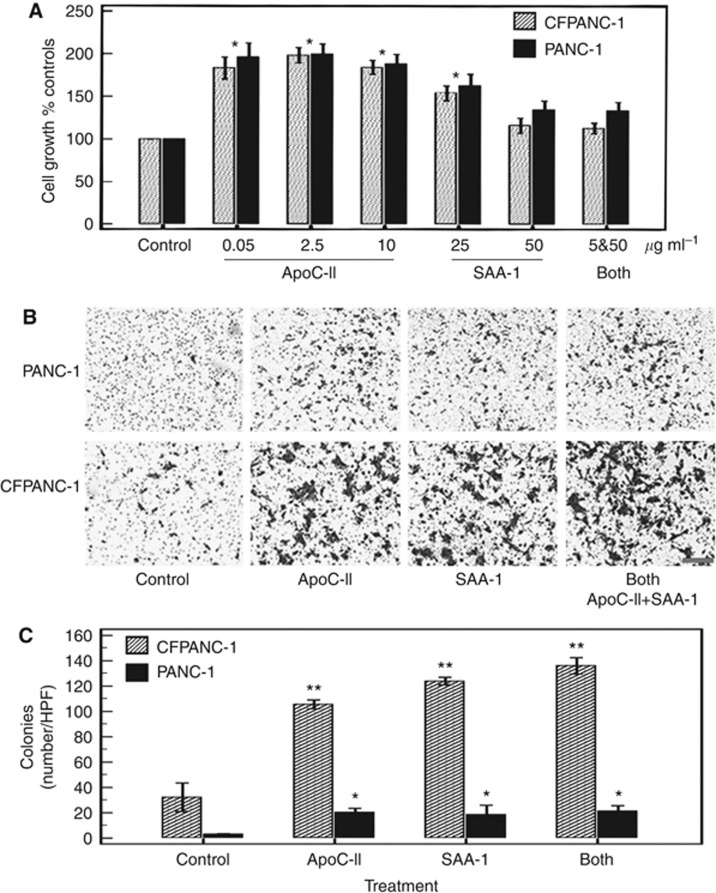
In-vitro studies of the influence of ApoC-II and SAA-1 on the growth of CFPAC-1 and PANC-I pancreatic cancer cell lines demonstrated a dose-dependent response to 0.05, and 2.5 μg ml−1 of purified ApoC-II and to 25 μg ml−1 of SAA-1 (Figure 5A, ANOVA, P<0.01) but not to 50 μg ml−1 of purified SAA-1 at 24 h. Furthermore, when CFPAC-1 and PANC-1 cells were pre-treated with recombinant ApoC-II or SAA-1 compared with control PBS, before seeding onto Matrigel-coated transwells there was significant increase in cell invasion (P<0.001) in both cell lines but there was no additive effect of combining SAA-1 and ApoC-II (Figure 5B and C).
Figure 5.
(A) The influence of ApoC-II and SAA-1 on cell growth (mean values±s.e.m.), as a percentage of controls after 24 h for CFPAC-I (shaded bars), and PANC-I (solid bars). Two-way analysis of variance showed no difference between cell lines. ApoC-II increased cell growth above that of controls (P<0.001 for both cell lines) and greater than the combined treatment with ApoC-II and SAA-1. SAA-1 increased cell growth at the lower dose of 25 μg ml−1 P<0.001, but not at the higher dose 50 μg ml−1, which was less stimulatory than each dose of ApoC-II (P<0.001). ‘*’ indicates different from controls, P<0.001. 5 and 50 indicates 5 μg ml−1 ApoC-II and 50 μg ml−1 SAA-1. (B) Micrograph of × 10 magnification transwell invasion assay for PANC-1 and CFPAC-1 cells treated with control solutions, ApoC-1, SAA-1 and both treatments. Bar =200 μm. (C) Cell counts per high power field at × 10 results for 3–4 fields (mean values±s.e.m.). Analysis of variance indicated a significant increase from controls for CFPAC-1 cells (**P<0.001) and for PANC-1 cells (*P<0.02). When treatments were combined, the apparent increase did not reach significance.
Discussion
This paper demonstrates that preoperative levels of ApoC-II are prognostic of long-term survival in patients undergoing resection for PDCa. The discovery of this protein was undertaken using the high through-put technique of SELDI-TOF MS and logistic regression analysis with sixfold cross-validation. These findings were verified by SELDI in a separate sample set from a different hospital and, were validated by ELISA in a prospective patient group where the patients with a good outcome had depressed levels of ApoC-II. Survival curve analysis by Cox regression of the validation-set samples confirmed that ApoC-II, CA 19-9 and lymph node involvement independently predict survival. Major-vessel involvement, which may be more easily assessed preoperatively with CT scanning (Manak et al, 2009) than lymph node involvement, was shown by Kaplan–Meier analysis to allow for a clear separation of patient outcomes when combined with ApoC-II and CA19-9. The influence of SAA-1, also identified on SELDI analysis, was not found to be significant in the validation set. Of interest, SAA-1 and CRP have previously been shown to be inflammatory markers with prognostic utility (Raynes and Cooper, 1983).
ApoC-II is mainly synthesised by the liver (Kardassis et al, 2003), and is an important component of LDL, where it helps to solubilise lipid and deliver it to peripheral tissues by its ability to activate lipoprotein lipase (LPL). After the lipid is released, LDL shrinks and ApoC-II splits off either to pass back into the circulation or be utilised by the cancer. Interestingly ApoC-II has been shown to be a putative substrate for the metalloproteinases MMP-14 and MMP-7 (Kim et al, 2006). ApoC-II is efficiently cleaved by MMP-7, which may account for the depressed levels of ApoC-II in our cases with better outcomes (Giannopoulos et al, 2008). Further, pancreatic cancer utilises lipid in preference to carbohydrate (Wang et al, 2009) for energy, which may add to the turnover of ApoC-II. Our finding that ApoC-II increases the growth and spread of pancreatic cancer cells in vitro may indicate a further role of ApoC-II where high levels predicted poorer survival. Thus, although the prognostic importance of ApoC-II has not been previously reported, there are plausible cellular mechanisms involving ApoC-II in the progress of pancreatic cancer. It is interesting that elevated levels of ApoC-II have been considered to be of diagnostic importance (Chen et al, 2007) and associated with pancreatic cancer-associated cachexia (Felix et al, 2011).
SAA-1, the second protein selected by SELDI was previously shown to be prognostic of outcome in a number of malignancies related to an induced systemic acute-phase reaction (Weinstein et al, 1984), but, when measured by ELISA in our validation set, it failed as a prognostic marker. This is an unexpected result given that a number of these patients required vascular resection. The cachexia of pancreatic cancer is associated with an increase in inflammatory cytokines and would be expected to increase SAA-1 (Argiles et al, 2009), but few patients with resectable pancreatic cancer have established cachexia (Aslani et al, 2010). Of interest it is a lipoprotein, which is associated with HDL (van der Hilst, 2011) and has a complex, highly conserved, role in acute inflammatory conditions (Manley et al, 2006), which accounts for its elevation in our preoperative patients who had recently had stents for obstructive jaundice (Wagholikar et al, 2003). Recently SAA-1 was included in a diagnostic panel for pancreatic cancer (Bunger et al, 2011). Similarly, the ratio of neutrophils to lymphocytes has intriguingly been associated with poor prognosis (Bhatti et al, 2010), particularly in patients undergoing chemotherapy (Chua et al, 2011), implying an effect on the body’s inflammatory response, although, again we were not able to confirm this.
Carbohydrate Antigen 19-9 level of >200 U ml−1 and lymph node involvement were independently prognostic of poor survival after pancreatic resection (Berger et al, 2004), as confirmed by this study. Although the prognostic value of CA 19-9 has been debated both for (Mehta et al, 2010) and against (Kondo et al, 2010), it does appear to indicate benefit for selection of patients for adjuvant therapy (Hallemeier et al, 2011). Multiple influences on survival include pathological criteria and nutritional and performance status (Schnelldorfer et al, 2008), but CA 19-9 may have further independent predictive value (Tsavaris et al, 2009). However, CA 19-9 results may be confusing because it can be elevated in the presence of biliary inflammation (Dogan et al, 2011) and is not produced in Lewis antigen-negative subjects (Vestergaard et al, 1999).
Surgical technique to achieve an R0 resection has an important role in achieving long-term survival (Waraya et al, 2009) where en-bloc resection allows for clearance of involved vessels (Schnelldorfer et al, 2008). This approach has been used in the current study (Samra et al, 2006). However, margin status continues to be a debated topic and there is a wide belief that the aggressive nature of PDCa over-rides the importance of margin status (Butturini et al, 2008). Major-vessel involvement seen on multi-slice computer tomography is predictive of survival (Manak et al, 2009) and may be used in the preoperative assessment of prognosis along with levels of ApoC-II and CA 19-9.
Our results led us to analyse whether ApoC-II and SAA-1 were involved in cancer cell proliferation and invasion. ApoC-II and to a lesser extent SAA-1 stimulated cell proliferation and invasion in two pancreatic cancer cell lines. The apolipoproteins appear to have important roles in the biology of pancreatic cancer. ApoA-II is also depressed in pancreatic cancer (Xue et al, 2010). Although ApoC-II has a different function from ApoA-II, their involvement is consistent with pancreatic cancer’s reliance on lipid metabolism. Further, RELN (Reelin) pathway signalling via the very low-density lipoprotein (VLDL) receptor, to which ApoC-II is known to bind, influences cell motility in pancreatic cancer (Sato et al, 2006). We speculate that ApoC-II levels may be involved with tumour progression.
In conclusion, this study shows that serum levels of ApoC-II, and CA 19-9 along with major vessel invasion, which can be determined preoperatively with a CT scan, allow for improved prediction of prognosis of a pancreatic resection. These prognostic indicators selected a small group of short-term survivors, who might have been better served by a palliative procedure, while the remaining patients benefitted from the surgery. Furthermore, these measures provide information about factors leading to a poor prognosis by highlighting the stimulation of cancer tissue growth by apolipoproteins. These factors are so strong that even after the inclusion of the influence of standard histopathological measures and CA 19-9, the blood level of ApoC-II remains a strong independent predictor of survival.
Acknowledgments
This work was supported by grants from the Cancer Surgery Research Foundation, a Cancer Council NSW Innovator Grant and FIMP (Fondazione Italiana Malattie del Pancreas). The research was facilitated by an initiative of the Australian Government as part of the National Collaborative Research Infrastructure Strategy. We thank Dr Sarah Smith for her editorial assistance.
Footnotes
Supplementary Information accompanies the paper on British Journal of Cancer website (http://www.nature.com/bjc)
This work is published under the standard license to publish agreement. After 12 months the work will become freely available and the license terms will switch to a Creative Commons Attribution-NonCommercial-Share Alike 3.0 Unported License.
Supplementary Material
References
- Akizuki E, Kimura Y, Nobuoka T, Imamura M, Nishidate T, Mizuguchi T, Furuhata T, Hirata K (2008) Prospective nonrandomized comparison between pylorus-preserving and subtotal stomach-preserving pancreaticoduodenectomy from the perspectives of DGE occurrence and postoperative digestive functions. J Gastrointest Surg 12(7): 1185–1192 [DOI] [PubMed] [Google Scholar]
- American joint Committee on cancer (2009) Cancer Staging Handbook. Springer: Chicago [Google Scholar]
- Argiles JM, Busquets S, Toledo M, Lopez-Soriano FJ (2009) The role of cytokines in cancer cachexia. Curr Opin Support Palliat Care 3(4): 263–268 [DOI] [PubMed] [Google Scholar]
- Aslani A, Gill AJ, Roach PJ, Allen BJ, Smith RC (2010) Preoperative body composition is influenced by the stage of operable pancreatic adenocarcinoma but does not predict survival after Whipple's procedure. HPB (Oxford) 12(5): 325–333 [DOI] [PMC free article] [PubMed] [Google Scholar]
- Berger AC, Meszoely IM, Ross EA, Watson JC, Hoffman JP (2004) Undetectable preoperative levels of serum CA 19-9 correlate with improved survival for patients with resectable pancreatic adenocarcinoma. Ann Surg Oncol 11(7): 644–649 [DOI] [PubMed] [Google Scholar]
- Bhatti I, Peacock O, Lloyd G, Larvin M, Hall RI (2010) Preoperative hematologic markers as independent predictors of prognosis in resected pancreatic ductal adenocarcinoma: neutrophil-lymphocyte vs platelet-lymphocyte ratio. Am J Surg 200(2): 197–203 [DOI] [PubMed] [Google Scholar]
- Bunger S, Laubert T, Roblick UJ, Habermann JK (2011) Serum biomarkers for improved diagnostic of pancreatic cancer: a current overview. J Cancer Res Clin Oncol 137(3): 375–389 [DOI] [PubMed] [Google Scholar]
- Butturini G, Stocken DD, Wente MN, Jeekel H, Klinkenbijl JH, Bakkevold KE, Takada T, Amano H, Dervenis C, Bassi C, Buchler MW, Neoptolemos JP (2008) Influence of resection margins and treatment on survival in patients with pancreatic cancer: meta-analysis of randomized controlled trials. Arch Surg 143(1): 75–83 [DOI] [PubMed] [Google Scholar]
- Cao L, Wang L, Wang G, Liu D, Yu J, Que R, Xie D (2010) Evaluation of the effectiveness of a chemoprevention model of pancreatic adenocarcinoma using protein chip technology. Int J Oncol 37(6): 1515–1520 [DOI] [PubMed] [Google Scholar]
- Castellanos EH, Cardin DB, Berlin JD (2011) Treatment of early-stage pancreatic cancer. Oncology (Williston Park) 25(2): 182–189 [PubMed] [Google Scholar]
- Chen J, Anderson M, Misek DE, Simeone DM, Lubman DM (2007) Characterization of apolipoprotein and apolipoprotein precursors in pancreatic cancer serum samples via two-dimensional liquid chromatography and mass spectrometry. J Chromatogr A 1162(2): 117–125 [DOI] [PMC free article] [PubMed] [Google Scholar]
- Chua W, Charles KA, Baracos VE, Clarke SJ (2011) Neutrophil/lymphocyte ratio predicts chemotherapy outcomes in patients with advanced colorectal cancer. Br J Cancer 104(8): 1288–1295 [DOI] [PMC free article] [PubMed] [Google Scholar]
- Distler M, Kersting S, Ruckert F, Dobrowolski F, Miehlke S, Grutzmann R, Saeger HD (2010) Palliative treatment of obstructive jaundice in patients with carcinoma of the pancreatic head or distal biliary tree. Endoscopic stent placement vs. hepaticojejunostomy. JOP 11(6): 568–574 [PubMed] [Google Scholar]
- Dogan UB, Gumurdulu Y, Golge N, Kara B (2011) Relationship of CA 19-9 with choledocholithiasis and cholangitis. Turk J Gastroenterol 22(2): 171–177 [DOI] [PubMed] [Google Scholar]
- Felix K, Fakelman F, Hartmann D, Giese NA, Gaida MM, Schnolzer M, Flad T, Buchler MW, Werner J (2011) Identification of serum proteins involved in pancreatic cancer cachexia. Life Sci 88(5-6): 218–225 [DOI] [PubMed] [Google Scholar]
- Firpo MA, Gay DZ, Granger SR, Scaife CL, DiSario JA, Boucher KM, Mulvihill SJ (2009) Improved diagnosis of pancreatic adenocarcinoma using haptoglobin and serum amyloid A in a panel screen. World J Surg 33(4): 716–722 [DOI] [PMC free article] [PubMed] [Google Scholar]
- Giannopoulos G, Pavlakis K, Parasi A, Kavatzas N, Tiniakos D, Karakosta A, Tzanakis N, Peros G (2008) The expression of matrix metalloproteinases-2 and -9 and their tissue inhibitor 2 in pancreatic ductal and ampullary carcinoma and their relation to angiogenesis and clinicopathological parameters. Anticancer Res 28(3B): 1875–1881 [PubMed] [Google Scholar]
- Gill AJ, Johns AL, Eckstein R, Samra JS, Kaufman A, Chang DK, Merrett ND, Cosman PH, Smith RC, Biankin AV, Kench JG (2009) Synoptic reporting improves histopathological assessment of pancreatic resection specimens. Pathology 41(2): 161–167 [DOI] [PubMed] [Google Scholar]
- Hallemeier CL, Botros M, Corsini MM, Haddock MG, Gunderson LL, Miller RC (2011) Preoperative CA 19-9 level is an important prognostic factor in patients with pancreatic adenocarcinoma treated with surgical resection and adjuvant concurrent chemoradiotherapy 8. Am J Clin Oncol 34(6): 567–572 [DOI] [PubMed] [Google Scholar]
- Hata S, Sakamoto Y, Yamamoto Y, Nara S, Esaki M, Shimada K, Kosuge T (2012) Prognostic impact of postoperative serum CA 19-9 levels in patients with resectable pancreatic cancer. Ann Surg Oncol 19(2): 636–641 [DOI] [PubMed] [Google Scholar]
- Jamieson NB, CR Carter, McKay CJ, Oien KA (2011) Tissue biomarkers for prognosis in pancreatic ductal adenocarcinoma: a systematic review and meta-analysis. Clin Cancer Res 17(10): 3316–3331 [DOI] [PubMed] [Google Scholar]
- Kardassis D, Roussou A, Papakosta P, Boulias K, Talianidis I, Zannis VI (2003) Synergism between nuclear receptors bound to specific hormone response elements of the hepatic control region-1 and the proximal apolipoprotein C-II promoter mediate apolipoprotein C-II gene regulation by bile acids and retinoids. Biochem J 372(Part 2): 291–304 [DOI] [PMC free article] [PubMed] [Google Scholar]
- Kim SY, Park SM, Lee ST (2006) Apolipoprotein C-II is a novel substrate for matrix metalloproteinases. Biochem Biophys Res Commun 339(1): 47–54 [DOI] [PubMed] [Google Scholar]
- Kondo N, Murakami Y, Uemura K, Hayashidani Y, Sudo T, Hashimoto Y, Nakashima A, Sakabe R, Shigemoto N, Kato Y, Ohge H, Sueda T (2010) Prognostic impact of perioperative serum CA 19-9 levels in patients with resectable pancreatic cancer. Ann Surg Oncol 17(9): 2321–2329 [DOI] [PubMed] [Google Scholar]
- Manak E, Merkel S, Klein P, Papadopoulos T, Bautz WA, Baum U (2009) Resectability of pancreatic adenocarcinoma: assessment using multidetector-row computed tomography with multiplanar reformations. Abdom Imaging 34(1): 75–80 [DOI] [PubMed] [Google Scholar]
- Manley PN, Ancsin JB, Kisilevsky R (2006) Rapid recycling of cholesterol: the joint biologic role of C-reactive protein and serum amyloid A. Med Hypotheses 66(4): 784–792 [DOI] [PubMed] [Google Scholar]
- Mehta J, Prabhu R, Eshpuniyani P, Kantharia C, Supe A (2010) Evaluating the efficacy of tumor markers CA 19-9 and CEA to predict operability and survival in pancreatic malignancies. Trop Gastroenterol 31(3): 190–194 [PubMed] [Google Scholar]
- Molina V, Visa L, Conill C, Navarro S, Escudero JM, Auge JM, Filella X, Lopez-Boado MA, Ferrer J, Fernandez-Cruz L, Molina R (2012) CA 19-9 in pancreatic cancer: retrospective evaluation of patients with suspicion of pancreatic cancer. Tumour Biol 33(3): 799–807 [DOI] [PubMed] [Google Scholar]
- Pine JK, Fusai KG, Young R, Sharma D, Davidson BR, Menon KV, Rahman SH (2009) Serum C-reactive protein concentration and the prognosis of ductal adenocarcinoma of the head of pancreas. Eur J Surg Oncol 35(6): 605–610 [DOI] [PubMed] [Google Scholar]
- Raynes JG, Cooper EH (1983) Comparison of serum amyloid A protein and C-reactive protein concentrations in cancer and non-malignant disease. J Clin Pathol 36(7): 798–803 [DOI] [PMC free article] [PubMed] [Google Scholar]
- Samra JS, Gananadha S, Gill A, Smith RC, Hugh TJ (2006) Modified extended pancreatoduodenectomy: en bloc resection of the peripancreatic retroperitoneal tissue and the head of pancreas. ANZ J Surg 76(11): 1017–1020 [DOI] [PubMed] [Google Scholar]
- Sato N, Fukushima N, Chang R, Matsubayashi H, Goggins M (2006) Differential and epigenetic gene expression profiling identifies frequent disruption of the RELN pathway in pancreatic cancers. Gastroenterology 130(2): 548–565 [DOI] [PubMed] [Google Scholar]
- Schnelldorfer T, Ware AL, Sarr MG, Smyrk TC, Zhang L, Qin R, Gullerud RE, Donohue JH, Nagorney DM, Farnell MB (2008) Long-term survival after pancreatoduodenectomy for pancreatic adenocarcinoma: is cure possible? Ann Surg 247(3): 456–462 [DOI] [PubMed] [Google Scholar]
- Tang D, Zhang JQ, Wang DR (2011) Long term results of pancreatectomy with portal-superior mesenteric vein resection for pancreatic carcinoma: a systematic review. Hepatogastroenterology 58(106): 623–631 [PubMed] [Google Scholar]
- Tsavaris N, Kosmas C, Papadoniou N, Kopteridis P, Tsigritis K, Dokou A, Sarantonis J, Skopelitis H, Tzivras M, Gennatas K, Polyzos A, Papastratis G, Karatzas G, Papalambros A (2009) CEA and CA-19.9 serum tumor markers as prognostic factors in patients with locally advanced (unresectable) or metastatic pancreatic adenocarcinoma: a retrospective analysis. J Chemother 21(6): 673–680 [DOI] [PubMed] [Google Scholar]
- Valkovskaya N, Kayed H, Felix K, Hartmann D, Giese NA, Osinsky SP, Friess H, Kleeff J (2007) ADAM8 expression is associated with increased invasiveness and reduced patient survival in pancreatic cancer. J Cell Mol Med 11(5): 1162–1174 [DOI] [PMC free article] [PubMed] [Google Scholar]
- van der Hilst JC (2011) Recent insights into the pathogenesis of type AA amyloidosis. Scientific World Journal 11: 641–650 [DOI] [PMC free article] [PubMed] [Google Scholar]
- Vestergaard EM, Hein HO, Meyer H, Grunnet N, Jorgensen J, Wolf H, Orntoft TF (1999) Reference values and biological variation for tumor marker CA 19-9 in serum for different Lewis and secretor genotypes and evaluation of secretor and Lewis genotyping in a Caucasian population. Clin Chem 45(1): 54–61 [PubMed] [Google Scholar]
- Wagholikar GD, Sikora SS, Pandey R, Prasad KK, Kumar A, Saxena R, Kapoor VK (2003) Morphological changes in bile ducts following preoperative biliary stenting. Indian J Gastroenterol 22(5): 166–169 [PubMed] [Google Scholar]
- Wang F, Kumagai-Braesch M, Herrington MK, Larsson J, Permert J (2009) Increased lipid metabolism and cell turnover of MiaPaCa2 cells induced by high-fat diet in an orthotopic system. Metabolism 58(8): 1131–1136 [DOI] [PubMed] [Google Scholar]
- Waraya M, Yamashita K, Katagiri H, Ishii K, Takahashi Y, Furuta K, Watanabe M (2009) Preoperative serum CA19-9 and dissected peripancreatic tissue margin as determiners of long-term survival in pancreatic cancer. Ann Surg Oncol 16(5): 1231–1240 [DOI] [PubMed] [Google Scholar]
- Weinstein PS, Skinner M, Sipe JD, Lokich JJ, Zamcheck N, Cohen AS (1984) Acute-phase proteins or tumour markers: the role of SAA, SAP, CRP and CEA as indicators of metastasis in a broad spectrum of neoplastic diseases. Scand J Immunol 19(3): 193–198 [DOI] [PubMed] [Google Scholar]
- Xue A, Scarlett CJ, Chung L, Butturini G, Scarpa A, Gandy R, Wilson SR, Baxter RC, Smith RC (2010) Discovery of serum biomarkers for pancreatic adenocarcinoma using proteomic analysis. Br J Cancer 103(3): 391–400 [DOI] [PMC free article] [PubMed] [Google Scholar]
- Xue A, Xue M, Jackson C, Smith RC (2009) Suppression of urokinase plasminogen activator receptor inhibits proliferation and migration of pancreatic adenocarcinoma cells via regulation of ERK/p38 signaling. Int J Biochem Cell Biol 41(8-9): 1731–1738 [DOI] [PubMed] [Google Scholar]
Associated Data
This section collects any data citations, data availability statements, or supplementary materials included in this article.


