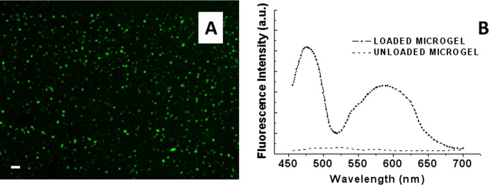
Figure 4.
Confocal microscopy image of the PMAA microgels microchannel loaded with TF1-antiCD4 and TF2-antiCD19 antibodies. (a) Fluorescence image of the double loaded microgels functionalized microchannel. (b) Fluorescence spectra of the immobilized microgels after the uptake (dotted line) and after the release process of labeled antibodies (straight line). The scale bar was 10 μm.

