Abstract
Dietary Supplements based on an extract from Irvingia gabonensis (African Mango, AM for abbreviation) seeds are one of the popular herbal weight loss dietary supplements in the US market. The extract is believed to be a natural and healthy way to lose weight and improve overall health. However, the chemical composition of African mango based-dietary supplements (AMDS) has never been reported. In this study, the chemical constituents of African mango seeds, African mango seeds extract (AMSE), and different kinds of commercially available African mango based dietary supplements (AMDS) have been investigated using an ultra high-performance liquid chromatography with high resolution mass spectrometry (UHPLC-HRMS) method. Ellagic acid, mono, di, tri-O methyl-ellagic acids and their glycosides were found as major components in African Mango seeds. These compounds may be used for quality control of African Mango extract and related dietary supplements.
Keywords: Irvingia gabonensis, African Mango, UHPLC-HRMS, Ellagic Acid, O methyl-ellagic acids
INTRODUCTION
Irvingia gabonensis (African Mango, or AM) belongs to the Irvingiaceae family. It is also known as wild mango, or bush mango (1), which is different from regular mango (Mangifera indica L., belongs to Anacardiacea). The mango-like fruits of AM are especially valued for their dietary-fiber, fat- and protein-rich seeds. The consumption of dried Irvingia gabonensis seeds is common in West African countries and for centuries, have been part of the local diets (2).
In recent years, African Mango based dietary supplements (AMDS) have appeared as a popular herbal weight loss dietary supplement in the United States. The labels of AMDS products sold in the US all claim the use of African mango seed extract (AMSE) as the major ingredient. AMSE is believed to be a natural and healthy way to lose weight and improve overall health. A randomized, double-blind, placebo-controlled study demonstrated significant differences between the treatment and placebo groups in weight and fat loss, as well as reductions in hip and waist circumference (3). AMSE combined with Veldt Grape (Cissus quadrangularis) resulted in significant reductions of six anthropomorphic and serological measurements (body weight, body fat, waist sizes; total plasma cholesterol, LDL cholesterol, and fasting blood glucose level) (4). In another study, AMSE showed positive effects on body weight and a variety of parameters characteristic of metabolic syndrome in a double blind, randomized, placebo controlled clinical trial investigating the anti-obesity and lipid profile modulating effects (5). It is believed that the high soluble fiber content may cause the lowering of plasma cholesterol, triglycerides, and glucose concentrations (6). However, detailed chemical analyses of the seeds of AM, AMSE, and AMDS have not been reported.
To advance our understanding of the health benefits of AMSE and/or AMDS, it is important to investigate their chemical composition, and to find marker compounds for the authentication of AMDS products. To date, the only report on analysis of “African Mango” was performed on the peel and flesh of the fruit, not on the seeds. The study was conducted using gas chromatography mass spectrometry (GC-MS) and 33 volatile compounds were reported including 8 monoterpene alcohols, 5 aldehydes, 4 acids, 7 esters, and 5 C13 norisoprenoids(7). Unfortunately, the authors confused the name of “African Mango” with mango growing in Africa, and incorrectly used the name “African Mango” in the paper as the research was actually done on mangos growing in Africa (Mangifera indica L.), not African Mango (Irvingia gabonensis). Other studies on mango (Mangifera indica L.) revealed that flavonol, carotenoids, tocopherols and xanthone glycosides were the major compounds from mango peel and flesh (8–11), while the major compounds from the seeds were shown to be benzophenone and gallotanins by LC-MSn methods (12–14).
Currently, AMDS sold in the U.S. market can be divided into 3 groups according to their labels: 1) AMSE only; 2) AMSE with green tea extracts; and 3) AMSE with other botanical extracts, such as berries, kelp, and caffeine as well as green tea extracts. Some manufacturers claim on their labels that Irvingia gabonensis seed fiber and flavonoids are major components in their products. Others claim mangiferin, a known constituent from mango, but not from AM, for their products. Clearly, there is a lot of confusion on the manufacturer and distributor sides.
In this study, the chemical constituents of AM (Irvingia gabonensis) seeds, mango (Mangifera indica L) seeds, AMSE, and different kinds of AMDS have been investigated using an ultra high-performance liquid chromatography high-resolution mass spectrometry (UHPLC-HRMS) method. Ellagic acid, mono, di, tri-O-methyl-ellagic acid and their related glycosides were found to be the major constituents in AM seeds, which may be used for future quality control and assurance for AMSE or AMDS as marker compounds. A wide variation in the constituents was found among AMSE products from China and AMDS products sold in the US.
MATERIALS AND METHODS
Chemicals and Materials
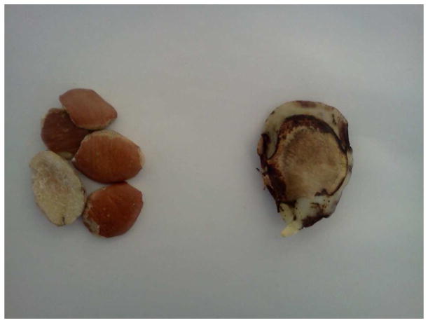
HPLC grade methanol, acetonitrile and formic acid were purchased from VWR International, Inc. (Clarksburg, MD). HPLC water was purchased from Sigma-Aldrich (St. Louis, MO). Authentic AM seeds and AMSE were kindly provided by Strategic Sourcing, Inc (Reading, PA), imported directly from Africa. The authentication vouchers of the samples were deposited in the Food Composition and Methods Development Laboratory, Beltsville Human Nutrition Research Center, Agricultural Research Service, United States Department of Agriculture. As shown in Figure 1, the AM seeds are quite different from regular mango seeds. Two AMSE commercial products were obtained from Changsha, Hunan Province, China, courtesy of the Hunan Food Test and Analysis Center (Changsha, Hunan, China). The label claims them to be 20:1 and 10:1 African Mango ethanol extract, respectively. Five AMDS commercial products were purchased from internet distributors in the US. The Mexican mango fruit (Mangifera indica L.) was purchased from a local supermarket.
Figure 1.
African Mango seeds (left) and Mexican mango (right) seeds.
Ellagic acid, 3-O-methyl-ellagic acid, quercetin 3-O-rhamnoside (quercitrin) and diosmetin were purchased from Chromadex Inc (Irvine, CA). Kaempferol 3-O-glucoside and caffeine were purchased from Sigma-Aldrich (St. Louis, MO).
Sample Preparation
The AM seeds were ground into powder and then passed through a 60 mesh sieve. Five hundred milligram seeds powder/seeds extract or dietary supplement sample equal to one serving size was extracted with 5.00 mL of methanol-water (60:40, v/v) with sonication for 20 min at room temperature. The slurry mixture was centrifuged at 5,000 g for 15 minutes. The supernatant was filtered through a 17 mm (0.20 μm) PVDF syringe filter (VWR Scientific, Seattle, WA, USA) and stored at 4°C before analysis. All analyses were done within 24 hours of extraction. The injection volume for all samples was 1 μL.
The UHPLC-HRMS Conditions
The UHPLC-HRMS system consisted of a LTQ Orbitrap XL mass spectrometer with an Accela 1250 binary Pump, a PAL HTC Accela TMO autosampler, an Accela PDA detector (Thermo Fisher Scientific, San Jose, CA), and a G1316A column compartment (Agilent, Santa Clara, CA). The separation was carried out on a Hypersil Gold C18 column (200 μm × 2.1 mm, 1.9 μm) (Thermo Fisher Scientific, San Jose, CA) with a flow rate of 0.3 mL/min. The mobile phase consisted of a combination of A (0.1% formic acid in water) and B (0.1% formic acid in acetonitrile). The linear gradient was from 4% to 20% B (v/v) at 40 min, to 35% B at 60 min, to 100% B at 61 min, and was then held at 100% B to 65 min. The column temperature was set at 50°C, and UV/Vis spectra were recorded from 200–700 nm. The high accurate mass measurements were carried out under both positive and negative mode. The MS conditions were set as follows: sheath gas at 70 (arbitrary units), auxiliary and sweep gas at 10 (arbitrary units), spray voltage at 4.5 kV for positive mode and 4 kV for negative mode, capillary temperature at 250 °C, capillary voltage at 40 V for positive mode and −50 V for negative mode, and tube lens at 150 V. For FTMS, the mass range is from 200 to 2000 m/z with a resolution of 15,000, AGC target value of 200,000 and 100,000 in full scan and FTMS/MS AGC target at 1e5, isolation width of 1 amu, and max ion injection time of 750 ms; the ion trap settings used were: AGC target value of 30,000 and 10,000 in full scan and MSn mode respectively, max ion injection time of 200 ms. The most intense ion was selected for the data-dependent scan with normalization collision energy at 30%. The formulas of compounds of interest were calculated based on their measured mass using the Xcalibur software (Thermo Fisher Scientific, San Jose, CA). The compounds of interest were identified or tentatively identified based on their respective calculated formulas, retention times, UV spectra, MS fragmentation patterns, the literature, and reference standards (when available).
RESULTS AND DISCUSSION
Tentative Identification of Constituents from African Mango Seeds
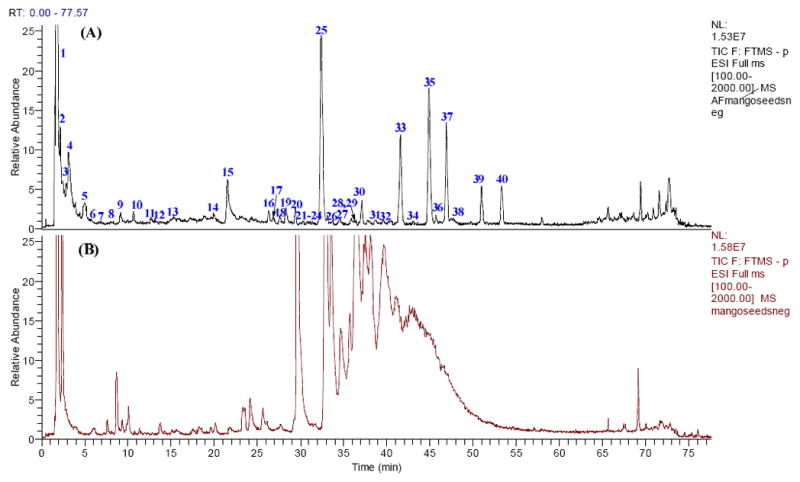
The UHPLC-PDA analysis revealed that most of the constituents in AM seeds have three UV maximum absorbance bands at 220–230 nm, 240–260 nm, and 350–370 nm. Forty one phenolic compounds were found in African mango seeds including ellagic acid, methyl-ellagic acid, ellagitannins and flavonol glycosides (Table 1). As shown in Figure 2, the chemical profile of AM seeds is quite different from that of mango seeds. The constituents identified from mango seeds are mostly flavonol, xanthone glycosides, benzophenone and gallotanins which is consistent with the literature(8–11). (Figure S1, Table S1).
Table 1.
UHPLC-HRMS data of constituents from African Mango seeds.
| Peak No | RT (min) | Formula | [M−H]− | Error (mmu) | UV (λmax, nm) | Main MS2~MS4 Product Ions | Tentative Identification |
|---|---|---|---|---|---|---|---|
| 1 | 1.81 | C12H22O11 | 341.1081 | −0.88 | 233 | 179(100), 161(20), 143(21), 131(7), 119(15), 113(17) | hexosyl-hexose |
| 2 | 1.87 | C6H8O7 | 191.0191 | −0.60 | N.D.b | 173(21), 111(100) | citric acid or its isomer |
| 3 | 2.15 | C20H18O14 | 481.0613 | −1.07 | 233 | 301(100), 275(12) | hexahydroxydiphenoyl (HHDP)-hexose |
| 4 | 3.28 | C34H24O22 | 783.0669 | −2.70 | N.D. | 765(3), 481(35), 301(100), 275(16) | di-HHDP-hexose |
| 5 | 4.9 | C34H24O22 | 783.0658 | −2.85 | N.D. | 765(3), 481(35), 301(100), 275(16) | di-HHDP-hexose |
| 6 | 5.64 | C34H24O22 | 783.0658 | −2.85 | N.D. | 481(35), 301(100), 275(16) | di-HHDP-hexose |
| 7 | 7.02 | C34H24O22 | 783.065 | −3.65 | 271 | 481(35), 301(100), 275(16) | di-HHDP-hexose |
| 8 | 8.16 | C34H24O22 | 783.0667 | −1.94 | 271 | 481(35), 301(100), 275(16) | di-HHDP-hexose |
| 9 | 9.16 | C21H32O10 | 443.1907 | −1.57 | 223, 275, 349 | 425(33), 375(56), 237(100), 219(67), 189(67), 161(56) | unknown |
| 10 | 10.66 | C18H18O8 | 361.0951 | 2.20 | 223, 271, 343 | 317(12), 293(3), 281(100), 237(27), | unknown |
| 11 | 11.33 | C33H24O22 | 771.0655 | −3.16 | 222, 281, 398 | 753(17), 481(100), 469(29), 379(6), 301(17), 289(27) | unknown ellagitannin |
| 12 | 11.89 | C33H24O22 | 771.0651 | −3.53 | 295 | 481(100), 469(75), 307(12), 301(29), 289(71), 263(25), | unknown ellagitannin |
| 13 | 15.35 | C18H28O9 | 387.1647 | −1.39 | 271, 349 | 370(12), 360(4), 343(6), 341(4), 207(100), 163(40) | unknown |
| 14 | 18.82 | C33H24O21 | 755.0698 | −3.91 | 255, 345 | 727(27), 453(89), 393(23), 301(51), 291(99), 247(100) | unknown ellagitannin |
| 15a | 21.51 | C14H6O8 | 300.9978 | −1.16 | 223, 253, 367 | 301(100), 300(14), 284(24), 257(59), 229(50), 185(22) | ellagic acid |
| 16 | 26.29 | C22H20O13 | 491.0817 | −1.444 | 222, 250, 358 | 476(19), 328(100), 313(9) MS3: 313(100) MS4: 298(100), 285(49) | di-O-methyl-ellagic acid hexoside |
| 17 a | 26.83 | C15H8O8 | 315.0135 | −1.09 | 222, 254, 354 | 300(100), MS3: 300(100), 283(16), 272(64), 271(31), 244(98), 243(31), 228(36), 216(20), 200(20) | MS3[491N.D.>328]: 313(100) methyl-ellagic acid |
| 18 | 27.61 | C15H8O8 | 315.0136 | −1.00 | 220, 250, 360 | 300(100), 272(13), 244(21), 200(10) | methyl-ellagic acid |
| 19 | 28.26 | C34H28O20 | 755.1072 | −2.88 | 220, 251, 364 | 711(15), 603(4), 453(7), 301(100), 284(3), 275(6) | galloyl-HHDP-ellagic acid |
| 20 | 28.94 | C34H28O20 | 755.1063 | −3.80 | N.D. | 711(9), 453(6), 301(100), 291(5), 275(10), 247(5) | galloyl-HHDP-ellagic acid |
| 21 | 29.38 | C25H16O15 | 555.0732 | 1.51 | 220, 250, 358 | 475(100) MS3: 460(23), 328(100), 313(7) MS4 313(100) | di-O-methyl-ellagic acid deoxyhexide with an 80 amu group |
| 22 a | 29.91 | C21H20O11 | 447.0919 | −1.43 | N.D. | 357(10), 327(19), 301(8), 285(75), 284(100), 255(15) | kaemferol 3-O-glucoside |
| 23 a | 30.34 | C21H20O11 | 447.0917 | −1.55 | N.D. | 301(100), 300(40) | quercetin 3-O-rhamnoside |
| 24 | 30.73 | C21H18O12 | 461.0708 | −1.78 | 220, 253, 349 | 315(100), MS3: 300(100), MS4: 300(100), 284(13), 272(17), 271(33), 244(33), 243(10), 228(20), 216(27), 200(17), | mono-O-methyl ellagic acid deoxyhexoside |
| 25 | 32.47 | C16H10O8 | 329.0290 | −1.30 | 220, 247, 363 | 314(100) MS3: 299 (100) MS4: 271(100) | di-O-methyl ellagic acid |
| 26 | 33.08 | C21H18O12 | 461.0712 | −1.30 | 220, 248, 364 | 446(17), 328(100), MS3 313(100) MS4 298(100), 285(43) | di-O-methyl ellagic acid-O-pentoside |
| 27 | 33.7 | C34H42O20 | 769.2166 | −3.06 | 220, 250, 349 | 607(100), 477(23), 315(14) | rhamnetin or isorhamnetin with one hexose group and one rhamnosyl-rhamnose |
| 28 | 35.91 | C41H30O24 | 905.1021 | −3.32 | N.D. | 603(55), 301(100) | di-HHDP-ellagic acid |
| 29 | 36.45 | C22H20O12 | 475.0867 | −1.47 | N.D. | 460(100), 329(8), 328(16), 327(9), 313(15), 299(7) | di-O-methyl-ellagic acid deoxyhexoside |
| 30 | 36.9 | C22H20O12 | 475.0869 | −1.29 | N.D. | 460(25), 328(100), 313(6), 313(100), 298(100),285(45) | di-O-methyl-ellagic acid deoxyhexoside |
| 31 | 37.85 | C30H26O17 | 657.1069 | −2.82 | 220, 244, 364 | 491(48), 329(100), 315(14), 314(31), 313(36), 299(13), | di-O-methyl-ellagic acid with one 166 amu group and one hexose goup |
| 32 | 39.57 | C16H10O8 | 329.0289 | −0.84 | N.D. | 314(100) | di-O-methyl-ellagic acid |
| 33 | 41.52 | C16H10O8 | 329.0292 | −0.57 | 223, 246, 375 | 314(100) MS3: 299(100,) MS4: 271(100) | di-O-methyl-ellagic acid |
| 34 | 43.01 | C34H42O20 | 769.217 | −2.14 | 239 | 697(3), 315(100), 300(25), 271(25), 255(5) | galloyl-HHDP-methyl-ellagic acid |
| 35 | 44.96 | C17H12O8 | 343.0449 | −0.50 | 222, 246, 362 | 328(100) 313(100) 298(100, MS4), 285(38, MS4) | tri-O-methyl-ellagic acid |
| 36 | 45.78 | C22H22O11 | 461.1074 | −0.97 | 220, 236, 341 | 315(100), 300(5) | mono-O-methyl-ellagic acid rhamnoside |
| 37 | 46.94 | C35H28O10 | 607.1649 | −3.95 | 220, 236, 347 | 315(100), 300(17), MS3: 300 (100), MS4: 271 (100), 255 (55) | mono-O-methyl-ellagic acid rhamnosyl- rhamnoside |
| 38 | 47.76 | C30H26O17 | 657.1071 | −2.66 | N.D.- | 343(100), 329(11), 328(28), 313(48), 285(3) | galloyl-tri-O-methyl-ellagic acid hexoside |
| 39 a | 49.75 | C16H12O6 | 299.0549 | −1.18 | N.D. | 284(100), 271(16), 227(5) | diosmetin |
| 40 | 53.3 | C17H12O8 | 343.0449 | −1.05 | 220, 236, 375 | 328(100) 313(100) 298(100), 285(40) | tri-O-methyl-ellagic acid |
In comparison with reference standards.
N.D.- Not Detected or very weak absorpance.
Figure 2.

The total ion chromatograms of African Mango (A) and Mexican mango (B) seeds.
Ellagic Acid, Mono, di, tri--Methyl-Ellagic Acids and Their Glycosides
Peaks 15, 25, 33, 35, 37, 39, and 40 were found to be the major constituents in the PDA and total ion chromatograms (Figure 2). Peak 15 was identified as ellagic acid. Its molecular ion was 300.9978, indicating the formula of C14H6O8 (−1.16 mmu). Its major product ions were m/z 257, 229, 284, 185, and 201, consistent with previous literature for ellagic acid. It was further confirmed by analysis of a reference standard. Peaks 25, 33, 35, 37, 39, and 40 displayed similar UV spectra to peak 15, suggesting they were ellagic acid derivatives. Peaks 25, 33, and 32 (minor contituent) all had a formula of C16H10O8, and the deprotonated ion of 25, 33 and 32 underwent the successive losses of CH3 radicals. Thus, two methyl groups existed in the structures. These three compounds were tentatively identified as di-O-methyl ellagic acids. The molecular ion of peaks 35 and 40 was ([M−H]−, C17H12O8) and the major product ions were m/z 328, m/z 313, and m/z 298 via the successive losses of CH3 radicals. Thus, three methyl groups exist in the structures of peaks 35 and 40. Hence these two compounds were tentatively identified as tri-O-methyl ellagic acids. Peaks 17 and 18 had the formula of C15H8O8 and the ion at m/z 300 was the predominate ion in the MS2 spectra. Further fragmentation of this ion showed an almost identical behavior of that of ellagic acid, thus these two compounds were tentatively identified as mono-methyl-ellagic acids.
Peak 16 had the [M−H]− ion at m/z 491 (C22H20O13). Its product ion was m/z 328 via the loss of 163 amu, corresponding to a cleavage of a glycosidic bond. The MS3 and MS4 spectra of the ion m/z 328 were almost identical with that of di-O-methyl ellagic acid. Thus, peak 16 was tentatively identified as di-O-methyl-ellagic acid hexoside. It is worth noting that the HRMS data can easily differentiate between a hexosyl (C6H10O5) and caffeoyl (C9H6O3) group, or between a deoxy-hexosyl and coumaroyl group. Similarly, Peak 24 was tentatively identified as methyl ellagic acid-O-deoxyhexose. Peaks 26 and 36 were tentatively identified as Di-O-methyl ellagic acid-O-pentoside. Peaks 29 and 30 were tentatively identified as Di-O-methyl-ellagic acid deoxyhexoside. Peak 41 had the deprotonated ion at m/z 607.1649 (C35H28 O10), and the loss of 292 Da unit was observed to produce the product ion at m/z 315. The successive fragmentation of m/z 315 was very similar to Peak 17 and 18 (methyl ellagic acid). Hence this compound was tentatively identified as methyl-ellagic acid-O-rhamnosyl-rhamnoside.
Ellagitannins
Ellagitannins are hydrolyzable tannins containing one or more hexahydroxydiphenoyl (HHDP) groups esterified to a sugar. The [M−H]− ion of peak 3 was 481.0613, indicating the formula of C20H18O14. The ion at m/z 301 was observed as the major product ion via the loss of 180 amu in the MS/MS spectrum. Hence, this compound was considered to contain one HHDP unit esterified to hexose. The loss of 180 amu corresponded to a hexosyl unit (162 amu) and H2O (18 amu) simultaneously.
Peaks 4, 5, 6, 7 and 8 showed the [M−H]− ion at 783 with the formula of C34H24O22. According to MS analysis, all of these five constituents likely contain HHDP-hexose in their makeup due to the presence of the product ions at m/z 481 (HHDP+hexose) and 303 (HHDP). These four compounds were characterized as di-HHDP-hexose. Peaks 11, 12, 15 were identified as unknown ellagitannins due to the characteristic neutral losses observed (loss of 302 amu unit, C14H6O8). Peaks 11 and 12 had the formula of C33H24O22, with a mass 14 amu less than that of Peak 4. These two compounds may lack one –CH2 group in the structure in comparison with Peaks 4 to 8.
Flavonoids
Peaks 22 and 23 were kaempferol 3-O-glucoside and quercetin 3-O-rhamnoside according to their HRMS measurements and UV spectra. Their identities were confirmed by reference standards. Peak 27 had a formula of C34H42O20, and the losses of one 162 and 292 group was observed in the MS2 to MS4 spectra, which correlated to one glucose group and one rhamnosyl rhamnose group. The aglycone part of peak 27 was C16H12O7, a rhamnetin or isorhamnetin. Peak 39 had a formula of C16H12O6, and the retention time and MS2 fragmentation was consistent with the reference standard of diosmetin, an O-methylated flavone.
Chromatographic comparisons with AMDSs
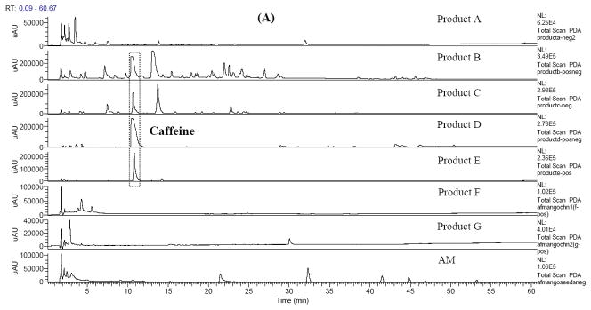
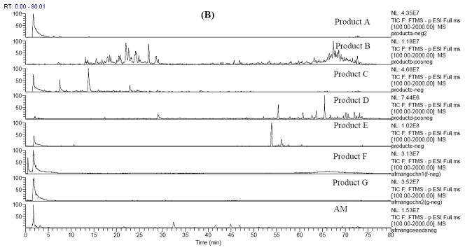
With the chemical information acquired with authentic AM seeds and AMSE, the UHPLC-HRMS chromatograms of AMDS were compared with that of African Mango seeds for quality evaluation. The 5 AMDS and 2 Chinese AMSE commercial products and their label claims are listed in Table S2. Huge variances were observed across the samples. Figure 3 shows the UV chromatograms and total ion chromatograms (TIC) in negative mode of products A to G and authentic AMSE. All commercial products displayed different TIC profiles from each other. Extracted ion chromatograms (EIC) were used to find specific peaks, if any, that might come from AMSE. Unfortunately, none of the products shared any common peaks with authentic AMSE in meaningful amounts. Product A claimed that it used AMSE only and 15 peaks were found in its TIC (Figure S2). Among them, six compounds were successfully identified by comparing the formula, multi-stage mass fragmentation, UV spectra, literature reports, and reference standards when available. The deprotonated ion [M−H]− of peak 8 is 421.0771, suggesting the chemical composition C19H17O11(−1.22 ppm). The [M−H-120]−, [M−H-90]−, and [M−H-18]− were observed as three major product ions in the MS2 spectra, which suggests that it is most probably a C-glycoside. Together with the UV maximum absorbance at 257 nm, 318 nm and 365 nm, it was unambiguously identified as mangiferin, a xanthone glycoside which has been extensively reported in mango (Mangifera indica L)(8, 11, 14), but not in AMSE. Peak 9 also showed a chemical composition C19H17O11 (−2.22 ppm), and was identified as isomangiferin by comparing the elution order and MS/MS data reported in the literatures (8, 11, 14). Peak 12 revealed a deprotonated ion [M−H]− at m/z 573.0879, suggesting a chemical composition of C26H21O15. A neutral loss of 152 amu (C7H4O4) was observed in the MS2 spectra. The MS3 and MS4 spectra were almost identical with that of mangiferin. So it can be concluded that a galloyl moiety was substituted in peak 12. This compound was then identified as galloylated mangiferin(8). Similarly, peak 13 was identified as galloylated isomangiferin(8). Peak 14 shows a [M−H]- ion at 300.9984 (C14H5O8, −1.34 ppm) and was identified as ellagic acid by comparing the MS/MS data and with reference standards(9). Peak 15 with the retention time at 31.97 min shows a [M−H]- ion at 201.0295 (C11H7O5, −1.81 ppm). The neutral losses of CO, H2O, and C2H2O were observed in the MS2 spectrum and are consistent with the fragmentation behavior of coumarin(15). Obviously, product A used mango seed extract instead of AMSE.
Figure 3.
The HPLC chromatogram (200–700 nm, A) and total ion chromatogram in negative mode (B) of Products A~G and African Mango Seeds Extract.
The labels of both products B and C claim the use of AM seed extract (4:1) with 200 mg green tea extract. However, no compounds related to either AMSE or xanthone glycosides (mangiferin related constituents) were detected.
Product D claims that maqui berry (Aristotelia Chilensisa), acai fruit (Euterpe oleracea), green tea extract, resveratrol, caffeine, apple cider vinegar, kelp (seaweeds) and grapefruit were added in addition to AMSE. The UV chromatogram of Product D (Figure 3A) shows a major peak with the retention time of 10.4 min, which constitutes more than 90% of the total peak area. The protonated ion [M+H]+ at m/z 195.0883 revealed the formula of C8H10N4O2. It was identified as caffeine and confirmed with a reference standard. No compounds from AMSE were detected in product D.
The label of product E claimed that it contained green tea extract, cranberry, raspberry, fibersol, citric acid, and stevia along with AMSE. First, no compound from AMSE was detected. In addition, the major groups of constituents did not have strong UV absorbance and were eluted from 45 min to 66 min. That means phenolic compounds (major components of green tea extract, cranberry, raspberry) are not a significant part of the product E.
Products F and G claimed to be pure AMSE. They were obtained directly from suppliers through Hunan Food Test and Analysis Center (Changsha, Hunan, China). The UHPLC-HRMS analysis did not show any detectable amount of components related to either AM or regular mango. If the AMDS samples B–E used AMSE as claimed in their labels, they might have used extracts similar to the products F and G and it might be the reason no AM related components were detected in these AMDS samples.
The chemical constituents of AM (Irvingia Gabonensis) seeds and related dietary supplements were studied with the developed UHPLC-PDA-HRMS method. It was found that ellagic acid, methyl-ellagic acids and their glycosides were the main compounds in the authentic AM seeds and AM seed extract. The chromatographic profiles of AM seeds are distinctly different from that of regular mango (Mangifera indica). Among the 5 representative AMDS purchased from the internet, only one sample contained trace constituents of regular mango seeds. The other 4 AMDS samples and the 2 AMSE samples from Chinese suppliers do not contain detectable amount of authentic AM seed extract. This study continues to demonstrate the urgency for the enforcement of FDA’s GMP requirements for the dietary supplement industry. For AMDS, the labels do not provide accurate information for the consumer. Proper standardization and quality control of raw materials and the herbal preparations should be carried out. Finally, it is worth noting that although the conclusion was based on the authentic samples from one source only, the differences between the authentic samples and commercial products were significant that the probable cause of the difference were not due to variability of growing conditions, harvesting times, and storage conditions.
Supplementary Material
Acknowledgments
This research is supported by the Agricultural Research Service of the United States Department of Agriculture and an Interagency Agreement with the Office of Dietary Supplements of the National Institutes of Health.
References
- 1.Lamorde M, Tabuti JR, Obua C, Kukunda-Byobona C, Lanyero H, Byakika-Kibwika P, Bbosa GS, Lubega A, Ogwal-Okeng J, Ryan M, Waako PJ, Merry C. Medicinal plants used by traditional medicine practitioners for the treatment of HIV/AIDS and related conditions in Uganda. J Ethnopharmacol. 2010;130:43–53. doi: 10.1016/j.jep.2010.04.004. [DOI] [PubMed] [Google Scholar]
- 2.Spindler AA, Akosionu N. Gross Composition and Crude and Dietary Fiber Content of Soups Made with African Mango Seeds. Nutr Rep Int. 1985;31:1165–1169. [Google Scholar]
- 3.Ngondi JL, Oben JE, Minka SR. The effect of Irvingia gabonensis seeds on body weight and blood lipids of obese subjects in Cameroon. Lipids Health Dis. 2005;4:12. doi: 10.1186/1476-511X-4-12. [DOI] [PMC free article] [PubMed] [Google Scholar]
- 4.Oben JE, Ngondi JL, Momo CN, Agbor GA, Sobgui CSM. The use of a Cissus quadrangularis/Irvingia gabonensis combination in the management of weight loss: a doubleblind placebo-controlled study. Lipids Health Dis. 2008;7 doi: 10.1186/1476-511X-7-12. [DOI] [PMC free article] [PubMed] [Google Scholar]
- 5.Ross SM. African mango (IGOB131): a proprietary seed extract of Irvingia gabonensis is found to be effective in reducing body weight and improving metabolic parameters in overweight humans. Holist Nurs Pract. 2011;25:215–7. doi: 10.1097/HNP.0b013e318222735a. [DOI] [PubMed] [Google Scholar]
- 6.Oben J, Kuate D, Agbor G, Momo C, Talla X. The use of a Cissus quadrangularis formulation in the management of weight loss and metabolic syndrome. Lipids Health Dis. 2006;5 doi: 10.1186/1476-511X-5-24. [DOI] [PMC free article] [PubMed] [Google Scholar]
- 7.Adedeji J, Hartman TG, Lech J, Ho CT. Characterization of Glycosidically Bound Aroma Compounds in the African Mango (Mangifera-Indica L) J Agr Food Chem. 1992;40:659–661. [Google Scholar]
- 8.Berardini N, Fezer R, Conrad J, Beifuss U, Carle R, Schieber A. Screening of mango (Mangifera indica L) cultivars for their contents of flavonol O- and xanthone C-glycosides, anthocyanins, and pectin. J Agric Food Chem. 2005;53:1563–70. doi: 10.1021/jf0484069. [DOI] [PubMed] [Google Scholar]
- 9.Ajila CM, Rao LJ, Rao UJ. Characterization of bioactive compounds from raw and ripe Mangifera indica L. peel extracts. Food Chem Toxicol. 2010;48:3406–11. doi: 10.1016/j.fct.2010.09.012. [DOI] [PubMed] [Google Scholar]
- 10.de Ornelas-Paz JJ, Yahia EM, Gardea-Bejar A. Identification and quantification of xanthophyll esters, carotenes, and tocopherols in the fruit of seven Mexican mango cultivars by liquid chromatography-atmospheric pressure chemical ionization-time-of-flight mass spectrometry [LC-(APcI(+))-MS] J Agric Food Chem. 2007;55:6628–35. doi: 10.1021/jf0706981. [DOI] [PubMed] [Google Scholar]
- 11.Schieber A, Berardini N, Carle R. Identification of flavonol and xanthone glycosides from mango (Mangifera indica L. Cv “Tommy Atkins”) peels by high-performance liquid chromatography-electrospray ionization mass spectrometry. J Agric Food Chem. 2003;51:5006–11. doi: 10.1021/jf030218f. [DOI] [PubMed] [Google Scholar]
- 12.Engels C, Ganzle MG, Schieber A. Fractionation of Gallotannins from mango (Mangifera indica L. ) kernels by high-speed counter-current chromatography and determination of their antibacterial activity. J Agric Food Chem. 2010;58:775–80. doi: 10.1021/jf903252t. [DOI] [PubMed] [Google Scholar]
- 13.Engels C, Knodler M, Zhao YY, Carle R, Ganzle MG, Schieber A. Antimicrobial activity of gallotannins isolated from mango ( Mangifera indica L) kernels. J Agric Food Chem. 2009;57:7712–8. doi: 10.1021/jf901621m. [DOI] [PubMed] [Google Scholar]
- 14.Berardini N, Carle R, Schieber A. Characterization of gallotannins and benzophenone derivatives from mango (Mangifera indica L. cv. ‘Tommy Atkins’) peels, pulp and kernels by highperformance liquid chromatography/electrospray ionization mass spectrometry. Rapid Commun Mass Spectrom. 2004;18:2208–16. doi: 10.1002/rcm.1611. [DOI] [PubMed] [Google Scholar]
- 15.Horai H, Arita M, Kanaya S, Nihei Y, Ikeda T, Suwa K, Ojima Y, Tanaka K, Tanaka S, Aoshima K, Oda Y, Kakazu Y, Kusano M, Tohge T, Matsuda F, Sawada Y, Hirai MY, Nakanishi H, Ikeda K, Akimoto N, Maoka T, Takahashi H, Ara T, Sakurai N, Suzuki H, Shibata D, Neumann S, Iida T, Funatsu K, Matsuura F, Soga T, Taguchi R, Saito K, Nishioka T. MassBank: a public repository for sharing mass spectral data for life sciences. J Mass Spectrom. 2010;45:703–14. doi: 10.1002/jms.1777. [DOI] [PubMed] [Google Scholar]
Associated Data
This section collects any data citations, data availability statements, or supplementary materials included in this article.


