Abstract
Angiosarcoma is a rare, aggressive malignancy of endothelial cells lining blood vessels. It poses therapeutic challenges since there is no standard established treatment. It is typically treated with resection and wide-field postoperative radiation therapy. Chemotherapy and radiation therapy have also been reported as initial therapies. Regardless of the treatment rendered, the risk of local regional failure and distant relapse remains high for this disease. We present the case of a patient who developed a well-differentiated angiosarcoma of the nose with bilateral malar extension. No commonly associated risk factors such as lymphedema, prior radiotherapy or chronic venous ulceration were present. Given her age, pre-existing renal condition and preference not to receive chemotherapy, systemic therapy was not utilized. Surgery was also refused by the patient due to the projected cosmetic deficit. The patient was ultimately treated with definitive radiotherapy, utilizing electrons to the central face, differential thickness bolus, an intraoral stent, eye shields, an aquaplast mask for immobilization and a wax-coated lead shield over the face in order to limit penumbra of the radiation beam. Right and left anterior 6-MV photons were used to tangentially treat the bilateral malar region in order to extend the field edges. At the time of this report, the patient remains disease free at nearly 2.0 years after radiotherapy. To the best of our knowledge, this represents only the second case in the literature reporting radiotherapy as a single modality treatment that resulted in complete remission of an angiosarcoma of the face.
Key Words: Angiosarcoma, Endothelial cells, Radiation therapy, Single modality treatment
Introduction
Angiosarcoma is a rare neoplasm of vascular endothelial cells. It accounts for 1–2% of all sarcomas [1, 2]. While it can occur in any part of the body, skin, soft tissue, breast and liver are most commonly affected. It has a predilection for skin and soft tissues in head and neck region, given the vascular density and exposure to ultraviolet radiation [2]. There are various subtypes of this disease and cutaneous angiosarcoma is the most common. Cutaneous angiosarcomas commonly occur in the face and scalp region; they account for about 60% of all angiosarcomas [3, 4]. Soft tissue angiosarcomas and breast angiosarcomas roughly account for about 25 and 8% of angiosarcomas, respectively [3, 4]. In the US, the incidence of angiosarcoma is reported to be higher amongst Caucasians compared to African Americans. It appears to be increasing among Caucasians during recent years [2]. Risk factors for angiosarcoma include exposure to agents such as thorotrast, vinyl chloride, insecticides containing arsenic, long-term anabolic steroid or estrogen therapy; morbid obesity; chronic venous ulceration; chronic lymphedema (Stewart-Treves syndrome); prior radiotherapy; renal transplantation; familial syndromes such as Klippel-Trenaunay syndrome, Maffucci syndrome, retinoblastoma, xeroderma pigmentosum, neurofibromatosis; pre-existing cancers such as germ cell tumors, vestibular schwannomas, leiomyomas, nerve sheath tumors; foreign bodies such as vascular graft material, surgical sponges, Dacron, plastic, steel [2].
In general, angiosarcomas have a poor prognosis. Prognosis depends upon factors such as depth of tumor invasion, tumor diameter, local regional spread, distant metastasis, positive margins on surgical tumor resection and tumor recurrence [2]. In the US, the overall 5-year survival is reported to be in the range of 10–45%. Given the rarity of this cancer, no standard treatment has been established. In addition, the multifocal nature of the disease, as well as different combinations of disease location and subtypes, makes the treatment challenging. In spite of limited retrospective and prospective nonrandomized data on the treatment of angiosarcoma, radical surgical resection followed by wide-field postoperative radiotherapy is considered an optimal treatment strategy for the majority of patients. The use of chemotherapy and radiation has also been reported [2]. Chemotherapy may be especially helpful for short-term palliation [5]. In regard to definitive treatment, the role of adjuvant chemotherapy remains unclear. Doxorubicin, paclitaxel, and subcutaneous interferon alpha-2a with oral 13-cis-retinoic acid have been reported to be used [2].
Case Report
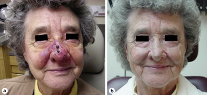
The patient is an 81-year-old Caucasian woman with a 15-pack year smoking history and no family or past medical history of cancer who presented to our multidisciplinary head and neck tumor board at the University of Wisconsin, Madison, Wisc., USA, with a large erythematous lesion on her nose with bilateral malar extension. The lesion also extended superiorly to the infraorbital region and into the lower eyelids; it further extended inferiorly into the nasal vestibule and into the nasal cavity. The lesion was characterized by multiple papules and eschars, most prominently over the bridge over her nose (fig. 1). Ten months prior to presentation, the patient developed red papules on her bilateral nasal ala. The initial biopsy was read as benign. However, the lesion on the right side of her nose continued to grow and bled intermittently over a 10-month period. She eventually underwent excision of a portion of the lesion elsewhere. A subsequent pathology report revealed an epithelioid hemangioma. This diagnosis was reviewed and confirmed at a second local hospital. The patient was managed with continued observation. However, the lesion progressed and this prompted her to undergo reevaluation of the lesion by a dermatologist. She underwent a repeat biopsy of the lesion and the pathology returned positive for angiosarcoma. The patient was referred to the University of Wisconsin for recommendations concerning her treatment options. The pathology was reviewed and the diagnosis of angiosarcoma was confirmed.
Fig. 1.
a Presentation of the patient with angiosarcoma prior to treatment. b Presentation of the patient nearly 2 years after treatment with radiation therapy as single modality.
At presentation, the patient's review of systems was essentially negative for any constitutional symptoms or those referable to head and neck. She denied any pain or discomfort associated with the lesion. She reported bilateral orbital swelling that developed over 2 months prior to her presentation, excessive lacrimation and intermittent bleeding from the lesion. Her past medical history was unremarkable for prior radiotherapy, chronic venous ulceration, lymphedema, or other causative factors of angiosarcoma. Of note, she donated one of her kidneys to her son in the past. She had a history of bilateral cataract removal, hypothyroidism, and diabetes mellitus type II at presentation. She had undergone chest and head CTs prior to presentation at our practice. Head CT demonstrated that the lesion extended into the nasal cavity, and a chest CT demonstrated a 1-cm pleural based pulmonary nodule in the posteromedial aspect of the left upper lobe. No prior films were available for comparison. The patient underwent a PET/CT scan that demonstrated a mildly diffuse FDG avidity associated with her known angiosarcoma without any evidence of local regional lymph nodes or distant metastasis. It did not reveal any abnormal FDG avidity associated with the patient's previously appreciated left upper lobe subpleural pulmonary nodule. Therefore, this abnormality was deemed to be nonspecific and most likely represented a prior granulomatous infection, given the presence of densely calcified mediastinal and right hilar lymph nodes. An MRI of the orbit, face, and neck was performed which demonstrated the angiosarcoma of the nose with bilateral malar extension and without evidence of perineural spread. Physical examination confirmed the radiographic findings. The tumor measured 7.0 cm cephalad to caudad, 12.0 cm in the lateral dimension and there was 3.0 cm extension along the floor of the nasal cavity, as measured from the nasal vestibule. There were no palpable regional lymph nodes.
Given the location and size of the tumor, surgery was deemed inappropriate. Considering the patient's age, medical condition, only one functional kidney and refusal to receive chemotherapy, systemic agents were not given. Therefore, the patient underwent definitive radiation therapy with electrons and photons. A total dose of 66 Gy in 33 fractions, utilizing 12 MeV electrons (custom bolus for uniform dosing) was delivered to the central face. A dose of 57.2 Gy in 29 fractions was delivered to the bilateral cheeks using 6 MV photons. The patient tolerated radiotherapy well with the expected side effects. At nearly 2.0 years following treatment, she remains free of disease recurrence with the only late complication consisting of bilateral trichiasis (fig. 1).
Discussion
The diagnosis and treatment of angiosarcoma presents unique challenges. Given the poor overall survival (OS) for this tumor, it is crucial to perform a thorough history and physical examination with a high index of clinical suspicion when evaluating skin and vascular lesions in the head and neck region. Mortality typically results from either extensive local disease or distant metastasis to organs such as lungs [6]. The reported patient had localized disease without distant metastasis. Her tumor was well-differentiated; however, in contrast to other sarcomas, grade is not helpful in predicting survival [6]. Furthermore, no correlation has been shown between local recurrence or survival and tumor characteristics such as ulcerated, diffuse, or nodular. Nevertheless, multifocal disease, positive surgical margins, size of the tumor (>5 cm of external diameter of the tumor), mitotic rate (>3 HPF), depth of invasion (>3 mm), local regional recurrence and distant metastases have been shown to correlate with poor outcomes [6]. In our patient, difficulty in making the diagnosis placed her at significant risk for reduced survival, especially given the size of her tumor. Often, cutaneous angiosarcomas will initially be perceived as benign. According to one study, clinical signs of disease exist for an average of 5.1 months prior to diagnosis of scalp angiosarcomas. In some cases, diagnosis may be delayed for as long as 1 year despite continued signs and symptoms of disease [7]. Expanding nodular or papule type lesions that bruise or bleed for a prolonged period of time should raise concerns about an underlying malignancy and should be promptly investigated [8].
A recent retrospective study reported on survival outcomes of 48 patients who were treated for angiosarcoma of face and scalp between 1987 and 2009 with either a single modality or combination of surgery, radiotherapy, chemotherapy, and immunotherapy [9]. The median follow-up for all 48 patients was 13.7 months (range 2.5–105.9). Surgery and radiotherapy were found to be significant favorable and independent prognostic factors for OS. Patients who underwent both surgery and radiotherapy (2-year OS: 45.8%) had a significantly more favorable OS (p < 0.0001) compared with patients treated with either surgery or radiotherapy (2-year OS: 11.1%) alone and patients who received no surgery or radiotherapy (2-year OS: 0%). These findings corroborate data from a literature review which suggests that the optimal treatment for angiosarcoma of head and neck is surgery followed by wide-field radiotherapy [5]. However, the tumor is often so extensive at diagnosis that complete surgical resection of the tumor may not be feasible. Even with optimal local regional treatment, the likelihood of a local recurrence in the radiation field or distant metastases through hematogenous spread is quite high [5]. Mendenhall et al. [5] reported 5-year local regional control from 40 to 50%, 5-year distant metastasis-free survival from 20 to 40%, and 5-year OS from 10 to 30%.
Data regarding impact of adjuvant chemotherapy on survival is limited. Available data suggests usefulness for palliation with progression-free survival rates ranging from 1 to 5 months [5]. Nevertheless, there are a few case reports that have reported complete or partial response of tumor to chemotherapy when delivered either as a single modality or in combination with surgery and/or radiation. Koontz et al. [10] reported two cases of nasal angiosarcoma that responded with complete remission to treatment with bevacizumab, radiotherapy, and surgical resection with response duration of 26 and 8.5 months for the two cases. A retrospective study by Schlemmer et al. [11] reported on treatment outcomes for 8 patients with angiosarcoma of scalp and face who were treated with paclitaxel with or without other modalities such as surgery and radiotherapy. The authors reported one case (1/8) of complete remission with response duration of 42 months and five cases of partial response (5/8) with mean response duration of 5.8 months. A retrospective study by Nagano et al. [12] reported treatment responses for 9 patients with cutaneous angiosarcoma treated with docetaxel with or without previous treatment by surgery and radiation. The authors reported two cases of complete remission (2/9) and four cases of partial remission (4/9); of these, only one case had a single modality treatment with paclitaxel, resulting in a partial response. There are few other case reports that report complete remission of cutaneous angiosarcoma of scalp and face treated with combined liposomal doxorubicin and radiotherapy; response duration ranged from 15 months to 4 years [2, 13, 14]. In summary, data on the role of chemotherapy in the definitive treatment of cutaneous angiosarcoma of face are limited and varied. To the best of our knowledge, there is only one documented case in the past literature that reports radiotherapy as a single modality treatment for angiosarcoma of the face. Gkalpakiotis et al. [15] reports durable complete remission of a well-differentiated exophytic angiosarcoma of the face that responded well to radiotherapy alone. The patient remained free of disease recurrence at 5 years after radiotherapy.
Given the size and location of the tumor, our patient was deemed ineligible for surgery. Aggressive surgical resection would have resulted in significant life-long morbidity with compromised cosmesis. From a definitive standpoint, combined chemotherapy and radiotherapy was the next available option. However, chemotherapy was refused by the patient and she had multiple relative contraindications. Therefore, the patient underwent successful treatment with definitive radiotherapy, using electrons and photons. Her tumor fully regressed, resulting in excellent cosmesis.
Conclusions
Definitive radiotherapy may be an effective treatment in a select group of patients with head and neck angiosarcoma in whom chemotherapy and surgery may not be practically feasible. A delay in the diagnosis of angiosarcoma in the head and neck region could present with significant treatment challenges due to increased tumor size, especially since surgery and postoperative radiotherapy is the mainstay therapy in many patients. Hence, it is very important to diagnose patients with angiosarcoma in a timely fashion. Given the propensity of angiosarcoma to develop metastases, a high index of clinical suspicion early in the clinical course is crucial in order to maximize patient survival.
References
- 1.Brennan MF, Alekitar KM, Maki RG. Soft tissue sarcomas. In: De Vita VT Jr, Hellman S, Rosenberg SA, editors. Cancer: Principles and Practice of Oncology. 6th ed. Philadelphia, PA: Lippincott Williams and Wilkins; 2001. pp. 1841–1891. [Google Scholar]
- 2.Al-Enezi M, Brassard A. Chronic venous ulceration with associated angiosarcoma. J Dermatol Case Rep. 2009;1:8–10. doi: 10.3315/jdcr.2009.1023. [DOI] [PMC free article] [PubMed] [Google Scholar]
- 3.Holden CA, Spittle MF, Jones EW. Angiosarcoma of the face and scalp, prognosis and treatment. Cancer. 1987;59:1046–1057. doi: 10.1002/1097-0142(19870301)59:5<1046::aid-cncr2820590533>3.0.co;2-6. [DOI] [PubMed] [Google Scholar]
- 4.Morales PH, Lindberg RD, Barkley HT., Jr Soft tissue angiosarcomas. Int J Radiat Oncol Biol Phys. 1981;7:1655–1659. doi: 10.1016/0360-3016(81)90188-7. [DOI] [PubMed] [Google Scholar]
- 5.Mendenhall WM, Mendenhall CM, Werning JW, Reith JD, Mendenhall NP. Cutaneous angiosarcoma. Am J Clin Oncol. 2006;29:524–528. doi: 10.1097/01.coc.0000227544.01779.52. [DOI] [PubMed] [Google Scholar]
- 6.Kharkar V, Jadhav P, Thakkar V, Mahajan S, Khopkar U. Primary cutaneous angiosarcoma of the nose. Indian J Dermatol Venereol Leprol. 2012;78:496–497. doi: 10.4103/0378-6323.98086. [DOI] [PubMed] [Google Scholar]
- 7.Pawlik TM, Paulino AF, Mcginn CJ, et al. Cutaneous angiosarcoma of the scalp: a multidisciplinary approach. Cancer. 2003;98:1716–1726. doi: 10.1002/cncr.11667. [DOI] [PubMed] [Google Scholar]
- 8.Lewis JM, Sondak VK: Angiosarcoma. Electronic Sarcoma Update Newsletter. 2005. http://sarcomahelp.org/learning_center/angiosarcoma.html
- 9.Ogawa K, Takahashi K, Asato Y, Yamamoto Y, Taira K, Matori S, Iraha S, Yagi N, et al. Treatment and prognosis of angiosarcoma of the scalp and face: a retrospective analysis of 48 patients. Br J Radiol. 2012 doi: 10.1259/bjr/31655219. E-pub ahead of print. [DOI] [PMC free article] [PubMed] [Google Scholar]
- 10.Koontz BF, Miles EF, Rubio MA, et al. Preoperative radiotherapy and bevacizumab for angiosarcoma of the head and neck: two case studies. Head and Neck. 2008;30:262–266. doi: 10.1002/hed.20674. [DOI] [PubMed] [Google Scholar]
- 11.Schlemmer P, Reichardt J, Verweij J, et al. Paclitaxel in patients with advanced angiosarcomas of soft tissue: a retrospective study of the EORTC soft tissue and bone sarcoma group. Eur J Cancer. 2008;44:2433–2436. doi: 10.1016/j.ejca.2008.07.037. [DOI] [PubMed] [Google Scholar]
- 12.Nagano T, Yamada Y, Ikeda T, Kanki H, Kamo T, Nishigori C. Docetaxel: a therapeutic option in the treatment of cutaneous angiosarcoma: report of 9 patients. Cancer. 2007;110:648–651. doi: 10.1002/cncr.22822. [DOI] [PubMed] [Google Scholar]
- 13.Lankester KJ, Brown RS, Spittle MF. Complete resolution of angiosarcoma of the scalp with liposomal daunorubicin and radiotherapy. Clin Oncol. 1999;11:208–210. doi: 10.1053/clon.1999.9046. [DOI] [PubMed] [Google Scholar]
- 14.Eiling S, Lischner S, Busch JO, Rothaupt D, Christophers E, Hauschild A. Complete remission of a radio-resistant cutaneous angiosarcoma of the scalp by systemic treatment with liposomal doxorubicin. Br J Dermatol. 2002;147:150–153. doi: 10.1046/j.1365-2133.2002.04726.x. [DOI] [PubMed] [Google Scholar]
- 15.Gkalpakiotis S, Arenberger O, Vohradnikova O, Arenbergerova M. Successful radiotherapy of facial angiosarcoma. Int J Dermatol. 2008;47:1190–1192. doi: 10.1111/j.1365-4632.2008.03813.x. [DOI] [PubMed] [Google Scholar]


