Abstract
Abstract
We retrospectively analyzed the outcomes of hematopoietic stem cell transplantation in 7 patients with primary immunodeficiency diseases treated at the National University Hospital, Singapore, over the period from December 1996 to January 2010. The primary immunodeficiency diseases managed were X-linked hyperimmunoglobulin M syndrome (n = 3), severe combined immunodeficiency (n = 1), leukocyte adhesion deficiency type 1 (n = 1), chronic granulomatous disease (n = 1), and Wiskott–Aldrich syndrome (n = 1). The age of the patients ranged from 5 months to 17 years. Conditioning regimen depended on the type of immunodeficiency, whereas supportive treatment was tailored for differing pretransplant conditions. Eight stem cell transplantations were performed for 7 patients. Donors were HLA-matched sibling donors for 2 patients and unrelated donors for the rest. At the median follow-up of 8.6 years (range 2.2–15.0 years) as of December 2011, 6 patients were alive and cured of their primary diseases.
Key Words: primary immunodeficiency, hematopoietic stem cell transplantation
Stem cell transplantation (SCT) is the curative treatment for severe primary immunodeficiency diseases (PIDs). If left untreated, severe PIDs are associated with high mortality risk and poor quality of life from serious infections in childhood. Overall survival rates for matched-related donor transplants ranged from 69 to 100% depending on the underlying disease type and promptness of the transplant.1–3 For matched-unrelated donor transplants, the increase in the donor pool together with improvements in supportive care and novel conditioning regimens to reduce risks of graft-versus-host disease (GVHD) has resulted in overall survival to up to 81%.1,4 In addition, early transplantation before the development of serious infections has proven beneficial.1–6 In our center, we have performed SCT for the following PIDs: hyperimmunoglobulin M (HIgM) syndrome, severe combined immunodeficiency (SCID), leukocyte adhesion deficiency type 1 (LAD1), chronic granulomatous disease (CGD), and Wiskott–Aldrich syndrome. This article aims to describe the experience over the period from 1996 to 2008 in treating 7 children with PIDs at the National University Hospital, Singapore.
METHODS
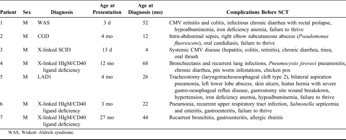
This was a retrospective study of 7 patients with PID who underwent SCT at Singapore's University Children's Medical Institute, National University Health System, from December 1996 to January 2010 (Table 1). There were 8 SCTs, which took place for these 7 patients, as 1 patient required a second transplant. All the SCTs for PIDs that had been performed in our center were included in this study description. We traced the patient files to determine the events pre-SCT and to assess survival rate and event-free survival after SCT. Ethics approval was obtained from the Institutional Review Board of the National University Hospital.
TABLE 1.
Clinical Features and Source of Graft of Patients With Primary Immunodeficiency

Stem Cell Transplants and Conditioning
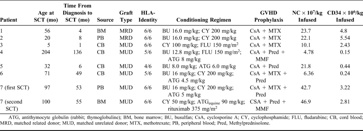
Two SCTs were HLA-matched, sibling donor transplants (1 bone marrow and 1 peripheral blood), 3 patients underwent unrelated transplants, which were fully HLA-matched at the antigen level (1 peripheral blood, 1 bone marrow, and 1 cord blood), and 3 patients had cord blood unrelated transplants, which were HLA-matched at 4 or 5 (out of 6) antigens (Table 2).
TABLE 2.
Characteristics of Graft Source and Conditioning Regimes Used

Except for patient 3, busulfan was used in all regimens in combination with cyclophosphamide, fludarabine, and antithymocyte globulin (rabbit). Only the patient with SCID (patient 3) underwent a reduced intensity preconditioning regimen comprising cyclophosphamide and fludarabine. All patients received GVHD prophylaxis with cyclosporine A and/or a combination of short-course methotrexate, methylprednisolone, and mycophenolate mofetil. The median total nucleated cell dose was 22.0 × 10⁷ per kilogram (range, 4.78–46.9 × 10⁷/kg), whereas the median CD34 cell dose was 2.62 × 10⁶ per kilogram (range, 0.15–5.54 × 10⁶/kg) (Table 2).
Supportive Treatment
Bacterial prophylaxis comprised oral bactrim until day −2 of SCT, which was restarted when the absolute neutrophil count was >1 × 10⁹/L. Antifungal prophylaxis comprised oral itraconazole solution for all patients. Weekly intravenous immunoglobulin (from day −5 to day +84 of SCT) was given for antiviral prophylaxis. Acyclovir was given to patients who were seropositive for herpes viruses. Other supportive therapy included granulocyte colony-stimulating factor.
Engraftment
Myeloid engraftment was taken as the first of 3 consecutive days with absolute neutrophil count >0.5 × 10⁹/L, red cell engraftment as the first of 7 days without transfusions, and platelet engraftment as the day when platelet count was >50 × 109/L without transfusions for 7 days prior.
RESULTS
The PIDs managed were X-linked HIgM syndrome (n = 3), SCID (n = 1), LAD1 (n = 1), CGD (n = 1), and Wiskott–Aldrich syndrome (n = 1) (Table 1). Genetic studies were done for all patients except patient 1 for whom it was not available. For patient 2 (CGD), chorionic villus sampling was done for the brother—mutation in the gp91-phox gene located on chromosome X (Xp21.1). Tissue sample of patient 3 (X-linked SCID) could not be genetically typed; however, the mother was heterozygous for a deleterious splice mutation at complementary DNA (cDNA) 868 in the IL2RG gene [cDNA mutation, 868(+5) splice del ga]. Patient 4 (HIgM syndrome) had an X-linked insertion deletion (indel) mutation in exon 3 on the CD40 ligand gene (cDNA mutation, 347-355delTATAATGTTinsG). Patient 5 (LAD1) had a 2490 base pair deletion on intron 8, exon 9, and intron 9 of the CD18 gene located on chromosome 21q22.3. Patient 6 and 7 (HIgM syndrome) are brothers and have the same X-linked deletion mutation on exon 5 in the CD40 ligand gene (“TA” del at nucleotide positions 775-776, 775-776delTA).
PIDs were diagnosed 4 to 56 months after presentation (median, 18 months), with the SCT performed at a median of 28.5 months after diagnosis (range, 0.7–135 months). The median age at SCT was 63.5 months (range, 5–204 months). Median time to myeloid engraftment was 17.5 days (range, 30–75 days). Median time to red cell engraftment was 25 days (range, 10–31 days). Median time to platelet engraftment was 41.5 days (range, 12–145 days) (Table 3).
TABLE 3.
Time to Engraftment for Each Cell Lineage

Of the 7 patients, there was 1 death (patient 5) after SCT. Pre-SCT morbidity for patient 5 was considerable. He was in poor nutritional state and suffered multiple infections and required a tracheostomy for upper airway obstruction. Post-SCT, he primarily experienced acute respiratory distress syndrome secondary to parainfluenza type 3 and Pseudomonas pneumonia as well as Escherichia coli sepsis, renal impairment requiring hemodialysis, and gastrointestinal bleed secondary to refractory thrombocytopenia. He underwent a cord blood unrelated transplant matched at 4 of 6 loci at the antigen level but failed to engraft.
For the surviving patients, all except one had sustained engraftment. Patient 7 had his first graft rejected before platelet engraftment was achieved. The second transplant using the same donor for this patient was successful, with platelet engraftment occurring after 145 days. GVHD prophylaxis was modified by replacing methotrexate with mycophenolate mofetil. The conditioning regimen for the second transplant, performed 57 days after the first transplant, was with cyclophosphamide 50 mg/kg, antithymocyte globulin 90 mg/kg, and rituximab 375 mg/m2 (Table 2).
Only 1 patient was reported to have GVHD (grade I) after transplant. Two patients had reactivated cytomegaloviral (CMV) infections. Patient 1, who entered transplant with CMV retinitis and colitis, received foscarnet and CMV hyperimmune globulin, although this was limited by secondary renal impairment. Patient 3, who also had CMV retinitis and hepatitis before transplant, received daily intravenous immunoglobulin and gancyclovir before transplant and foscarnet after transplant. Other posttransplant infections included lung infections from a variety of causes including Pseudomonas, Aspergillus, parainfluenza, and fungi.
Follow-up and Outcome
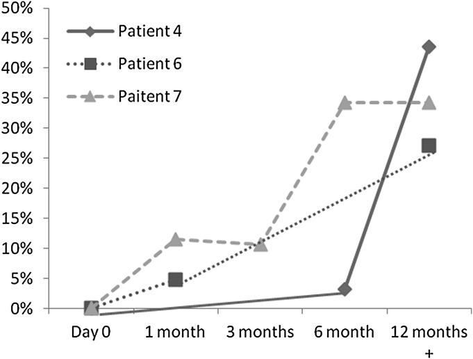
Patients were followed up at a median time of 8.6 years (range, 2.2–15.0 years) based on the last follow-up (Table 3). All surviving patients were well, infection free, and leading a good quality of life. One patient had been discharged from follow-up 15 years post-SCT. Chimerism studies demonstrated donor engraftment at 21 days, 6 months, 1 year, and 2 years after SCT (Table 4). None of the surviving patients required immunological replacement 1 year after SCT, and all had been given hepatitis B and measles, mumps, and rubella vaccinations with good response in specific IgG levels 2 years after SCT. IgG levels measured at 1 year from SCT for those with HIgM syndrome and SCID improved from pretransplant (Table 4). For patient 2 with CGD, the nitroblue tetrazolium test also improved from 6% (pretransplant) to 50% (22 days after transplant). Patient 4, 6, and 7 with HIgM syndrome secondary to CD40 ligand deficiency, and there were increased CD40 ligand levels after SCT (Fig. 1).
TABLE 4.
Summary of Chimerism Studies Demonstrating Donor Engraftment

FIGURE 1.

CD40 ligand expression (percentage positive on stimulated CD3 cells) in patients with HIgM syndrome (CD40 ligand deficiency).
DISCUSSION
Although SCTs have been considered the definitive treatment for PIDs, studies continue to be performed to demonstrate efficacy and determine what factors may improve survivability. Advances in conditioning regimens have allowed the survivability rates for matched related donor and matched unrelated donor to be almost on par, 69 to 100% compared with 42 to 81%, respectively. The survival improved with earlier timing of the transplant and varied with the type of PID being treated (SCID having a higher survivability compared with other PIDs).1,7
Although it has been demonstrated by Buckley2 that overall survival for SCTs improves greatly if performed before the patient is 3.5 months old for patients with SCID, most studies have been unable to perform a SCT within that time frame.6,8,9 Our center was no exception with 1 patient diagnosed with SCID at 4 months and entering transplant at 5 months of age. Patients with other PIDs were transplanted at a median age of 63.5 months (range, 5–204 months). Our median age for diagnosis was also quite late, at a median of 35 months (range, 4–68 months). Consequently, we had a high rate (4 of 7 patients) for pulmonary infections pretransplant among our patients, a factor associated with reduced survival. Although our patient numbers were small, we had 1 fatality out of 7 subjects who had significant infections before transplant. This further emphasizes the need for early diagnosis of PIDs and early transplantation for improved outcomes.
CONCLUSIONS
Our small series of patients adds to the experience of others and demonstrates the importance of SCT in severe PIDs as a curative strategy. Despite significant pre-SCT morbidity from the underlying PID in our patients (eg, systemic CMV infection, pneumonias, and bronchiectasis), the outcome of our patients, with the exception of 1 death, was satisfactory.
REFERENCES
- 1.Dvorak CC, Cowan MJ. Hematopoietic stem cell transplantation for primary immunodeficiency disease. Bone Marrow Transplant. 2008;41:119–126. doi: 10.1038/sj.bmt.1705890. [DOI] [PubMed] [Google Scholar]
- 2.Buckley RH. Transplantation of hematopoietic stem cells in human severe combined immunodeficiency: longterm outcomes. Immunol Res. 2011;49:25–43. doi: 10.1007/s12026-010-8191-9. [DOI] [PMC free article] [PubMed] [Google Scholar]
- 3.Gennery AR, Cant AJ. Advances in hematopoietic stem cell transplantation for primary immunodeficiency. Immunol Allergy Clin North Am. 2008;28:439–456. doi: 10.1016/j.iac.2008.01.006. [DOI] [PubMed] [Google Scholar]
- 4.Grunebaum E, Mazzolari E, Porta F, Dallera D, Atkinson A, et al. Bone marrow transplantation for severe combined immune deficiency. JAMA. 2006;295:508–518. doi: 10.1001/jama.295.5.508. [DOI] [PubMed] [Google Scholar]
- 5.Petrovic A, Dorsey M, Miotke J, Shepherd C, Day N. Hematopoietic stem cell transplantation for pediatric patients with primary immunodeficiency diseases at All Children's Hospital/University of South Florida. Immunol Res. 2009;44:169–178. doi: 10.1007/s12026-009-8111-z. [DOI] [PubMed] [Google Scholar]
- 6.Bhattacharya A, Slatter MA, Chapman CE, Barge D, Jackson A, et al. Single centre experience of umbilical cord stem cell transplantation for primary immunodeficiency. Bone Marrow Transplant. 2005;36:295–299. doi: 10.1038/sj.bmt.1705054. [DOI] [PubMed] [Google Scholar]
- 7.Porta F, Forino C, De Martiis D, Soncini E, Notarangelo L, et al. Stem cell transplantation for primary immunodeficiencies. Bone Marrow Transplant. 2008;41(suppl 2):S83–S86. doi: 10.1038/bmt.2008.61. [DOI] [PubMed] [Google Scholar]
- 8.Sato T, Kobayashi R, Toita N, Kaneda M, Hatano N, Iguchi A, et al. Stem cell transplantation in primary immunodeficiency disease patients. Pediatr Int. 2007;49:795–800. doi: 10.1111/j.1442-200X.2007.02468.x. [DOI] [PubMed] [Google Scholar]
- 9.Tsuji Y, Imai K, Kajiwara M, Aoki Y, Isoda T, et al. Hematopoietic stem cell transplantation for 30 patients with primary immunodeficiency diseases: 20 years experience of a single team. Bone Marrow Transplant. 2006;37:469–477. doi: 10.1038/sj.bmt.1705273. [DOI] [PubMed] [Google Scholar]


