Abstract
Multiple biliary hamartomas (MBH), also known as von Meyenberg complexes, are rare benign malformations of the intrahepatic bile ducts. It is thought that MBH result from ductal plate malformations involving the small interlobular bile ducts. Proliferation of bile ducts’ cuboidal epithelium in fibrous stroma subsequently leads to duct dilation. Macroscopically, MBH appear as well-defined subcapsular or parenchymal grayish-white nodules less than 0.5 cm in diameter without true capsulation in an abundant fibrous stroma. MR imaging is considered the best imaging modality for demonstrating MBH. We herein reported MR imaging and MR cholangiopancreatography of a patient with typical MBH appearance. Multiple tiny T1 hypointense and T2 hyperintense lesions of uniform distribution in the hepatic parenchyma were revealed. At MR cholangiopancreatography, the lesions were also hyperintense. Maximum intense projection (MIP) of MR cholangiopancreatography showed uniformly distributed hyperintense lesions appearing as ‘‘starry sky’’ configuration.
Key Words: Multiple biliary hamartomas, magnetic resonance imaging, cholangiopancreatography
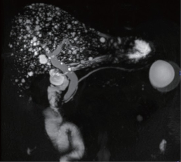
A 57-year-old male presented to our hospital due to recurrent unspecific abdominal pain for several years. Physical examinations were unremarkable. Laboratory examinations were normal except slight elevation of bilirubin 27 mmol/L (normal range, 1.7-21 mmol/L) and alanine aminotransferase (ALT) 51 U/L (normal range, 0-40 U/L). MR imaging depicted multiple small cysts of hypointense on T1 weighted images (Figure 1 A) and hyperintense on T2 weighted images (Figure 1 B), which were of diffused distribution in the parenchyma of the liver. Maximum intense projection (MIP) of MR cholangiopancreatography demonstrated small cysts scattered uniformly within contour of the liver to form “starry sky” configuration (Figure 2). A diagnosis of multiple biliary hamartomas was suggested due to the typical MR imaging features.
Figure 1.
A: T1 weighted MR image shows multiple small hypointense round lesions scattered in the liver; B: T2 weighted MR image demonstrates these lesions are hyperintense
Figure 2.

MIP of MR cholangiopancreatography depicts “starry sky” configuration as these cysts uniformly distributed in the liver
Acknowledgements
Disclosure: The authors declare no conflict of interest.


