Abstract
Digital subtraction angiography (DSA) is currently the gold standard for diagnosing the residue or recurrence of aneurysm after treatment, especially in the presence of metal coils. However, DSA is an invasive procedure which may cause additional trauma and economic burden to patients. Spectral CT imaging, as a newly introduced CT imaging mode, produces monochromatic image sets that is able to reduce beam-hardening and other metal-related artifacts, and has found its use in several clinical applications including brain imaging to reduce beam-hardening artifacts. In this study, we describe a case of spectral CT imaging in follow-up of the metal coils treatment and detection of a small leaf of residual aneurysm after metal coils treatment.
Key Words: Residual aneurysm, DSA, spectral CT
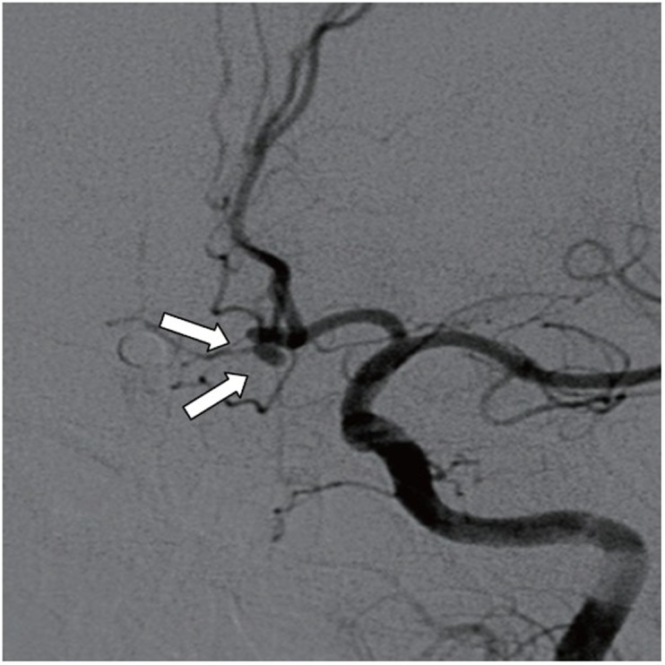
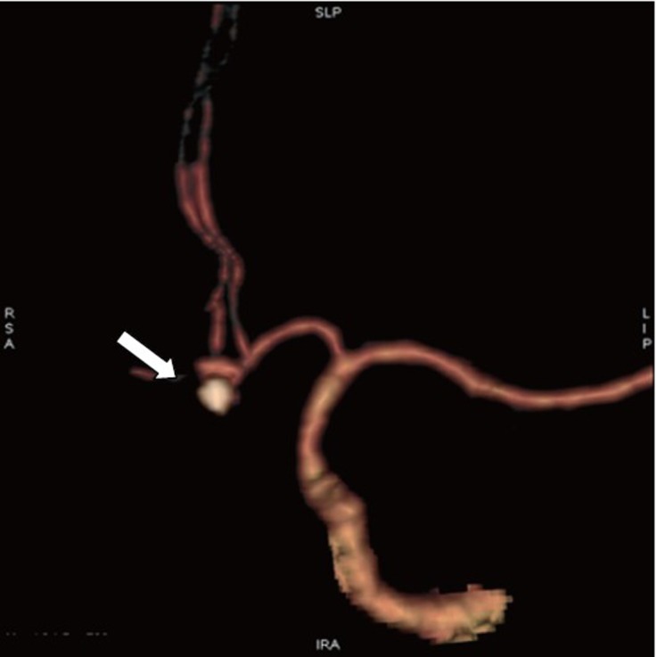
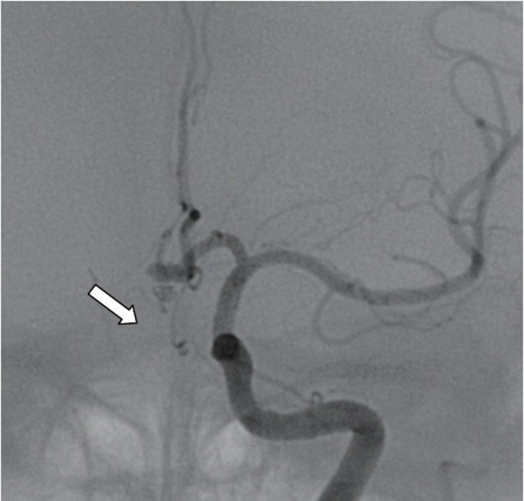
A 48-year-old woman presented to our hospital with a headache lasting for 22 days. A conventional head CT imaging indicated a subarachnoid hemorrhage. One week later, digital subtraction angiography (DSA) revealed a lobulated aneurysm existing in the anterior communicating artery (Figure 1). The diameter of the neck of the aneurysm was about 2.98 mm, and the size of aneurysm was about 4.33 mm × 2.54 mm (Figure 1). The patient refused a surgical clipping of the aneurysms, therefore embolization was performed. Because the neck of the lobulated aneurysm was relatively broad and the coil could not be stabilized, a small leaf of the aneurysm was left. Six months later, a spectral CT angiography was performed. The monochromatic images at 100 keV photon energy obtained with spectral CT indicated that the metal coils in the bigger aneurysm were still intact and in place with no metal artifacts in the vicinity of the metal coils. The residual small aneurysm could also be clearly displayed (Figure 2). The follow-up DSA reaffirmed the existence of the small residual aneurysm (Figure 3). The size and pattern of the residual aneurysm were consistent between the DSA and spectral CT images.
Figure 1.

DSA shows an anterior communicating artery lobulated aneurysm
Figure 2.

Follow-up spectral CT angiography shows the metal coil for the relatively bigger lobulated aneurysm is in place, the remnants of a small leaf of aneurysm and its wide neck are clearly visible, and there is no metal artifact in the vicinity of it
Figure 3.

DSA reaffirms the existence of a small aneurysm and the patterns are consistent with the findings of the spectral CT angiography
Conventional CT scans have not been routinely used in reviewing the aneurysm which is filled with the metal coils because the hyperdense metal artifacts could interfere with the observer’s views. On the other hand, spectral CT, with its ability to generate a set of monochromatic images ranging from 40 to 140 keV and incorporate advanced metal artifact reduction sequence (MARS), can eliminate beam-hardening artifacts and minimize metal artifacts. Spectral CT effectively eliminated the radial metal artifacts by the metal coils in the aneurysm and clearly show the fine structure around the metal. As the spectral CT is introduced to clinical application, this new scanning method might become a new alternative to DSA in evaluating aneurysm after metal coils treatment.
Acknowledgements
The authors wish to thank Dr. Li Jianying for his technical support in understanding the dual energy spectral CT imaging mode and editing the manuscript. We thank Dr. Xin Zhen, Dr. Biyun Xu, Dr. Yun Shen, Na Gao and Dr. Jia Wang for technical assistance. Dr. Li Zhang and Ling Zhang for spiritual power and encouragement.
Disclosure: We would like to submit the enclosed manuscript entitled “Residual aneurysm after metal coils treatment detected by spectral CT”, which we wish to be considered for publication in “Quantitative Imaging in Medicine and Surgery”. No conflict of interest exits in the submission of this manuscript, and manuscript is approved by all authors for publication. I would like to declare on behalf of my co-authors that the work described was original research that has not been published previously, and not under consideration for publication elsewhere, in whole or in part. All the authors listed have approved the manuscript that is enclosed.


