Abstract
de Quervain’s disease is a commonly encountered problem; its management is multimodal, and often, there is recurrence which is commonly associated with anatomical variation in the first dorsal compartment of the wrist. Our purpose was to find out the anatomical variation of the first dorsal compartment of the wrist in the general population to assess the anatomical basis of de Quervain’s disease and its recurrence. In this cadaveric study, 86 wrists in 46 patients were dissected to search out the first dorsal compartment of the wrist and its content tendons, presence of septa in the compartment, and insertion of the tendons. Supernumerary tendons in the first dorsal compartment were seen in 74.41 % of cases. The most commonly found tendon arrangement was two abductor pollicis longus (APL) and one extensor pollicis brevis (EPB). In all cases, there was a fixed insertion of APL to the base of the first metacarpal. Among other sites, the most common site of insertion of APL is the trapezium, which was 56.14 %. Variations of EPB with respect to number, site of insertion, thickness, and bilaterality were also found. The presence of septations was found in 37.20 % of dissected cadaveric wrists. We had found supernumerary tendons or slips in the first dorsal compartment very commonly. The presence of a septum was less frequently found. So, it may be concluded that there is immense anatomical variation present in the first dorsal compartment of the wrist, supernumerary tendons/tendon slips are commonly found, there is a variation of insertions present in the population, septum/aberrant compartment are also present, and bilateral variations are present in the population. These variations may be responsible for recurrence and unilateral affection in de Quervain’s disease.
Electronic supplementary material
The online version of this article (doi:10.1007/s12593-012-0073-z) contains supplementary material, which is available to authorized users.
Keywords: First dorsal compartment of the wrist, Abductor pollicis longus, Extensor pollicis brevis, Septum, Aberrant compartment
Introduction
Stenosing tenosynovitis of the first dorsal compartment of the wrist or de Quervain’s disease is a commonly encountered debilitating condition of the wrist. The common mode of treatment is conservative, but recurrence is seen in 15–20 % of cases in this management. Several factors are responsible for the recurrence. Literature suggests that the anatomical variations in the first dorsal compartment of the wrist like supernumerary tendon slips, aberrant compartments or presence of septa, and variation of insertion of tendon slips are encountered to be important causative factors for this problem [1–10]. Abnormal septation of the compartment was first reported by Finkelstein in 1930 [8, 9].
Many anatomical and clinical studies [3–20] were showing the variations of the first dorsal compartment of the wrist and its contents, but a specific relation among the variations was not established. The variations of insertion of the abductor pollicis longus which underscore the potential that extrafriction might have some role in the etiology of de Quervain’s Disease [18]. But, detailed information about the insertions is lacking to establish a definite relationship. To predict the recurrence of de Quervain’s disease, it is important to have clear knowledge about the presence of aberrant tendons and aberrant compartment and their relation.
We had done a cadaveric study of 86 specimen wrists. The purpose of our study was to find out the anatomical variation in the general population of India and to provide detailed information of the contents of the first dorsal compartment of the wrist that will help to access the anatomical basis of de Quervain’s disease so that it may help in the treatment plan.
Method
This cadaveric study was performed in cadavers that had been posted for postmortem examination in the Department of Forensic and State Medicine of Burdwan Medical College, Burdwan, from June 2010 to May 2011 after taking proper permission and ethical clearance from the authorities. Only unknown cadavers and death due to road traffic accidents were examined. Any wrist that was damaged by injury was excluded. We had performed all the dissections under supervision of faculty members of Forensic Medicine. Eighty-six cadaveric wrists were studied and searched for the presence of septation in the first dorsal compartment of the wrist and the relationship of tendons and there insertions.
Extra/aberrant tendons were defined as those which had separate definite origin and separate insertion. Additional tendon slips were part of tendons having a separate insertion site. It was very much difficult to differentiate aberrant from additional tendon slips in wrist dissection in our study, as the origin was not studied. So, aberrant tendons were counted as tendon slips. Tendons and tendon slips of the first dorsal compartment of the wrist were divided functionally into two groups. Tendons and tendon slips that move with first metacarpophalangeal joint movement were considered in extensor pollicis brevis (EPB), and other tendons or slips were considered as abductor pollicis longus (APL). Septation of the first extensor compartment of the wrist was defined as any division between the tendons or slips extending from the extensor retinaculum to the periosteum of the radius.
We had dissected 86 wrists. Bilateral dissection was done in 40 cadavers (80 wrists) and unilateral dissection was done in six wrists (due to damage to the other hand in road traffic accident). So, we examined 44 right and 42 left wrists.
Result
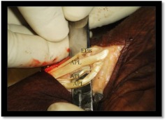
The findings of our study were recorded, analyzed, and shown in the tables and pie charts. We had found 22 wrists having two tendons in first dorsal compartment of wrist. In them one had two APLs and no EPB and the others had one APL with one EPB. Supernumerary tendons were commonly found (Fig. 1). The most common pattern found in our study was two APLs and one EPB in 48 cases (Table 1).
Fig. 1.
Radial view of the left wrist of a cadaver showing supernumerary tendons in the first dorsal compartment of the wrist joint
Table 1.
Pattern of tendon slips in the first dorsal compartment of the wrist
| No. of EPBs | 1 EPB | 2 EPBs | Total | |||||||
|---|---|---|---|---|---|---|---|---|---|---|
| 1 APL | 2 APLs | 1 APL | 2 APLs | 3 APLs | 4 APLs | 1 APL | 2 APLs | 3 APLs | ||
| Number of wrists | 0 | 1 | 21 | 48 | 2 | 1 | 12 | 1 | 0 | 86 |
| Number of wrists with septation | 0 | 0 | 5 | 19 | 2 | 0 | 5 | 1 | 0 | 32 |
APL abductor pollicis longus, EPB extensor pollicis brevis
The insertion of the APL was in the radial side of the first metacarpal base in all cases. In our study, among other sites, the most common site of insertion of the APL tendon slips was the trapezium (56.14 %, 32 in 143 tendons or tendon slips), and it was mostly found if there were two tendon slips of APL present. It was found that if there was a single APL tendon, then they were inserted in the base of the first metacarpal (Table 2 and Chart 1).
Table 2.
Pattern of insertion of abductor pollicis longus
| Insertion sites | 1 APL | 2 APLs | 3 APLs | 4 APLs | Total |
|---|---|---|---|---|---|
| Radial side, 1st Mc base | 33 | 50 | 2 | 1 | 86 |
| In APB | 0 | 16 | 1 | 1 | 18 |
| In trapezium | 0 | 29 | 2 | 1 | 32 |
| Opponens pollicis | 0 | 4 | 1 | 0 | 5 |
| CMC joints | 0 | 1 | 0 | 1 | 2 |
| Total | 33 | 100 | 6 | 4 | 143 |
APL abductor pollicis longus, Mc metacarpal, APB abductor pollicis brevis, CMC carpometacarpal
EPB was numerically symmetrical in 62.5 % of cadavers in which data of both sides were taken. The presence of a septum was symmetrical in both sides in 67.5 % (27 in 40 cases). In most of the cases, EPB tendon slips were inserted in the first phalanx. In most cases, there was one EPB (Table 3).
Table 3.
Pattern of insertion of extensor pollicis brevis
| 1 EPB | 2 EPBs | Total | |
|---|---|---|---|
| Proximal phalanx | 66 | 13 | 79 |
| Distal phalanx | 6 | 8 | 14 |
| Both | 0 | 5 | 5 |
| Total | 72 | 26 | 98 |
EPB extensor pollicis brevis
Thirty-two of 86 wrists had septations. We found in most of the cadaveric wrists that EPB tendon slips were placed deeper in the compartment and they were mostly encountered in an aberrant compartment. APB tendon slips were placed superficially. Most of the septations were obliquely directed. Septations were commonly bilateral (28 in 40 cases) (Table 4).
Table 4.
Relation of presence of septation with side orientation
| No. of cadavers | Only right (4) | Only left (2) | Bilateral cases (40) | ||
|---|---|---|---|---|---|
| Unilateral | Bilateral | ||||
| Septations present | 1 | 1 | Rt | Lt | Rt + Lt |
| 1 | 1 | 28 | |||
Rt right, Lt left
Discussion
We had found supernumerary tendons in the first dorsal compartment of the wrist in 74.41 % of cases. Other studies are corroborating this [2, 9–11]. So, it can be counted as a rule rather than an exception, and its mere presence is not responsible for the recurrence of de Quervain’s disease. The most commonly found tendon arrangement was two APLs and one EPB. So, finding two tendons in the compartment after release may not be indicating complete release.
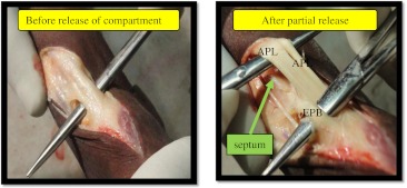
Septums were infrequent, so their presence should be analyzed for recurrence. The presence of septations was seen in 37.20 % of cadavers in our study. It is nearly similar to the study of Lomis [9] and Leslie [20]. Multiple tendon slips and septum were found in a high proportion of cases. So, release of the first extensor compartment and the finding of tendons do not ensure complete release. To ensure complete release, it should reach up to the periosteum of the radius and release all the components of the compartments, so that these can be pulled freely (Fig. 2). In conservative management by steroid injection, it has been suggested that both subcompartments should be injected to prevent recurrence [20, 21].
Fig. 2.
Radial view of the wrist showing tendons present in the compartment before and after partial release
There was variation of insertion of APL that may have an effect in increasing the tendon volume and producing potential extra friction. APL tendon slips were inserted in the first metatarsal base in all the cases. Among other sites of insertion, they were seen most commonly to insert into the trapezium, followed by merging with abductor pollicis brevis (Fig. 3). Different tendinous insertions of the APL have different innervation patterns that produce more friction as described by Dos Remédios et al. [18]. Commonly associated insertion in the trapezium may be associated with trapeziometacarpal arthritis as seen in previously done studies [11, 12].
Fig. 3.
Different sites of insertion of APL
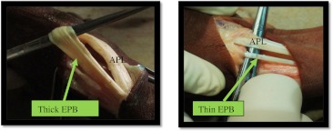
We found in most of the cases that EPBs were in deeper and medial compartment. As it moves more than APL during thumb movement, it may be responsible for more friction. The EPB compartment often causes failed injection and surgical release as it is smaller and deeper in location [15, 16]. EPB tendon slips had a different diameter in our study; its thickness may be responsible for increase volume and cause recurrence (Fig. 4). Previously, it was not studied. The numbers of EPB tendon slips were symmetrical in 62.5 % of cases, so there is chance of asymmetry in 37.5 %. This difference in number and thickness may be responsible in determining laterality (unilateral or bilateral) and recurrence in one hand following treatment.
Fig. 4.
Different diameters of EPB
We had found variation of tendon arrangement and their insertion and presence of septa when comparing both sides in bilateral cases. They may be responsible for recurrence in one hand after injection though the other hand becomes cured. Bilateral variation was not studied previously.
So, it is concluded that there is immense anatomical variation present in the first dorsal compartment of the wrist, supernumerary tendons/tendon slips are commonly found, there is variation of insertion present in the population, septum/aberrant compartment is also present, and bilateral variations are present in the population. These variations are probably responsible for recurrence and unilateral affection in de Quervain’s disease. Ultrasound studies can detect the variation with high sensitivity and specificity [13], so we should have clear anatomical knowledge of region. The availability of multiple tendons or slips makes this compartment a source of tendons in a tendon transfer procedure [17]. The boon of our study is due to its cadaveric design. Our study does not involve formalin-fixed cadavers (this causes tissue shrinkage and could be problematic for measurement of anatomical structures). A limitation of our study was that a wider dissection of the muscles to show the origin cannot be done for medicolegal purpose.
Electronic supplementary material
(DOC 234 kb)
References
- 1.Anson BJ. An atlas of human anatomy. 2. Philadelphia: Saunders; 1963. [Google Scholar]
- 2.Baba MA. The accessory tendon of the abductor pollicis longus muscle. Anat Rec. 1954;119:541–547. doi: 10.1002/ar.1091190410. [DOI] [PubMed] [Google Scholar]
- 3.Bahm J, Szabo Z, Foucher G. The anatomy of de Quervain's disease. A study of operative findings. Int Orthop. 1995;19(4):209–211. doi: 10.1007/BF00185223. [DOI] [PubMed] [Google Scholar]
- 4.Bouchlis G, Bhatia A, Asfazadourian H, Touam C, Vacher C, Oberlin C. Distal insertions of abductor pollicis longus muscle and arthritis of the first carpometacarpal joint in 104 dissections. Ann Chir Main Memb Super. 1997;16(4):326–338. doi: 10.1016/S0753-9053(97)80047-6. [DOI] [PubMed] [Google Scholar]
- 5.Brown GD, 3rd, Roh MS, Strauch RJ, Rosenwasser MP, Ateshian GA, Mow VC. Radiography and visual pathology of the osteoarthritic scaphotrapezio-trapezoidal joint, and its relationship to trapeziometacarpal osteoarthritis. J Hand Surg. 2003;28(5):739–743. doi: 10.1016/S0363-5023(03)00258-2. [DOI] [PubMed] [Google Scholar]
- 6.Brunelli GA, Brunelli GR. Anatomical study of distal insertion of the abductor pollicis longus. Concept of a new musculo-tendinous unit: the abductor carpi muscle. Ann Chir Main Memb Super. 1991;10(6):569–576. doi: 10.1016/S0753-9053(05)80330-8. [DOI] [PubMed] [Google Scholar]
- 7.Eaton RG, Littler JW. Ligament reconstruction for the painful thumb carpometacarpal joint. J Bone Joint Surg. 1973;55(8):1655–1666. [PubMed] [Google Scholar]
- 8.Finkelstein H. Stenosing tendovaginitis at the radial styloid process. J Bone Joint Surg. 1930;12:509–540. [Google Scholar]
- 9.Lomis LK. Variations of stenosing tenosynovitis at the radial styloid process. J Bone Joint Surg. 1951;33-A:340–346. [PubMed] [Google Scholar]
- 10.Muckart RD. Stenosing tendovaginitis of abductor pollicis longus and extensor pollicis brevis at the radial styloid (de Quervain's disease) Clin Orthop. 1964;33:201–208. [PubMed] [Google Scholar]
- 11.Zancolli EA. The trapeziometacarpal joint. Tenotomy of the accessory tendons in early osteoarthritis. Hand Clin. 2001;17(1):13–43. [PubMed] [Google Scholar]
- 12.Saehle T, Sande S, Finsen V. Abductor pollicis longus tendon interposition for arthrosis in the fi rst carpometacarpal joint: 55 thumbs reviewed after 3 (1–5) years. Acta Orthop Scand. 2002;73(6):674–677. doi: 10.1080/000164702321039660. [DOI] [PubMed] [Google Scholar]
- 13.Rousset P, Vuillemin-Bodaghi V, Laredo JD, Parlier-Cuau C. Anatomic variation in the first extensor compartment of the wrist: accuracy of US. Radiology. 2010;257(2):427–433. doi: 10.1148/radiol.10092265. [DOI] [PubMed] [Google Scholar]
- 14.Yuasa K, Kiyoshige Y. Limited surgical treatment of de Quervain's disease: decompression of only the extensor pollicis brevis subcompartment. J Hand Surg [Am] 1998;23(5):840–843. doi: 10.1016/S0363-5023(98)80160-3. [DOI] [PubMed] [Google Scholar]
- 15.Zingas C, Failla JM, Holsbeeck MV. Injection accuracy and clinical relief of de Quervain's tendinitis. J Hand Surg. 1998;23(1):89–96. doi: 10.1016/S0363-5023(98)80095-6. [DOI] [PubMed] [Google Scholar]
- 16.Nayak SR, Hussein M, Krishnamurthy A, et al. Variation and clinical significance of extensor pollicis brevis: a study in South Indian cadavers. Chang Gung Med J. 2009;32(6):600–604. [PubMed] [Google Scholar]
- 17.Cho KO. Translocation of the abductor pollicis longus tendon. A treatment for chronic subluxation of the thumb carpometacarpal joint. J Bone Joint Surg Am. 1970;52(6):1166–1170. [PubMed] [Google Scholar]
- 18.Remédios C, Chapnikoff D, Wavreille G, Chantelot C, Migaud H, Fontaine C. The abductor pollicis longus: relation between innervation, muscle bellies and number of tendinous slips. Surg Radiol Anat. 2005;27(3):243–248. doi: 10.1007/s00276-004-0286-3. [DOI] [PubMed] [Google Scholar]
- 19.Bianchi S, Martinoli C, Abdelwahab IF. High-frequency ultrasound examination of the wrist and hand. Skeletal Radiol. 1999;28(3):121–129. doi: 10.1007/s002560050488. [DOI] [PubMed] [Google Scholar]
- 20.Leslie BM, Ericson WB, Jr, Morehead JR. Incidence of a septum within the first dorsal compartment of the wrist. J Hand Surg [Am] 1990;15(1):88–91. doi: 10.1016/S0363-5023(09)91111-X. [DOI] [PubMed] [Google Scholar]
- 21.Sawaizumi T, Nanno M, Ito H. De Quervain's disease: efficacy of intra-sheath triamcinolone injection. Int Orthop. 2007;31(2):265–268. doi: 10.1007/s00264-006-0165-0. [DOI] [PMC free article] [PubMed] [Google Scholar]
Associated Data
This section collects any data citations, data availability statements, or supplementary materials included in this article.
Supplementary Materials
(DOC 234 kb)


