Abstract
Distal radius fractures are a common disorder in industrialized nations associated with osteoporosis, with a reported incidence of two fractures per thousand patients per year. We performed a retrospective study comparing two sets of 40 patients, with fracture of the distal radius treated with Penning external fixator, compared to 40 patients treated with fixed-angle volar-locking plate (Plate Depuy ® DVR), with the objective of finding differences between both treatment methods in anatomical values, functional outcomes and complication rates. All fractures were classified according to the AO classification. Postero-anterior and lateral radiographs of the wrist were taken after fracture, after surgery and at 6 months after surgery. We also assessed functional outcome. Minimum follow up was of 10 months. We compared complications between both groups. In the group of patients treated with fixed-angle volar-locked plate, radiological results are found to be closer to the anatomical references. Final outcomes revealed similar functional scores between both groups. The complications rate was statistically higher in the group of patients who underwent external fixation. In the fixed-angle volar-locked plate group, most of complications were related to patient discomfort due to the volar-locking plate.
Keywords: External-fixator, Radius, Plate, Fracture, Complications
Introduction
In the last century there is a progressive increase in the average age of the population in industrialized nations, and it is logical to see a rise in the prevalence of distal radius fractures. This added to the bone characteristics of this aging population group and a collapse in three dimensions leading to loss of radial inclination, radial height, palmar tilt and increase in ulna variance and the increased complexity of the fractures, which could be a result of higher energy trauma, makes distal radius fractures complicated to treated. Fractures of the distal radius are currently the most prevalent osteoporotic fracture [1]. There are many treatment options for these injuries. Since the introduction of distal radius fixed-angle volar-locking plates, there is a growing trend in the use of them for the treatment of these fractures, although most of these fractures are still being treated conservatively [2].
We conducted a retrospective analysis of two groups of patients with distal radius fracture, comparing a group treated using a Penning dynamic external fixator with or without Kirschner wires, with a group treated using fixed angle volar locking plates (DVR ® Anatomic Volar Plating System, DePuy Companies). The objective of this paper is to see if there is any difference in the radiological (anatomic) and functional outcomes, and the complication rate.
Patients and Methods
Between 2004 and 2009, 520 patients with distal radius fractures were treated surgically (most of them by closed reduction and Kirschner wires), of which 113 were suitable for this study. The inclusion criteria were fractures with more than 2 mm of articular incongruity, those whose reduction could not be maintained by closed methods or open fractures. Of the 113 patients, 33 were rejected because of incomplete data, short duration of follow-up, or unwillingness to participate in the study. Most of the excluded patients, were due to missing or poor quality radiographs. As a result, there were 40 fractures in each of the groups treated with Penning External Fixator (Pennig, Orthofixs), (EF), and fixed angle volar locking late (Depuy ® DVR Board) (DVR).
In the emergency department an initial closed reduction was performed in all patients. Surgery was performed when possible. Postoperative, the external fixator was dinamized 40 (35–64) days after the surgery, and maintained 60 (49–98) days. Patients treated in the DVR group were immobilized for 14 (8–21) days with a cast, and then mobilization was allowed.
The average age of the patients were 45 years in the EF (range 17–77) group compared to 52 years (range 17–79) in the DVR group. All patients in the EF group were operated within 24 h from the fracture but two patients, compared to a mean delay to take surgery of three days in the DVR group. There were 21 women and 19 men in the EF group and 24 women and 16 men in the DVR group. Patients were classified according to the AO classification. (Table 1)
Table 1.
AO Classification distribution
| EF | A | B | C1 | C2 | C3 |
| EXELLENT | 0 | 3 | 4 | 5 | 0 |
| GOOD | 0 | 1 | 4 | 12 | 0 |
| REGULAR | 0 | 1 | 1 | 7 | 1 |
| POOR | 0 | 0 | 0 | 0 | 1 |
| DVR | A | B | C1 | C2 | C3 |
| EXELLENT | 2 | 0 | 2 | 4 | 0 |
| GOOD | 2 | 2 | 1 | 15 | 1 |
| REGULAR | 2 | 0 | 1 | 3 | 4 |
| POOR | 0 | 0 | 0 | 1 | 0 |
This table shows the distribution of fractures regard to the AO classification, in relation to the Lindström scale
The radiological evaluation of the fractures was done on admission, after surgery, at 6 weeks and finally at six months. It was made by simple true antero-posterior and lateral x-ray projections, where we measured volar tilt, radial inclination and ulnar variance. The two main authors made all measurements with a goniometer over plain radiographies [1, 3]. We accepted as anatomical values an average volar tilt of 11° angle with a range of 0° angle to 22° angle, the mean radial inclination as 23° angle with a range 19° angle to 29° angle, and ulnar variance as −0.9 mm with a range of −4.2 mm to 2.3 mm [4]. A Friedman statistical test was also used to show variations through the time (angular modifications and ulnar variance) in both groups.
We recorded those patients with a gap bigger than 2 mm in fractures type B and C in both groups.
Functional outcome was evaluated 10 months from the surgery by clinical Lindtröm scale that values function, symptoms, deformity and mobility by a grade scale (Table 2), and with the visual analogical scale (VAS) in both series at the end of follow-up [5]. The minimum follow up period was of 10 months, with an average of 15 months. All complications during the study were noted
Table 2.
Lindstöm scale
| Lindstöm scale | |
|---|---|
| Excellent | Function of the wrist unimpaired. No subjective symptoms. No deformity. Loss of dorsiflexion or palmar flexion not exceeding 15° accepted. |
| Good | Function of the wrist unimpaired. Negligible subjective symptoms. Deformity accepted if not producing subjective symptoms |
| Fair | Function of the wrist less satisfactory for activities requiring special strength or extreme movements, that must be avoided. Most pre-injury activities possible. Loss of motion, even if marked, accepted if not associated with subjective symptoms |
| Poor | Working capacity diminished or general way of life affected |
This table shows the explained Lindström scale
Results
Radiological outcomes in both groups during the study were noted in Table 3. We find changes in radiological values over the time, in both series, in particular in the ulnar variance, where we appreciate a progressive shortening of the radius through the follow-up period (p < 0,05).
Table 3.
Radiological outcomes
| EXTERNAL FIXATOR | |||
| RADIAL INCLINATION | VOLAR TILT | ULNAR VARIANCE | |
| FRACTURE | 10,6 | −11,5 | 3,0 |
| POSTOPERATIVE | 19,6 | −1,1 | 10,9 |
| 6 MONTHS | 18,3 | 0,7 | 10,0 |
| DVR | |||
| RADIAL INCLINATION | VOLAR TILT | ULNAR VARIANCE | |
| FRACTURE | 17,2 | 6,7 | 5,1 |
| POSTOPERATIVE | 20,0 | 9,5 | 1,0 |
| 6 MONTHS | 19,1 | 7,6 | 0,9 |
| ANATOMIC | 23 | 11 | −0,9 |
This table shows radiological average values in both groups, and in the bottom the considered anatomical values
Compared to anatomical values, there were no differences between groups in radial angulation and ulnar variance (p > 0,05), but there was a statistical difference (p < 0,05) in volar tilt for the DVR group (7,6°) compared to the EF group (0,8°) using the values taken as reference (11°) [4].
From the 40 patients included in the DVR group, only 34 had an AO B-C fracture (articular involvement), and from these. we only noted 1 patient with an articular gap of more than 2 mm, whereas in the EF group 7 patients had a postoperative gap higher than 2 mm.
We noted complications in 5 of the 40 patients from the DVR and 9 of the 40 in the EF group. In the DVR Group there were one case of Complex Regional Pain Syndrome I (CRPS I), there was an infection (treated with antibiotics) and there were needed 4 extractions of the volar-locking plate, 1 due to pain and 3 because of discomfort. Complications detected in the EF Group were four cases of CRPS I, four osteitis (an inflammatory reaction with no evidence of suppuration, and fast recovery with NAIDs), one patient with a painful scar, one lesion of the cutaneous branch of the radial nerve, and one patient suffered a fully loss of reduction (not acceptable with a volar tilt worse than the preoperative value, so another reduction was performed). One patient in the EF group developed a compartmental syndrome (diagnosed by disproportionate pain, pain with finger extension, and with an increase of compartment pressure measured with a the Stryker Intra-compartmental pressure monitor system. A fasciotomy of all of the compartments of the hand and forearm was performed to the patient). Analysis reveals that the rate of complication was significantly higher in the EF group (p < 0,05) (Table. 4).
Table 4.
Complications in both groups
| DVR | EF | |
|---|---|---|
| CRPS I | 1 | 4 |
| Osteitis | 0 | 4 |
| Radial cutaneous branch injury | 0 | 1 |
| Compartmental syndrome | 0 | 1 |
| Infection | 1 | 0 |
| Plate extraction | 4 | 0 |
| Scar complications | 0 | 1 |
| TOTAL COMPLICATIONS | 6 | 11 |
| PATIENTS WITH COMPLICATIONS | 5 | 9 |
This table illustrates all complications recorded in both groups
All of the complications had favourable resolution except the compartment syndrome that resulted in a claw hand with functional limitation.
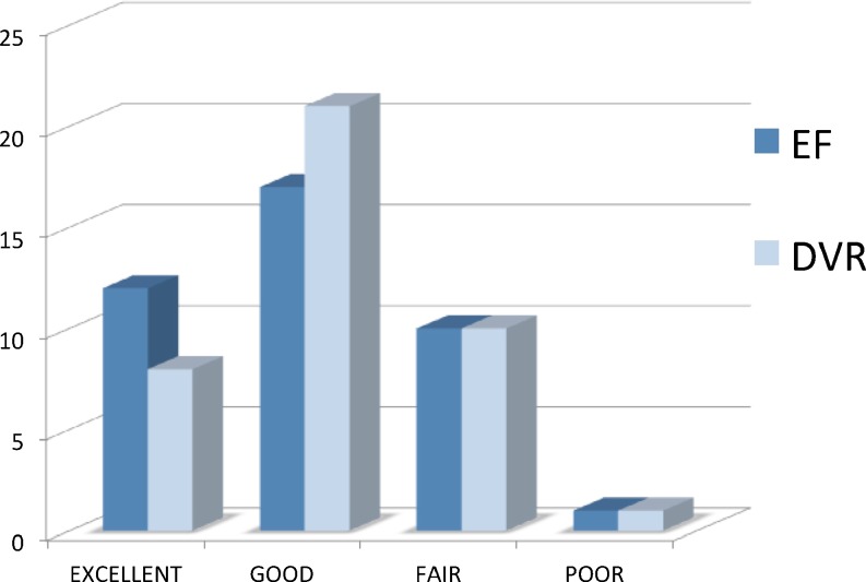
At the end of the study the clinical Lindtröm scale outcomes were: In the EF group were 12 excellent results, 17 good results, 10 fair results and there were 1 poor result (corresponding to the compartment syndrome). In the DVR group there were 8 excellent results, 21 good results, 10 fair results and 1 poor result (Fig. 1). We also recorded VAS outcome in both groups, with the same median value of 6/10. There were no significant differences between functional outcome scales at the end of the follow-up (p > 0,05 in both comparisons).
Fig. 1.
Lindström functional scale in both groups. This figure displays functional outcome
Discussion
There is currently moderate evidence that the surgical treatment of distal radius fractures has better outcomes in cases of open fractures, displaced fractures, angular and longitudinal collapsed, and in those fractures with joint steps more than 2 mm [1, 6, 9, 11]. The goal of surgical treatment is to achieve an anatomical reduction and promote early resumption of usual activities. Fixed-angle volar-locking plates and some nonbridging external fixators, give enough three dimension support for most distal radius fractures to begin early mobilization, which should justify a better score compared to other fixation techniques [1, 5, 6].
From a functional point of view in our study we haven’t observed differences between both groups, although there is scientific evidence that short-term outcomes in patients undergoing fixed-angle volar-locking plates have earlier recovery [8–10].
Both fixed-angle volar-locking plates and external fixators are supported techniques for the treatment of distal radius fractures susceptible for surgery [1, 11, 18]. There is a theoretical advantage in the use of fixed-angle volar-locking plates with regard to achieve a better anatomic reduction.
Since today there is no a correlation between anatomical reduction and functional outcomes in older patients. Is there also no correlation in young patients [1, 3, 7, 11–13]. We noted in our series radiological values closer to those anatomical in the DVR group, with better articular reduction, but without significance. Krishanan et al. in their series of intra-articular distal radius fractures treated with external fixator, show similar outcomes with statistically no difference between a bridging and non-bridging external fixator, with good range of movement in both groups, but with a significant grip strength loss [14].
Complications due to both surgical procedures are expected, and most when diagnosed, usually have a favourable ending. We found a higher rate of complications in fractures treated with external fixator. The most common complication in the group of patients treated with fixed-angle volar-locking plates was discomfort caused by the osteosynthesis material requiring removal. Have been described in patients treated with surgical with fixed-angle volar-locking plates median nerve damage and tendon injuries but we have not been registered similar complications in our serie [15]. In the case of the compartment syndrome in the group of patients treated with external fixation, some authors report a greater incidence in closed fractures treated with external fixation, although there is no scientific evidence of this relationship in distal radius fractures [16–18]. It is possible that an excess of traction added to a prolonged anaesthesia of the hand prevent from an early diagnose of the complication.
This analysis suggests that DVR is superior to EF with regard to complication rates and anatomical outcomes, but has not enough evidence to demonstrate functional superiority. This is also a retrospective study with small heterogeneous groups so prospective bigger randomized trials comparing the two groups would be necessary to find out functional differences between both groups.
References
- 1.Court-Brown ChM, Caesar M (2006) Epidemiology of adult fractures: a review. Injury 37(8):691–697, Epub 2006 Jun 30 [DOI] [PubMed]
- 2.Chung KC, Shauver MJ, Birkmeyer JD (2009 Aug) Trends in the United States in the treatment of distal radial fractures in the elderly. J Bone Joint Surg Am 91(8):1868–1873 [DOI] [PMC free article] [PubMed]
- 3.Othman AY (2009 May) Fixation of dorsally displaced radius fractures with volar plate. J Trauma 66(5):1416–1420 [DOI] [PubMed]
- 4.Schuind FA, Linscheid RL, An KN, Chao EY (1992 Oct) A normal data base of posteroanterior roentgenographic measurements of the wrist. Journal J Bone Joint Surg Am 74(9):1418–29 [PubMed]
- 5.Mirza A, Jupiter JB, Reinhart MK, Meyer P (2009) Fractures of the distal radius treated with cross-pin fixation and a nonbridging external fixator, the CPX system: a preliminary report. J Hand Surg Am 34:603–616 [DOI] [PubMed]
- 6.Windolf M, Schwieger K, Ockert B, Jupiter JB, Gradl G (2010) A novel non-bridging external fixator construct versus volar angular stable plating for the fixation of intra-articular fractures of the distal radius–a biomechanical study. Injury 41:204–209 [DOI] [PubMed]
- 7.Lindström A (1959) Fractures of the distal radius: a Clinical and statistical study of end results. Acta Orthop Scand 41(suppl):1–118 [PubMed]
- 8.Lichtman DM et al (2010) Treatment of distal radius fractures. J Am Acad Orthop Surg 18(3):180–189 [DOI] [PubMed]
- 9.Kapoor H, Agarwal A, Dhaon BK (2000) Displaced intra-articular fractures of distal radius: a comparative evaluation of results following closed reduction, external FIxation and open reduction with internal Fixation Injury. Int J Care Injured 31:75–79 [DOI] [PubMed]
- 10.Margaliot Z, Haase SC, Kotsis SV, Kim HM, Chung KC (2005) A meta- analysis of outcomes of external fixation versus plate osteosynt- hesis for unstable distal radius fractures. J Hand Surg Eur 30:1185–1221 [DOI] [PubMed]
- 11.Cecilia D et al (1997) Intra-articular comminuted fracture of the distal radius treated by external fixation. Rev Ortp Traumatol 41(Supl 1):58–63, Spanish
- 12.Leung F et al (2008 Jan) Comparasion of external and percutaneous pin fixation with plate fixation for intra-articular distal radial fractures. A randomized study. J Bone Joint Surg Am 90(1):16–22 [DOI] [PubMed]
- 13.Stevenson I et al (2009 Sep) Displaced distal radial fractures treated using volar locking plates: maintenance of normal anatomy. J Trauma 67(3):612–616 [DOI] [PubMed]
- 14.Krishnan J, Wigg AE, Walker RW, Slavotinek J (2003) Intra-articular fractures of the distal radius: a prospective randomised controlled trial comparing static bridging and dynamic non-bridging external fixation. J Hand Surg Br 28:417–421 [DOI] [PubMed]
- 15.Berglund LM, Messer TM (2009) Complicactions of volar plate fixation for managing distal radius fractures. J Am Acad Orthop Surg 17:369–377 [DOI] [PubMed]
- 16.Egol KA, Bazzi J, McLaurin TM, Tejwani NC (2008) The effect of knee-spanning external fixation on compartment pressures in the leg. J Orthop Trauma 22(10):680–685 [DOI] [PubMed]
- 17.McQueen MM, Gaston P, Court-Brown CM (2000) Acute compartment syndrome. Who is at risk? J Bone Joint Surg Br 82(2):200–203 [PubMed]
- 18.Suárez L et al (2009) Functional and radiological outcomes in distal radius fractures treated with a volar plate versus an external fixator. Rev Ortp Traumatol 53:98–105, Spanish


