Abstract
The cytokinin group of plant hormones regulates aspects of plant growth and development, including the release of lateral buds from apical dominance and the delay of senescence. In this work the native promoter of a cytokinin synthase gene (ipt) was removed and replaced with a Cu-controllable promoter. Tobacco (Nicotiana tabacum L. cv tabacum) transformed with this Cu-inducible ipt gene (Cu-ipt) was morphologically identical to controls under noninductive conditions in almost all lines produced. However, three lines grew in an altered state, which is indicative of cytokinin overproduction and was confirmed by a full cytokinin analysis of one of these lines. The in vitro treatment of morphologically normal Cu-ipt transformants with Cu2+ resulted in delayed leaf senescence and an increase in cytokinin concentration in the one line analyzed. In vivo, inductive conditions resulted in a significant release of lateral buds from apical dominance. The morphological changes seen during these experiments may reflect the spatial aspect of control exerted by this gene expression system, namely expression from the root tissue only. These results confirmed that endogenous cytokinin concentrations in tobacco transformants can be temporally and spatially controlled by the induction of ipt gene expression through the Cu-controllable gene-expression system.
The discovery of the plant hormone group the cytokinins and their involvement in aspects of plant growth and development, such as cell division (Skoog and Miller, 1957), delayed senescence (Richmond and Lang, 1957), and the release of lateral buds from apical dominance (Sachs and Thimann, 1964), has led to the attempted manipulation of these processes by altering the endogenous cytokinin content in plant tissues. Since the major gene(s) involved in cytokinin production in plants has not yet been isolated, a number of groups have utilized the ipt gene from the plant pathogenic bacterium Agrobacterium tumefaciens. This gene encodes the enzyme isopentenyl transferase, which catalyzes the rate-limiting step of the cytokinin biosynthetic pathway (Akiyoshi et al., 1984; Barry et al., 1984), in which Δ2-isopentenyl PPi is condensed with AMP to form isopentenyl AMP. The introduction of the ipt gene into the plant genome results in elevated levels of cytokinin in the transformed tissue (Smigocki and Owens, 1988; Beinsberger et al., 1991; Yusibov et al., 1991; McKenzie et al., 1994), together with associated morphological changes.
Plants transformed with highly expressing ipt genes produce shoots that do not elongate, show a severe lack of apical dominance, have small, rounded leaves, and are not able to form roots (Schmülling et al., 1989; Smigocki, 1991; Hewelt et al., 1994). Whole plants transformed with ipt genes with weaker or controlled expression also display the effects of cytokinin overproduction, the most consistent of these being the release of lateral bud growth from apical dominance (Medford et al., 1989; Smart et al., 1991; Smigocki, 1991; Van Loven et al., 1993; Hewelt et al., 1994; Faiss et al., 1997). This aspect of development is also inducible by exogenous cytokinin application (Sachs and Thimann, 1964). Additionally, a decrease in root production by ipt transformants has been repeatedly observed (Medford et al., 1989; Smigocki, 1991; Van Loven et al., 1993), and the leaves of ipt transformants exhibit delayed senescence (Smart et al., 1991; Li et al., 1992; Hewelt et al., 1994; Gan and Amasino, 1995; Faiss et al., 1997). Delayed leaf senescence has also been observed following the exogenous application of cytokinin (Richmond and Lang, 1957). Other reported effects of endogenous cytokinin overproduction include the production of the defense-related genes for extensin, chitinase, and PR1 (Memelink et al., 1987), and increased tuber formation from potato plants (Ooms and Lenton, 1985).
The altered morphology observed in plant tissues transformed with the native ipt gene has been clearly associated with a marked increase in cytokinin content (Budar et al., 1986; Smigocki and Owens, 1988; Beinsberger et al., 1991; Yusibov et al., 1991; McKenzie et al., 1994). Additional work has been carried out using the CaMV 35S promoter to drive ipt gene expression (Smigocki and Owens, 1988, 1989). This showed that the CaMV 35S promoter increased ipt expression over that of the native promoter. However, the constitutive expression of the ipt gene, through either its native promoter or the CaMV 35S promoter, prevented normal plant development, thereby precluding the study of cytokinin overexpression within the whole plant.
To study the effects of cytokinin overproduction on normal plant tissue, a number of groups have linked regulatable promoters to the ipt gene. The most commonly used promoters have been those regulated by heat shock, and several groups have obtained whole plants using such promoters to control ipt gene transcription (Medford et al., 1989; Schmülling et al., 1989; Smart et al., 1991; Smigocki, 1991; Van Loven et al., 1993). These plants were able to form normal roots but were often smaller and displayed a greater degree of axillary bud growth than control plants under both inductive and noninductive conditions. Hormone analyses indicated that even under non-heat-shock conditions transformed plants often contained higher levels of cytokinin than did control plants. When heat shock was carried out, cytokinin levels increased further, but this was not necessarily accompanied by further morphological changes (Medford et al., 1989; Smigocki, 1991). Thus, it seems that the heat-shock promoters allow sufficient expression from the ipt gene under noninductive conditions to alter plant morphology. Moreover, heat shock itself may affect plant growth, and plant treatment prior to heat shock may induce gene expression (Van Loven et al., 1993).
In moving away from the use of heat-shock promoters, a number of groups have utilized promoters that allow temporal or spatial gene expression. These have included promoters controlled by the external environment, e.g. light (Beinsberger et al., 1991), wounding (Smigocki et al., 1993), tetracycline (Redig et al., 1996; Faiss et al., 1997), and those related to a particular tissue or developmental state, such as fruit-specific (Martineau et al., 1994), hormone-specific (Li et al., 1992), or senescence-specific (Gan and Amasino, 1995) promoters. Generally, a higher level of control over ipt gene expression has been gained with these promoters than has been provided by the heat-shock promoters. However, in many cases cytokinin production still appears to be dependent on treatment type as well as on tissue type. In fact, Faiss et al. (1997) suggested that the cytokinins are active only in the tissue in which they are synthesized. Their conclusion is derived in part from grafting experiments in which they showed that there is no influence over apical dominance or senescence in wild-type tissue grafted onto transgenic rootstock. The use of a tightly controlled, inducible promoter activated specifically in the roots would not only avoid the possible compounding effect of the graft union but would also pinpoint the key areas of cytokinin control.
In the work reported here we controlled the expression of the ipt gene in tobacco (Nicotiana tabacum L. cv tabacum) using the Cu-inducible gene expression system (Mett et al., 1993, 1996). This system is activated directly by Cu, allowing the defined expression of genes to which it is linked, and has been shown to provide tight control over the expression of a GUS reporter gene, such that GUS expression occurred only from the root tissue of tobacco transformants in the presence of 50 μm CuSO4 (Mett et al., 1996). This method of control was considered ideal for supplying additional cytokinin to the plant, since increased cytokinin production in the roots might enhance the cytokinin supplied naturally to the other plant organs via the xylem. The provision of excess cytokinin in the appropriate physiological context could be expected to provide a particularly useful model system. When this system was used, ipt gene expression occurred in tobacco transformants in the presence of Cu2+ but did not occur in its absence. The controlled expression of the ipt gene resulted in increased cytokinin levels, the breaking of apical dominance, and delayed leaf senescence.
MATERIALS AND METHODS
PCR
Two primers were designed that flanked the coding and termination regions of a previously isolated ipt gene sequence (McKenzie et al., 1994). PCR was performed using a Perkin Elmer-Cetus kit according to the manufacturer's instructions.
Vector Construction
The vector pMMACEipt was constructed in three steps: (a) The promoterless ipt gene was cloned into the EcoRI/XbaI sites of the plasmid pUC119/4MT3 (Mett et al., 1996) following the four copies of the metal responsive element. (b) A second NotI site was introduced at the end of the ipt termination sequence allowing the NotI fragment (containing the metal-responsive elements and the promoterless ipt gene) to be cloned into the pACE-in-ART vector (Mett et al., 1996). (c) The NotI fragment was cloned into the NotI site of the binary vector pACE-in-ART, creating pMMACEipt. A similar vector, pGA643/ACE1.6/4MT-40GUS (Mett et al., 1996), was used for control transformations. This vector contained the GUS gene instead of the ipt gene.
Southern Analysis
Genomic DNA was prepared from the leaves of young, tissue-culture-grown tobacco (Nicotiana tabacum L. cv tabacum) using a cetyltrimethylammonium bromide method based on that described by Akama et al. (1992). The DNA was digested overnight with EcoRI, subjected to gel electrophoresis, and blotted to a Zeta Probe membrane (Bio-Rad) by capillary transfer (Southern, 1975). The membrane was probed with the promoterless ipt gene, which was labeled with [32P]dCTP (3000 Ci mmol−1).
Plant Transformation
Leaf discs of tobacco were transformed by co-cultivation with Agrobacterium tumefaciens (LBA4404) containing either pMMACEipt or pGA643/ACE1.6/4MT-40GUS, using standard protocols (Shillito and Saul, 1988). Plants were regenerated on solid Murashige-Skoog medium containing 150 mg L−1 kanamycin, but without CuSO4. Plants that produced a positive result in the neomycin phosphotransferase test (Herrera-Estrella and Simpson, 1988) were subcultured every 4 to 6 weeks and multiplied to produce clonal lines.
Gene Induction
Cu Treatment in Vitro
Tobacco transformants were treated with CuSO4 (to a final concentration of 5, 10, or 50 μm) via the application of a liquid stock solution to the growth medium. This was allowed to soak in and the plants were maintained, without subculture, while they were monitored for physiological change.
Cu Treatment in Vivo
A completely randomized design was used for the in vivo experiment with the treatment structure consisting of Cu2+ applied to two tobacco strains (Cu-GUS and Cu-ipt), each having two to four independently transformed lines and each line having four to seven clonal plants. The experimental unit was a single plant. Each plant was removed from the solid Murashige-Skoog medium and placed in sterilized pumice. The plants were grown during a 16-h photoperiod with a nominal photon flux density of 700 μmol m−2 s−1 and a day/night temperature regime of 24/21°C. They were watered when required with Murashige-Skoog medium and exposed to Cu2+ from the beginning of the experiment. Root tissue was removed throughout the experiment for GUS analysis. Thirty days after planting out, the following variables were measured for each plant: plant height, number of leaves, number of lateral buds (greater than 3 cm in length), length of lateral bud (measured from the base to the tip of the largest leaf), number of leaves in each lateral bud (not including those protecting the meristem), and position of each lateral bud (node number). The SAS Institute (Cary, NC) computer software package was used to fit a general linear model to each variable, and the effects due to strain and line (within each strain) were tested for significance.
Fluorogenic GUS Assay
Fluorogenic GUS assays were performed as described by Jefferson (1987). Protein content was determined according to the work of Bradford (1976), and GUS activity was expressed in picomoles per minute per milligram of protein.
Cytokinin Analysis
Harvested tissue was weighed and placed in modified Bieleski solution (Jameson et al., 1987) at −20°C until needed. The tissue was homogenized on ice, and internal standards ([3H]ZRTA, [3H]iPATA, and [14C]AMP [Amersham]; 30,000 cpm each) were added before storage at 4°C for 48 h. The extract was centrifuged, the supernatant removed, and the pellet resuspended in Bieleski solution for 24 h. The extract was centrifuged again and the second supernatant was combined with the first.
The cytokinins were purified by passage through linked columns of polyvinylpolypyrrolidone (Sigma; Palni et al., 1983), DEAE-cellulose (DE52, Whatman), and octadecyl silica (Bondesil, Analytichem International, Boston, MA; Jameson and Morris, 1989), which had been preconditioned with 10 mm ammonium acetate (pH 6.5; H. Zhang, personal communication). The columns were washed with ammonium acetate and the polyvinylpolypyrrolidone column was discarded. The nucleotides were eluted from the DEAE-cellulose column with 1 m acetic acid, and the free bases, ribosides, and glucosides were eluted from the octadecyl silica column with methanol.
Bulk separation of the free base/riboside fraction from the glucosides was achieved using normal-phase HPLC on an Alphasil 5NH2 column (250 × 4.6 mm, HPLC Technology, Cheshire, UK). The individual cytokinin forms were separated using reverse-phase HPLC on an octadecyl silica column (5 μm, 250 × 4.6 mm; Altex, Berkley, CA). Both separations were described by Lewis et al. (1996).
Before HPLC separation the nucleotide and glucoside fractions were converted to their riboside and/or free base forms (Lewis et al., 1996) and additional cytokinin standards ([3H]ZRTA and [3H]iPATA [10,000 cpm]) were added.
Two antibody clones were used for radioimmunoassay: clone 16, which had good affinity for hydroxylated cytokinins such as Z, DZ, ZR, DZR, and Z9G, and clone 12, which cross-reacted with iP, iPA (Trione et al., 1985), and iP-9-G (Lewis et al., 1996). Radioimmunoassays were carried out as described by Jameson and Morris (1989). The antibodies were diluted in radioimmunoassay buffer so that 50 μL bound 50% of the [3H]trialcohol in the absence of competitive antigen. Nonspecific binding was low for all assays. Aliquots from each HPLC fraction were evaporated to dryness and 5000 cpm of [3H]ZRTA or [3H]iPATA was added with the radioimmunoassay buffer. Fractions 1 to 60 (which contained the hydroxylated forms) were assayed with clone 16, and fractions 51 to 80 (which contained the nonhydroxylated forms) were assayed with clone 12. Standard curves of ZR (clone 16) and iPA (clone 12) were conducted in triplicate with every radioimmunoassay.
RESULTS
Morphology of Tobacco Transformants in Tissue Culture
Thirty-one independent transformants (Cu-ipt plants) were produced following transformation of tobacco with the ipt gene linked to the Cu-controllable promoter. Transformations using similar vectors containing the GUS reporter gene in place of the ipt gene produced 11 independent transformants (Cu-GUS plants). The transformants were propagated in tissue culture to produce clonal lines.
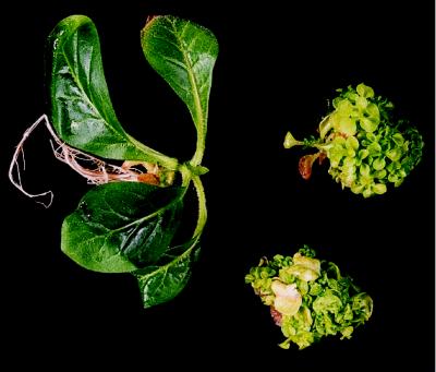
Twenty-eight of the Cu-ipt lines grew in vitro in a manner identical to the Cu-GUS controls. The growth pattern of these plants, which consisted of a single, apically dominant shoot with no visible lateral buds, is shown in Figure 1, left. These plants produced abundant root systems within 4 weeks of growth in the presence of kanamycin.
Figure 1.
Morphological comparison of tobacco lines ID8, ID9, and IR19. Left, Morphologically normal line ID8 21 d after subculture. Top right, Morphologically aberrant line ID9 21 d after subculture. Bottom right, Morphologically aberrant line IR19 21 d after subculture. The plants were grown in tissue culture on solid Murashige-Skoog medium containing 150 mg L−1 kanamycin but no CuSO4.
Three of the Cu-ipt lines grew in a manner consistent with aberrant cytokinin expression: ID9 initially produced an apically dominant shoot but failed to form roots and produced callus at the base of its stem. Shoot elongation was limited and lateral buds appeared in the axils of the leaves. Although these buds did not elongate, additional buds continued to emerge. This pattern continued until ID9 became a mass of miniature leaves growing on a lump of callus (Fig. 1, top right). This phenotype was stable in culture. IR19 was similar to ID9 but grew wrinkled leaves that produced epiphyllic shoots and had a tendency toward early necrosis (Fig. 1, bottom right). IR13 also showed reduced apical dominance, grew callus from the base of its stem, and produced small leaves (not shown).
Southern Analysis of the Tobacco Transformants
Southern analyses were carried out on five of the independent transformants. The analysis included both morphologically normal and morphologically aberrant lines. Genomic DNA was digested with EcoRI, which is known to have no restriction sites within the T-DNA of pMMACEipt, an area approximately 5 kb in size. The results confirmed the presence of the ipt gene in all of the lines analyzed (data not shown).
Comparison of Cytokinin between Normal and Aberrant Transformants
The cytokinin content of the aberrant line ID9 was analyzed to confirm that its altered morphology was correlated with cytokinin overproduction. A line that was morphologically normal in tissue culture and shown via Southern analysis to be transformed (ID8) was included in the analysis for comparison.
Line ID9 showed a marked increase in cytokinin content over line ID8 (Table I). In ID9 tissue ZR and iPA, ZOG and ZROG, and ZNT were detected. The most abundant cytokinin in line ID9 was ZR at 134.4 pmol g−1 fresh weight, making up 42% of the total cytokinin detected. The combined glucosides ZOG and ZROG (96.4 and 46.9 pmol g−1 fresh weight, respectively) contributed an equivalent amount, with smaller quantities of ZNT and iPA being detected. By comparison, line ID8 produced only trace quantities of DZR, iPA, ZOG, and ZNT. None of these was detected at a level large enough to allow accurate quantification (Table I). In addition to the cytokinin forms shown in Table I, the experimental system would have detected Z, DZ, iP, Z9G, iP9G, DZOG, DZROG, DZNT, and iPNT if they had been above the detection limit of the assay.
Table I.
Cytokinins detected in the leaf tissue of Cu-ipt tobacco lines ID8 and ID9 under noninductive conditions
| Cytokinin | ID8 | ID9 |
|---|---|---|
| pmol g−1 fresh wt | ||
| ZR | nda | 134.4 |
| DZR | Traceb | Trace |
| iPA | Trace | 9.6 |
| ZOG | Trace | 96.4 |
| ZROG | nd | 46.9 |
| ZNT | Trace | 28.0 |
| Total | Trace | 315.3 |
Values have been corrected for losses during purification and for differential cross-reactivity of the antibodies. The detection limit for ZR with clone 16 was 0.6 pmol and for iPA with clone 12 it was 0.8 pmol.
nd, Not detected.
Trace, Quantities detected were below the limit for accurate quantification of 1 pmol g−1 fresh weight.
Physiological Changes in Morphologically Normal Plants in Tissue Culture following ipt Gene Induction
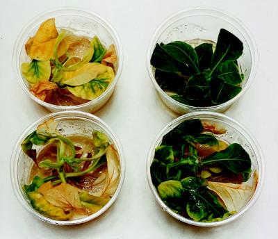
Two in vitro experiments were performed on morphologically normal transformants to assess the impact of cytokinin production on plant physiology. In the first experiment we investigated the morphological alteration of Cu-ipt and Cu-GUS plants following treatment with varying concentrations of CuSO4. Following 97 d of treatment with 5, 10, or 50 μm CuSO4, the leaves of the Cu-ipt plants were clearly greener than those of the Cu-GUS plants. This trend was most obvious in plants treated with 50 μm CuSO4 (Fig. 2). The second in vitro experiment compared the cytokinin profile of Cu-ipt plants from the same line treated with either 50 μm CuSO4 or water. Following 3 months of treatment, most of the plants were not senescent. However, leaf tissue was harvested for cytokinin analysis based on the senescence time frame observed in the first experiment.
Figure 2.
Comparison of leaf senescence in Cu-GUS and Cu-ipt tobacco transformants following treatment with 50 μm CuSO4. Cu-GUS (left) and Cu-ipt (right) plants growing in vitro following 97 d of treatment with 50 μm CuSO4.
Cytokinin analysis showed that one of the Cu-ipt lines produced a marked increase in cytokinin content following CuSO4 treatment compared with uninduced tissue of the same line (Table II). In the CuSO4-treated tissue the cytokinin free bases showed the largest increase, with Z measuring 27.5 pmol g−1 fresh weight and iP measuring 40.5 pmol g−1 fresh weight. DZR showed the largest increase of any single cytokinin form (52.2 pmol g−1 fresh weight), and a smaller quantity of ZOG (11.8 pmol g−1 fresh weight) was also observed. By comparison, only a trace of iP was detected in uninduced tissue (Table II). In addition to the cytokinin forms shown in Table II, the experimental system would have detected DZ, ZR, iPA, Z9G, iP9G, DZOG, ZROG, DZROG, ZNT, DZNT, and iPNT had these been above the detection limit of the assay.
Table II.
Comparison of the cytokinins detected in the leaf tissue of Cu-ipt tobacco line ID13 following in vitro treatment with water (−CuSO4) or Cu (+CuSO4).
| Cytokinin | −CuSO4 | +CuSO4 |
|---|---|---|
| pmol g−1 fresh wt | ||
| Z | nda | 27.5 |
| DZR | nd | 52.2 |
| iP | Traceb | 40.5 |
| ZOG | nd | 11.8 |
| Total | Trace | 132.0 |
Values have been corrected for losses during purification and for differential cross-reactivity of the antibodies. The detection limit for ZR with clone 16 was 0.6 pmol and for iPA with clone 12 it was 0.8 pmol.
nd, Not detected.
Trace, Quantities detected were below the limit for accurate quantification of 1 pmol g−1 fresh weight.
Morphological Changes in Whole Plants following ipt Gene Induction
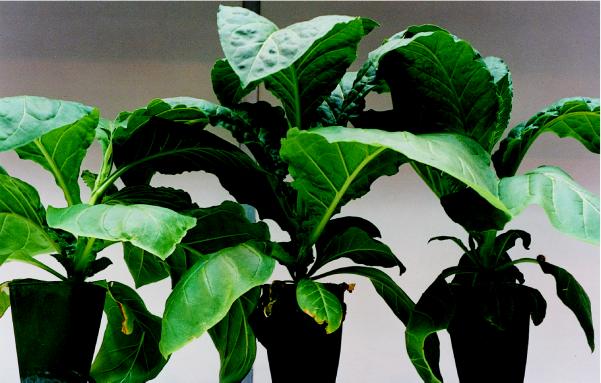
To determine the effect of a controlled endogenous cytokinin increase on whole plant morphology, we treated morphologically normal Cu-ipt and Cu-GUS strains with CuSO4 in vivo. Six independently transformed lines were included: two Cu-GUS control lines and four Cu-ipt lines. Each line was represented by 4 to 7 plants, resulting in a total of 31 plants over the whole experiment. After being removed from tissue culture, all plants required an initial recovery period of 1 week, after which they began rapid stem elongation and leaf expansion. After 17 d of exposure to Cu2+, the Cu-ipt plants began to display lateral bud growth. This continued until the conclusion of the experiment (Fig. 3), when the growth pattern of each plant was measured.
Figure 3.
Comparison of growth patterns in Cu-GUS and Cu-ipt tobacco transformants following treatment with Cu. Left, Representative plant from Cu-GUS line GD11 following 30 d of treatment with Cu. Middle and right, Representative plants from Cu-ipt line ID8 following 30 d of treatment with Cu.
There were a number of significant differences in the growth pattern of the Cu-ipt plants compared with the control plants, which are given in Table III. The Cu-ipt plants had a greater number of lateral buds (P < 0.0001), a greater lateral bud length (P < 0.001), a greater number of leaves per lateral bud (P < 0.01), and a greater leaf number per plant (P < 0.0001). Stems had also appeared on some of the lateral buds of the Cu-ipt plants (6 of 59), whereas they were completely absent from the controls. Measurements that were not significantly different between the Cu-ipt and Cu-GUS plants included plant height and the node number from which the lateral buds grew.
Table III.
Growth patterns of Cu-GUS and Cu-ipt tobacco from the in vivo experiment following 30 d of exposure to Cu
| Strain | Whole
Plant
|
Lateral Buds
|
Node No.a | |||
|---|---|---|---|---|---|---|
| Height | Leaves | No. | Length | Leaves | ||
| cm | no. | cm | no. | |||
| Cu-GUS | 14.5 (0.76)a | 8.6 (0.47)a | 1.2 (0.25)a | 4.5 (0.64)a | 2.1 (0.17)a | 4.1 (0.57)a |
| Cu-ipt | 14.9 (0.62)a | 11.8 (0.39)b | 3.3 (0.21)b | 7.0 (0.52)b | 2.7 (0.12)b | 5.6 (0.41)a |
| P > 0.05 | P < 0.0001 | P < 0.0001 | P = 0.0009 | P = 0.0083 | P > 0.05 | |
Data presented are the estimates of the least-squares means of the morphological characteristics measured. ses are in parentheses. Those figures in each column followed by a different letter are significantly different at P ≤ 0.05.
The node from which axillary buds were released (nodes were numbered upward from the base).
During the experiment root samples were taken from the Cu-GUS lines for analysis of GUS expression to confirm that the Cu-controllable promoter was directing expression in the roots. These plants displayed strong GUS expression (1376.0–5298.2 pmol min−1 mg−1 protein) in the root tissue.
DISCUSSION
We regarded the Cu-controllable gene expression system as an attractive candidate for use in the control of ipt gene expression, mostly because of the tight temporal control it exhibits (Mett et al., 1993). Because even small increases in endogenous cytokinin concentration have been shown to have a significant effect on plant growth and development (Medford et al., 1989; Smart et al., 1991; Smigocki, 1991), it was important that the lowest possible background be maintained before gene induction. Another advantage of using this system comes from the previous observation that, when its expression was controlled by the 46-bp TATA sequence of the CaMV 35S promoter, GUS activity was detected only in the roots of tobacco plants (Mett et al., 1996). Therefore, the expression of the introduced ipt gene could be expected to occur in the roots and to supplement the naturally originating cytokinin.
A Grossly Aberrant Morphology Results from Uncontrolled Cytokinin Expression
Despite the fact that most plant lines grew normally, three lines (ID9, IR13, and IR19) showed marked morphological differences in tissue culture when compared with controls. Morphological changes included the formation of small, rounded leaves that did not expand, breaking of apical dominance, lack of root formation, callus formation at the stem base, and dwarfing (Fig. 1). These features are indicative of cytokinin overproduction and have been repeatedly described by groups working with ipt genes (Medford et al., 1989; Smigocki, 1991; Beinsberger et al., 1992; Li et al., 1992; Hewelt et al., 1994). Additional features displayed only by line IR19 have also been previously described, including the production of wrinkled leaves that showed premature necrosis and epiphyllic shoot production (Estruch et al., 1991; Li et al., 1992; Hewelt et al., 1994).
The morphological changes we observed in these lines indicated that the ipt gene sequence was present and functional. The cytokinin profile of one of the aberrant lines (ID9) confirmed that the concentrations of five cytokinin forms had increased over those observed in a morphologically normal line (ID8; Table I). The presence of the physiologically active ribosides in line ID9 was not unexpected in light of its altered morphology. However, of particular interest was the presence of the O-glucosides. Regarded as storage forms, the O-glucosides are probably used to reduce the concentration of active cytokinin within the plant (for review, see Jameson, 1994). This seems especially likely in the case of ZROG, since ZR was detected as the most prominent cytokinin in ID9 tissue. Redig et al. (1996) also suggested that O-glucoside conjugation occurs in ipt-expressing tissue. As in the work reported here, they detected little conversion to the N-glucoside forms.
The increase in ZNT seen in line ID9 reinforces the importance of analyzing all of the cytokinin forms. ZNT is one of the first forms produced following the formation of iPNT by the enzyme isopentenyl transferase and may act as a source for the formation of ZR by phosphatase breakdown. The use of modified Bieleski solution (Jameson et al., 1987) for tissue storage following harvest was expected to stop the nonspecific cleavage of nucleotides by phosphatase enzymes. However, the adjustment of sample pH using concentrated ammonia before its application to chromatography columns is suspected to have caused some breakdown of the nucleotides (H. Zhang, personal communication), which would have added to the ZR pool detected in this tissue.
The cytokinin analysis of line ID9 confirmed that the ipt gene was still able to produce a functional isopentenyl transferase, although the expression of the ipt gene was uncontrolled in lines ID9 and IR19. This could have been due to position effect and/or copy number (Gendloff et al., 1990). The presence of plant lines that were transformed with the ipt gene but were morphologically indistinguishable from the Cu-GUS controls indicated that in these lines expression of the ipt gene was under tight control.
Controlled Cu-ipt Expression in Tissue Culture Results in Delayed Leaf Senescence
The cytokinins are believed to be involved in the regulation of senescence in leaves. The exogenous application of cytokinin to leaf tissue has been shown to delay its senescence (Richmond and Lang, 1957; Noodén et al., 1979), and cytokinin levels have been observed to decline in senescing leaf tissue (Singh et al., 1992). A number of groups working with ipt genes have described delayed leaf senescence in their transformants (Smart et al., 1991; Hewelt et al., 1994; Gan and Amasino, 1995).
To determine the influence of endogenously produced cytokinin over leaf senescence in our transformants and the level of control exerted by the Cu-controllable promoter over ipt gene expression, Cu-ipt and Cu-GUS lines were treated in vitro with either CuSO4 or water. Following CuSO4 treatment one line displayed a clear increase in cytokinin content (Table II). A total of 132.0 pmol g−1 fresh weight was detected, compared with only trace amounts of cytokinin following water treatment. This result provided conclusive evidence that the expression of the ipt gene was under the tight control of the Cu-controllable system to which it had been fused.
The increase in the concentration of Z following CuSO4 application is particularly interesting. The work of Singh et al. (1992) showed that Z was the most abundant cytokinin in nonsenescent tobacco leaves and that its concentration declined when senescence began to occur. As suggested in reference to the overexpressing line ID9, the detection of the O-glucoside group ZOG in the Cu-treated tissue may be indicative of an attempt to lower the levels of the very active Z base.
Despite the number of groups that have observed delayed leaf senescence as a major physiological response to ipt gene expression, only Smart et al. (1991) undertook detailed cytokinin analysis of this tissue. This is unfortunate since a detailed comparison of the cytokinin levels and forms required to delay senescence would be of interest, especially in systems that show tightly controlled morphology, such as that reported by Gan and Amasino (1995).
In their analysis of tobacco transformed with an ipt gene controlled by a heat-shock promoter, Smart et al. (1991) treated areas of leaves attached to the plant with 42°C for 2 h. Analysis of the treated regions and comparison with untreated regions showed marked increases in Z (13.4-fold), ZR (8-fold), iP (7.8-fold), DZ (8.1-fold), and DZR (7.5-fold). The nucleotide forms ZNT, iPNT, and DZNT also increased, and a small increase was observed in iPA. Unfortunately, the glucoside forms were not included in the analysis by Smart et al. (1991). The concentration of Z (120.6 pmol g−1 fresh weight) was markedly higher than that reported here, the concentration of iP was similar, and the riboside DZR was markedly lower (3.0 pmol g−1 fresh weight). It is possible that these differences could be due to differences in the gene-induction method (heat shock versus Cu application), plant growth conditions (in vivo versus in vitro), or the tissue of origin of the cytokinins (potentially every cell versus root tissue only). Nevertheless, all of the forms seen to increase in our work were also observed to increase by Smart et al. (1991). The most significant difference between the two sets of data is the amount of cytokinin production by transformed but uninduced leaf material. Smart et al. (1991) detected a significant increase in the concentration of cytokinin in tissue that had not been heat shocked. It is possible that some of the increase may have been due to cytokinin transport from heat-shocked areas to non-heat-shocked areas. However, the observation of morphological changes in untreated whole plants indicates that some expression occurred in the absence of heat shock. The increase in total cytokinin was 13-fold that seen in untransformed leaf tissue (averaged over all groups detected). Even when more tightly controlled promoters have been used to control ipt gene expression, e.g. tetracycline-inducible promoter (Redig et al., 1996; Faiss et al., 1997), some cytokinin production has been reported in the noninduced state. In contrast, cytokinins from the Cu-ipt line could not be detected in uninduced leaf tissue (except for a trace level of iP), and this pattern was identical to that of the Cu-GUS controls (data not shown).
Morphological alteration due to ipt gene induction was clearly observable in the first tissue culture experiment. Tobacco from the Cu-ipt and Cu-GUS lines were treated with 5, 10, or 50 μm CuSO4 and visually monitored for morphological changes. At the end of 3 months, all of the Cu-ipt plants showed delayed leaf senescence compared with the Cu-GUS controls. However, this response was most obvious in plants treated with 50 μm CuSO4 (Fig. 2), which is consistent with the previous observation of Mett et al. (1993) that the quantity of CuSO4 applied influences the level of gene expression.
Controlled Cu-ipt Expression in Whole Plants Results in a Break in Apical Dominance
Morphological alteration was also observed in the in vivo experiment. Four independently transformed Cu-ipt lines were included, with each line represented by at least four clonal replicates. Measurements made during the experiment allowed accurate comparison between the growth patterns of the lines, with particular attention paid to lateral bud growth. The results show that the Cu-ipt lines had significantly different morphology compared with Cu-GUS controls (Table III) after 30 d of exposure to Cu2+. Whereas the controls displayed strong apical dominance, the Cu-ipt plants displayed a clear release of lateral buds from apical dominance (Fig. 3). This was confirmed by significant increases in lateral bud number, lateral bud length, lateral bud leaf number, and total plant leaf number (Table III), and the presence of stems on some of the lateral buds.
With the exception of Van Loven et al. (1993), to our knowledge there are no other reports that document the extensive physiological data required for the statistical analysis of growth patterns of ipt-transformed plants. Using their data to analyze the effect of endogenous cytokinin production on plant growth when ipt expression was controlled by a heat-shock promoter, Van Loven et al. (1993) showed that heat shock itself interfered with whole plant growth, reducing both internode length and stem diameter. Furthermore, they suggested that in vitro cultivation, a stressful situation for plant growth, activated the heat-shock promoter, thereby inducing transient cytokinin production before heat shock. This may explain the pre-heat-shock expression of ipt genes in the work of Medford et al. (1989), Smart et al. (1991), and Smigocki (1991). In light of the above complications, recording physiological data for statistical analysis appears to be necessary.
The release of lateral bud growth from apical dominance is the most consistent morphological effect displayed by whole plants transformed with ipt genes (Medford et al., 1989; Smart et al., 1991; Smigocki, 1991; Van Loven et al., 1993; Hewelt et al., 1994; Faiss et al., 1997). This aspect of development is also inducible by exogenous cytokinin application (Sachs and Thimann, 1964). Under normal conditions lateral bud growth in tobacco occurs only after flowering and at the most apical buds. However, in ipt transformants lateral bud release has been observed to occur in vegetative plants, either around the middle nodes (Van Loven et al., 1993) or from the base to mid-node (Hewelt et al., 1994), a phenomenon that is shown in Figure 3.
Spatial ipt Gene Expression Results in Controlled Physiological Changes
A number of other morphological changes have been observed by various groups working with the ipt gene. These include the reduction of root mass (Medford et al., 1989; Smigocki, 1991; Van Loven et al., 1993), reduction of plant height (Medford et al., 1989; Smart et al., 1991; Smigocki, 1991; Van Loven et al., 1993), increased stem thickening (Ainley et al., 1993; Van Loven et al., 1993), and chlorosis and wrinkling of the leaves (Ainley et al., 1993). None of these changes was observed in the work reported here; the ipt transformed plants appeared to be identical to controls except in respect to apical dominance and leaf senescence. This is particularly interesting considering the spatial control exerted by the Cu-controllable gene expression system over gene expression (Mett et al., 1996). Cytokinin production from the plants studied here was targeted to the roots, which are believed to be a key site of cytokinin biosynthesis in the whole plant (Letham, 1994). Conversely, in many of the plants previously transformed with inducible ipt genes (e.g. those controlled by heat-shock promoters), expression was not targeted to a particular tissue but occurred throughout the plant, resulting in the aberrant morphology described above. In fact, many of the features described by these groups, such as wrinkling and chlorosis of the leaves, reduction of plant height, and reduction of root mass, were seen in our experiments only in lines that did not have controlled ipt expression (ID9, IR13, and IR19).
Our data support the classic notion of root-derived cytokinin influencing leaf senescence and apical dominance. However, Faiss et al. (1997) utilized the bacterial tetracycline repressor-operator complex in association with the ipt gene, and concluded that released apical dominance and delayed senescence were a consequence of localized cytokinin biosynthesis and were not due to enhanced cytokinin production by induced root tissue. Although increased ZR (> 10-fold) was noted in the transpiration stream, the subsequent break in apical dominance and delayed leaf senescence of whole plants were ascribed to tetracycline movement from the roots. To distinguish between enhanced cytokinin export from the roots and the movement of tetracycline, reciprocal grafts were carried out between wild-type and transgenic tissue. Neither lateral shoot growth nor senescence of wild-type material was affected by the presence of induced transgenic rootstock. However, the presence of massive shoot proliferation on the ipt rootstock itself may have created a large metabolic sink. This could have arisen because of a lack of cytokinin translocation through the graft union. Faiss et al. (1997) noted that cytokinin levels in the xylem were normal beyond the graft union.
In this work we produced transgenic tobacco carrying the ipt gene under the control of the Cu-inducible gene expression system. The temporal and spatial levels of control exerted by this system have allowed the production of physiologically normal plants that can be induced to produce more cytokinin at a desired time. This system will be used to study the role of the endogenous cytokinins in plant growth and development and to resolve the issue of cytokinins as long-distance signals.
ACKNOWLEDGMENTS
The authors wish to thank Leesa Lochhead for technical assistance and Dr. Nihal De Silva for statistical expertise. The cytokinin antibodies were a generous gift to P.E.J. from Prof. R.O. Morris (University of Missouri, Columbia).
Abbreviations:
- CaMV
cauliflower mosaic virus
- DZ
dihydrozeatin
- DZNT
DZ nucleotide
- DZOG
DZ-O-glucoside
- DZR
DZ riboside
- DZROG
DZ riboside-O-glucoside
- iP
isopentenyladenine
- iP9G
isopentenyl-9-glucoside
- iPA
isopentenyl adenosine
- iPATA
iPA trialcohol
- iPNT
isopentenyl nucleotide (isopentenyl AMP)
- ipt
isopentenyl transferase gene
- Z
zeatin
- Z9G
Z-9-glucoside
- ZNT
Z nucleotide (Z riboside-5′-monophosphate)
- ZOG
Z-O-glucoside
- ZR
Z riboside
- ZROG
Z riboside-O-glucoside
- ZRTA
Z riboside trialcohol
LITERATURE CITED
- Ainley MW, McNeil KJ, Hill JW, Lingle WL, Simpson RB, Brenner ML, Nagao RT, Key JL. Regulatable endogenous production of cytokinins up to “toxic” levels in transgenic plants and plant tissues. Plant Mol Biol. 1993;22:13–23. doi: 10.1007/BF00038992. [DOI] [PubMed] [Google Scholar]
- Akama K, Shiraishi H, Ohta S, Nakamura K, Okada K, Shimura Y. Efficient transformation of Arabidopsis thaliana: comparison of the efficiencies with various organs, plant ecotypes and Agrobacterium strains. Plant Cell Rep. 1992;12:7–11. doi: 10.1007/BF00232413. [DOI] [PubMed] [Google Scholar]
- Akiyoshi DE, Klee H, Amasino RM, Nester EW, Gordon MP. T-DNA of Agrobacterium tumefaciens encodes an enzyme of cytokinin biosynthesis. Proc Natl Acad Sci USA. 1984;81:5994–5998. doi: 10.1073/pnas.81.19.5994. [DOI] [PMC free article] [PubMed] [Google Scholar]
- Barry GF, Rogers SG, Fraley RT, Brand L. Identification of a cloned cytokinin biosynthetic gene. Proc Natl Acad Sci USA. 1984;81:4776–4780. doi: 10.1073/pnas.81.15.4776. [DOI] [PMC free article] [PubMed] [Google Scholar]
- Beinsberger SEI, Valcke RLM, Clijsters HMM, De Greef JA, Van Onkelen HA. Effects of enhanced cytokinin levels in ipt transgenic tobacco. In: Kamínek M, Mok DWS, Zazímalová E, editors. Physiology and Biochemistry of Cytokinins in Plants. The Hague, The Netherlands: SPB Academic Publishing; 1992. pp. 77–82. [Google Scholar]
- Beinsberger SEI, Valcke RLM, Deblaere RY, Clijsters HMM, De Greef JA, Van Onckelen HA. Effects of the introduction of Agrobacterium tumefaciens T-DNA ipt gene in Nicotiana tabacum L. cv. Petit Havana SR1 plant cells. Plant Cell Physiol. 1991;32:489–496. [Google Scholar]
- Bradford MM. A rapid and sensitive method for the quantitation of microgram quantities of protein utilizing the principle of protein-dye binding. Anal Biochem. 1976;72:248–254. doi: 10.1016/0003-2697(76)90527-3. [DOI] [PubMed] [Google Scholar]
- Budar F, Deboeck F, Van Montagu M, Hernalsteens J-P. Introduction and expression of the octopine T-DNA oncogenes in tobacco plants and their progeny. Plant Sci. 1986;46:195–206. [Google Scholar]
- Estruch JJ, Prinsen E, Van Onckelen H, Schell J, Spena A. Viviparous leaves produced by somatic activation of an inactive cytokinin-synthesizing gene. Science. 1991;254:1364–1367. doi: 10.1126/science.254.5036.1364. [DOI] [PubMed] [Google Scholar]
- Faiss M, Zalubìlová J, Strnad M, Schmülling T. Conditional transgenic expression of the ipt gene indicates a function for cytokinins in paracrine signaling in whole tobacco plants. Plant J. 1997;12:401–415. doi: 10.1046/j.1365-313x.1997.12020401.x. [DOI] [PubMed] [Google Scholar]
- Gan S, Amasino RM. Inhibition of leaf senescence by autoregulated production of cytokinin. Science. 1995;270:1986–1988. doi: 10.1126/science.270.5244.1986. [DOI] [PubMed] [Google Scholar]
- Gendloff EH, Bowen B, Buchholz WG. Quantitation of chloramphenicol acetyl transferase in transgenic tobacco plants by ELISA and correlation with gene copy number. Plant Mol Biol. 1990;14:575–583. doi: 10.1007/BF00027503. [DOI] [PubMed] [Google Scholar]
- Herrera-Estrella L, Simpson J. Foreign gene expression in plants. In: Shaw CH, editor. Plant Molecular Biology: A Practical Approach. Oxford, UK: IRL Press; 1988. pp. 131–160. [Google Scholar]
- Hewelt A, Prinsen E, Schell J, Van Onckelen H, Schmülling T. Promoter tagging with a promoterless ipt gene leads to cytokinin-induced phenotypic variability in transgenic tobacco plants: implications of gene dosage effects. Plant J. 1994;6:879–891. doi: 10.1046/j.1365-313x.1994.6060879.x. [DOI] [PubMed] [Google Scholar]
- Jameson PE. Cytokinin metabolism and compartmentation. In: Mok DWS, Mok MC, editors. Cytokinins Chemistry, Activity and Function. Boca Raton, FL: CRC Press; 1994. pp. 113–128. [Google Scholar]
- Jameson PE, Letham DS, Zhang R, Parker CW, Badenoch-Jones J. Cytokinin translocation and metabolism in lupin species. I. Zeatin riboside introduced into the xylem at the base of Lupinus angustifolius stems. Aust J Plant Physiol. 1987;14:695–718. [Google Scholar]
- Jameson PE, Morris RO. Zeatin-like cytokinins in yeast: detection by immunological methods. J Plant Physiol. 1989;135:385–390. [Google Scholar]
- Jefferson RA. Assaying chimeric genes in plants: the GUS gene fusion system. Plant Mol Biol Rep. 1987;5:387–405. [Google Scholar]
- Letham DS. Cytokinins as phytohormones—sites of biosynthesis, translocation, and function of translocated cytokinin. In: Mok DWS, Mok MC, editors. Cytokinins: Chemistry, Activity and Function. Boca Raton, FL: CRC Press; 1994. pp. 57–80. [Google Scholar]
- Lewis DH, Burge GK, Schmierer DM, Jameson PE. Cytokinins and fruit development in the kiwifruit (Actinidia deliciosa). I. Changes during fruit development. Physiol Plant. 1996;98:179–186. [Google Scholar]
- Li Y, Hagen G, Guilfoyle TJ. Altered morphology in transgenic tobacco plants that overproduce cytokinins in specific tissues and organs. Dev Biol. 1992;153:386–395. doi: 10.1016/0012-1606(92)90123-x. [DOI] [PubMed] [Google Scholar]
- Martineau B, Houck CM, Sheehy RE, Hiatt WR. Fruit-specific expression of the A. tumefaciens isopentenyl transferase gene in tomato: effects on fruit ripening and defence-related gene expression in leaves. Plant J. 1994;5:11–19. [Google Scholar]
- McKenzie MJ, Jameson PE, Poulter RTM. Cloning an ipt gene from Agrobacterium tumefaciens: characterisation of cytokinins in derivative transgenic plant tissue. Plant Growth Regul. 1994;14:217–228. [Google Scholar]
- Medford JI, Horgan R, El-Sawi Z, Klee HJ. Alterations of endogenous cytokinins in transgenic plants using a chimeric isopentenyl transferase gene. Plant Cell. 1989;1:403–413. doi: 10.1105/tpc.1.4.403. [DOI] [PMC free article] [PubMed] [Google Scholar]
- Memelink J, Hoge JHC, Schilperoot RA. Cytokinin stress changes the developmental regulation of several defence-related genes in tobacco. EMBO J. 1987;6:3579–3583. doi: 10.1002/j.1460-2075.1987.tb02688.x. [DOI] [PMC free article] [PubMed] [Google Scholar]
- Mett VL, Lochhead LP, Reynolds PHS. Copper-controllable gene expression system for whole plants. Proc Natl Acad Sci USA. 1993;90:4567–4571. doi: 10.1073/pnas.90.10.4567. [DOI] [PMC free article] [PubMed] [Google Scholar]
- Mett VL, Podivinsky E, Tennant AM, Lochhead LP, Jones WT, Reynolds PHS. A system for tissue-specific copper-controllable gene expression in transgenic plants: nodule-specific antisence of aspartate aminotransferase-P2. Transgenic Res. 1996;5:105–113. doi: 10.1007/BF01969428. [DOI] [PubMed] [Google Scholar]
- Noodén LD, Kahanak GM, Okatan Y. Prevention of monocarpic senescence in soybeans with auxin and cytokinin: an antidote for self-destruction. Science. 1979;206:841–843. doi: 10.1126/science.206.4420.841. [DOI] [PubMed] [Google Scholar]
- Ooms G, Lenton JR. T-DNA genes to study plant development: precocious tuberisation and enhanced cytokinins in A. tumefaciens transformed potato. Plant Mol Biol. 1985;5:205–212. doi: 10.1007/BF00020638. [DOI] [PubMed] [Google Scholar]
- Palni LMS, Summons RE, Letham DS. Mass spectrometric analysis of cytokinins in plant tissues. V. Identification of the cytokinin complex of Datura innoxia crown gall tissue. Plant Physiol. 1983;72:858–863. doi: 10.1104/pp.72.3.858. [DOI] [PMC free article] [PubMed] [Google Scholar]
- Redig P, Schmülling T, Van Onckelen H. Analysis of cytokinin metabolism in ipt transgenic tobacco by liquid chromatography-tandem mass spectrometry. Plant Physiol. 1996;122:141–148. doi: 10.1104/pp.112.1.141. [DOI] [PMC free article] [PubMed] [Google Scholar]
- Richmond AE, Lang A. Effect of kinetin on protein content and survival of detached Xanthium leaves. Science. 1957;125:650–651. [Google Scholar]
- Sachs T, Thimann KV. Release of lateral buds from apical dominance. Nature. 1964;201:939–940. [Google Scholar]
- Schmülling T, Beinsberger S, De Greef J, Schell J, Van Onckelen H, Spena A. Construction of a heat-inducible chimeric gene to increase the cytokinin content in transgenic plant tissue. FEBS Lett. 1989;249:401–406. [Google Scholar]
- Shillito RD, Saul MW. Protoplast isolation and transformation. In: Shaw CH, editor. Plant Molecular Biology: A Practical Approach. Oxford, UK: IRL; 1988. pp. 161–186. [Google Scholar]
- Singh S, Letham DS, Palni LMS. Cytokinin biochemistry in relation to leaf senescence. VII. Endogenous cytokinin levels and exogenous applications of cytokinins in relation to sequential leaf senescence of tobacco. Physiol Plant. 1992;86:388–397. [Google Scholar]
- Skoog F, Miller C. Chemical regulation of growth and organ formation in plant tissues cultured in vitro. Symp Soc Exp Biol. 1957;11:118–131. [PubMed] [Google Scholar]
- Smart CM, Scofield SR, Bevan MW, Dyer TA. Delayed leaf senescence in tobacco plants transformed with tmr, a gene for cytokinin production in Agrobacterium. Plant Cell. 1991;3:647–656. doi: 10.1105/tpc.3.7.647. [DOI] [PMC free article] [PubMed] [Google Scholar]
- Smigocki AC. Cytokinin content and tissue distribution in plants transformed by a reconstructed isopentenyl transferase gene. Plant Mol Biol. 1991;16:105–115. doi: 10.1007/BF00017921. [DOI] [PubMed] [Google Scholar]
- Smigocki AC, Neal JW, McCanna I, Douglass L. Cytokinin-mediated insect resistance in Nicotiana plants transformed with the ipt gene. Plant Mol Biol. 1993;23:325–335. doi: 10.1007/BF00029008. [DOI] [PubMed] [Google Scholar]
- Smigocki AC, Owens LD. Cytokinin gene fused with a strong promoter enhances shoot organogenesis and zeatin levels in transformed plant cells. Proc Natl Acad Sci USA. 1988;85:5131–5135. doi: 10.1073/pnas.85.14.5131. [DOI] [PMC free article] [PubMed] [Google Scholar]
- Smigocki AC, Owens LD. Cytokinin-to-auxin ratios and morphology of shoots and tissues transformed by a chimeric isopentenyl transferase gene. Plant Physiol. 1989;91:808–811. doi: 10.1104/pp.91.3.808. [DOI] [PMC free article] [PubMed] [Google Scholar]
- Southern EM. Detection of specific sequences among DNA fragments separated by gel electrophoresis. J Mol Biol. 1975;98:503–517. doi: 10.1016/s0022-2836(75)80083-0. [DOI] [PubMed] [Google Scholar]
- Trione EJ, Krygier BB, Banowetz GM, Kathrein JM. The development of monoclonal antibodies against the cytokinin zeatin riboside. J Plant Growth Regul. 1985;4:101–109. [Google Scholar]
- Van Loven K, Beinsberger SEI, Valcke RLM, Van Onckelen HA, Clijsters HMM. Morphometric analysis of the growth of Phsp70-ipt transgenic tobacco plants. J Exp Bot. 1993;44:1671–1678. [Google Scholar]
- Yusibov VM, Il PC, Andrianov VM, Piruzian ES. Phenotypically normal transgenic T-cyt tobacco plants as a model for the investigation of plant gene expression in response to phytohormonal stress. Plant Mol Biol. 1991;17:825–836. doi: 10.1007/BF00037064. [DOI] [PubMed] [Google Scholar]


