Abstract
Background
The bacterial pathogen Salmonella causes worldwide disease. A major route of intestinal entry involves M cells, providing access to B cell-rich Peyer’s Patches. Primary human B cells phagocytose Salmonella typhimurium upon recognition by the specific surface Ig receptor (BCR). As it is unclear how Salmonella disseminates systemically, we studied whether Salmonella can use B cells as a transport device for spreading.
Methodology/Principal Findings
Human primary B cells or Ramos cell line were incubated with GFP-expressing Salmonella. Intracellular survival and escape was studied in vitro by live cell imaging, flow cytometry and flow imaging. HEL-specific B cells were transferred into C57BL/6 mice and HEL-expressing Salmonella spreading in vivo was analyzed investigating mesenteric lymph nodes, spleen and blood. After phagocytosis by B cells, Salmonella survives intracellularly in a non-replicative state which is actively maintained by the B cell. Salmonella is later excreted followed by reproductive infection of other cell types. Salmonella-specific B cells thus act both as a survival niche and a reservoir for reinfection. Adoptive transfer of antigen-specific B cells before oral infection of mice showed that these B cells mediate in vivo systemic spreading of Salmonella to spleen and blood.
Conclusions/Significance
This is a first example of a pathogenic bacterium that abuses the antigen-specific cells of the adaptive immune system for systemic spreading for dissemination of infection.
Introduction
Salmonella enterica is a Gram-negative, enteric pathogen responsible for diseases that lead to significant morbidity and mortality [1]. After oral uptake, the bacterium crosses the intestinal epithelium via transcytosis of specialized M cells [2] or via luminal capture by sampling dendritic cells [3], [4]. They are eventually internalized by macrophages, dendritic cells and neutrophils in the lamina propia [5], [6]. Cellular entry in non-phagocytic cells is actively induced by the bacterium through an array of effector proteins that orchestrate uptake by manipulating the host’s cellular machinery [7]. Salmonella directs host cells during infection to alter the actin cytoskeleton allowing formation of macropinocytic ruffles and entry of the relatively large pathogen into host cells. Salmonella introduces bacterial effector proteins in the host cytosol via the Salmonella Type III Secretion System (TTSS). Salmonella can infect most cell types to form an intracellular vacuole called the Salmonella-containing vacuole (SCV). Here, another set of effectors is introduced into the host cytosol for vacuole maintenance and interference with the endosomal system to obtain nutrients and to prevent maturation and fusion with lysosomes [8], [9]. This involves the Akt-AS160-Rab14 cascade and PAK4 [10]. Salmonella replicates in an expanding SCV [11], [12] and may thus escape detection by the immune system [13], [14]. Although Salmonella replicates in the phagosomes, it remains unclear how the bacteria are released from the infected cell. Obvious mechanisms would involve apoptosis or necrosis of the infected cell, but such is not established.
When Salmonella has passed the intestinal epithelium, it spreads via mesenteric lymph nodes to liver, bone marrow and spleen where replication continues [15] and disease ensues. How Salmonella reaches these organs is unclear. So far, dendritic cells, macrophages, neutrophils and CD18-expressing phagocytes have been implicated [4], [16]. Neutrophils however exhibit efficient bactericidal mechanisms [6], [17] that render these cells less favorite as vehicles for systemic bacterial dissemination. Similar to HIV (reviewed in [18]), dendritic cells and macrophages may act as pathogen carriers for spreading of the infection, but are unlikely to cause spreading beyond mesenteric lymph nodes. CD11B+ and CD11c+ phagocytes harboring Salmonella were reported to be present in blood 5 min after oral inoculation, but it remains to be elucidated how these cells relate to DCs, macrophages or B cells [4], [19].
We recently showed that primary human antigen-specific B cells are able to internalize Salmonella after recognition by the B cell receptor (BCR) [20]. As Salmonella gains immediate access to the B cell-rich areas of the Peyer’s Patches after intestinal invasion, it may be that B cells mediate spreading of Salmonella infection as well. Here we show that Salmonella can indeed use antigen-specific B cells as transport vehicle for spreading within the host. Salmonella survives intracellularly in a non-replicative state that is actively maintained by the B cell. Ultimately, Salmonella is excreted by the B cell followed by reinfection and replication in other cell types. Adoptive transfer of B cells with transgenic BCRs that specifically recognize hen egg lysozyme (HEL)-expressing Salmonella showed that Salmonella-specific B cells contribute to the in vivo systemic dissemination of Salmonella in mice after oral administration of the bacteria. The antigen-specific B cells thus act as antigen-specific reservoirs and transport vehicles to release Salmonella at distant sites for further infection. These data provide the first example of the use of antigen-specific B cells by a bacterial pathogen for spreading infection in a situation analogous to the involvement of innate cells in spreading of HIV.
Materials and Methods
Ethics Statement
All human donors used in this study provided written informed consent in accordance with the protocol of the local institutional review board, the Medical Ethics Committee of Sanquin Blood Supply (Amsterdam, The Netherlands), and the Medical Ethics Committee of Sanquin approved the study. All mice experiments were performed in the European Institute of Oncology, Milan. All experimental procedures using mice were performed according to the Principles of Laboratory Animal Care guidelines (directive 86/609/EEC) and approved by the Italian Ministry of Health.
Mice
C57BL/6 mice (6–8 weeks old) were purchased from Harlan (Udine, Italy). BCR-HEL VDJ knock-in mice (a kind gift of Dr. J. Cyster, University of California, San Francisco) were bred under specific pathogen-free conditions at Charles River Laboratories. All experiments were performed in accordance with the guidelines established in the Principles of Laboratory Animal Care (directive 86/609/EEC).
Evaluation of Salmonella Spreading in vivo
CD43- naive B cells were purified from spleens of BCR-HEL VDJ knock-in mice with CD43 (Ly-48) Microbeads (Miltenyi Biotec, Bologna, Italy) according to the manufacturer’s instructions. Purity was more than 94%, with more than 75% of the B cells expressing the HEL-BCR. 2*105-106 cells were injected intravenously into C57BL/6 mice (14 mice per group) one day before oral infections. For evaluation of bacterial colonization experiments mice received 6*106 CFUs of HEL surface-expressing S. typhimurium SL1344 while transferred B cells were labeled with CFSE (2,5 µM) to monitor their spreading. 0, 24 and 72 hours after infection blood, spleens, mesenteric lymph nodes and liver were collected and processed; a fixed number of cells was lysed with 0.5% sodium-deoxycholate and plated onto TB-agar plates for CFU counting 12 hr later. In spleens and mesenteric lymph nodes the presence of CFSE positive B cells was assessed by FACS at the different time points.
Antibodies and Bacterial Strains
mAb anti-human IgM (MH15, Sanquin, Amsterdam, The Netherlands) was mixed with rat anti-mouse IgG1 antibody (RM161.1, Sanquin) and mAb anti-S. typhimurium LPS (1E6, Biodesign International, Kennebunk, ME) to generate BCR-LPS tetrameric antibody complexes. F(ab)2 fragments of MH15 were generated by standard pepsin digestion. Fluorescent secondary antibodies and Texas Red-phalloidin were from Molecular Probes (Leiden, The Netherlands). GFP-S. typhimurium SL1344 has been described [21]. The S. typhimurium strain 14028 containing the lux operon of P. luminescens (luxCDABE) was a kind gift from S. Vesterlund [22] and K. Nealson. Exponentially grown bacteria were washed with PBS, incubated with BCR-LPS tetrameric antibody complexes for 30 min at RT, and washed twice to remove unbound antibodies. Surface HEL-expressing S. typhimurium SL1344 was generated by electroporating bacteria with a pVUB4 vector (kindly provided by P. Cornelis, Flanders Institute for Biotechnology, Brussels, Belgium [23]) in which inactive HEL-encoding gene was cloned in frame with the one encoding for OprI protein from P. aeruginosa under the control of LacZ promoter. HEL expression was induced by the addition of 1 mmol/L isopropyl-L-thio-B-D-galactopyranoside to exponentially growing bacteria [24].
Lymphocyte Isolation, Infections and Cell Lines
Human B cells were isolated from peripheral blood of from a buffycoat obtained from healthy donors (Sanquin). This Isolation of human B cells from peripheral blood and culturing of the Ramos B cell line and NIH3T3 fibroblasts expressing human CD40L (3T3-CD40L) have been described [20], purity of peripheral B cells was determined by FACS analysis and was always >99%. B lymphocytes with viable uncoated bacteria and Ramos cells with viable anti-IgM coated bacteria were incubated at 20 bacteria per cell for 40 min at 37°C without antibiotics while tumbling. Next, cells were washed four times and cultured for 1 h in media containing 100 µg/ml gentamicin (Invitrogen) to eliminate non-phagocytosed bacteria. Cells were cultured in RPMI 1640 medium with 5% FCS, p/s, 2 mM L-Glutamine, 50 µM 2-mercaptoethanol, 20 µg/ml human apo-transferrin ((Sigma-Aldrich) depleted for human IgG with prot-G sepharose) and 10 µg/ml gentamicin.
Live Cell Imaging and EM Analyses
Wide field microscopy was performed at 37°C using 6-well plates (coated with Poly-L Lysine) and a Zeiss Axiovert 200 M microscope equipped with a FluorArc fluorescence lamp, motorized scanning stage, 63× LD Achroplan objective; NA 0.75 and climate chamber. Images were acquired using a Zeiss AxioCam MRm Rev.2 CCD in combination with the manufacturer’s AxioVision software. All experiments presented were repeated several times on different days, and results were consistent and reproducible. Further image processing was performed using the ImageJ software package.
For EM, cells were allowed to take up anti-IgM coated bacteria for 4 hr before fixation in a mixture of paraformaldehyde (4%) and glutaraldehyde (0.5%). After embedding in a mixture of methyl cellulose and uranyl acetate, ultrathin sections were stained and analyzed with a Philips CM electron microscope.
Intracellular Survival and Growth Assays
Human primary B cells were incubated in parallel experiments with either GFP- or Lux-expressing Salmonella. The percentage of living cells and GFP levels were determined using a FACS Calibur (Becton Dickinson). Bioluminescence was measured for 5s in a luminometer (Berthold). Bacterial growth was determined by dividing the relative bioluminescence signal by the relative number of GFP+ living B cells, resulting in the amount of light produced per bacteria containing B cell. For induction of apoptosis, cells were treated with 0.1 µM Edelfosine (Biomol) [25].
Plating Assay
For enumeration of intracellular surviving bacteria, freshly isolated primary B cells were incubated with anti-IgM coated GFP-Salmonellae and Ramos cells with uncoated Salmonellae as a control, washed and cultured in medium with 10 µg/ml gentamicin as described above. At the indicated time points cells were analyzed by FACS for GFP expression. In parallel, cells were washed with PBS and lysed in 0.1% Triton X-100 (Merck) for 10 min on ice, washed with PBS and a dilution series was plated onto LB agar plates. Plates were incubated overnight at 37°C and colonies were counted.
Flow Imaging Analysis
Primary B cells were incubated with anti-BCR coated, GFP-expressing Salmonella for 1 h in medium without antibiotics, and after washing cultured in the presence of gentamycin. B cell membrane was stained with CD20-PerCP/Cy5.5 (BD) and cells were analysed by ImagestreamX (Amnis) at indicated time points. Analyses were performed using IDEAS software (Amnis). Single cells were gated on Salmonella positivity, and only cells and Salmonella detected in the focal plane were selected for further analysis. Extracellular and intracellular Salmonella were discriminated by deltacentroid-xy analysis, measuring the distance of Salmonella to the center of the cell. The numbers of intracellular Salmonella were analyzed using the spot-count feature.
Bacterial Excretion Assay
To visualize bacterial excretion, human primary B cells were incubated with uncoated GFP-Salmonella and followed using wide field microscopy in medium containing anti-LPS antibodies coupled to TexasRed. To quantify excretion, cells were stained with DAPI (Sigma-Aldrich) to exclude dead cells and anti-LPS coupled to APC and fixed with 3.7% formaldehyde before analysis using a LSR II (Becton Dickinson). For the increase in LPS levels, the initial level at time point 0 was set to 1. The percentage of excreted bacteria was calculated as the loss of GFP+/LPS- B cells compared to time point 0. To discriminate between bacterium and B cell-induced excretion, cells were cultured in medium containing 10 µg/ml tetracycline to arrest intracellular bacteria (bacteriostatic capacity was verified using lux-Salmonella in Ramos cells).
Statistical Analysis
Kaplan-Meier plots and long-rank tests were used to assess survival differences of adoptively transferred mice after virulent S. typhimurium infection. Statistic calculations were performed by JMP 5.1 software (SAS, Cary, NC).
Results
Salmonella Survives in Human Primary Antigen-specific B Cells in a Nonreplicative State
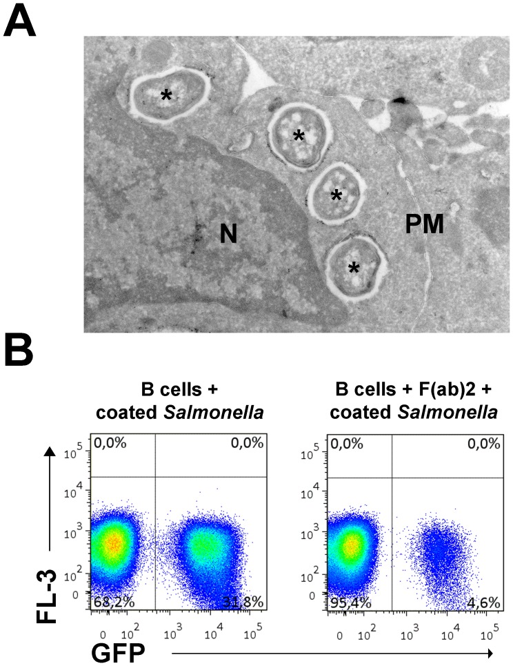
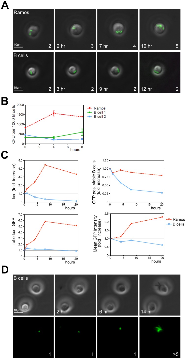
We recently demonstrated that primary antigen-specific B cells phagocytose Salmonella typhimurium when the bacteria are recognized by the antigen-specific BCR [20]. We now studied the fate of internalized Salmonella in more detail. To enhance the number of primary B cells that phagocytose bacteria for some of our experiments, we coated GFP-expressing Salmonella with anti-LPS and anti-IgM tetrameric antibody complexes. These complexes bridge the bacterium and the BCR on B cells, leading to BCR crosslinking and uptake of Salmonella. Electron microscopy of primary human B cells incubated with Salmonella indeed demonstrate the occurrence of phagocytosis as it shows bacteria residing in phagosomes (Figure 1A). Pre-incubation of primary B cells with F(ab)2 fragments of the anti-IgM antibody before exposure to anti-IgM coated GFP-Salmonella dramatically reduces the amount of GFP-Salmonella positive B cells, demonstrating that phagocytosis of Salmonella by B cells is mediated by the BCR (Figure 1B). To study the fate of Salmonella in B cells over extended periods of time, time-lapse imaging of GFP-tagged Salmonella containing B cells was performed using wide-field microscopy to limit phototoxicity. Phagocytosed GFP-Salmonella replicated in the Ramos B cell line (Figure 2A, top panel; see also Video S1). Interestingly, no multiplication was detected in human primary B cells (Figure 2A, bottom panel; see also Video S2). This was confirmed by counting of CFU when Salmonella was retrieved at various time points from Ramos and primary B cells after uptake of anti-IgM coated Salmonella (Figure 2B). These observations show an increase of Salmonella numbers over time in Ramos and a steady number of Salmonella bacteria in primary B cells. To quantify these observations with more sophisticated methods, we performed parallel experiments to compare GFP-Salmonella (detected by flow cytometry) with light producing lux-Salmonella (detected by luminometry). Light production by lux-Salmonella is dependent on metabolic activity of the bacteria and is thus a marker for bacterial viability [22], while the GFP signal only indicates the cellular presence of the bacteria. GFP-Salmonella was used to determine the number of infected B cells at the different time points. Figure 2B shows that the GFP-Salmonella phagocytosed by Ramos cells expanded intracellularly. Over a time course of 10 h, the lux activity increased strongly (Figure 2C, top left panel), while the number of Ramos cells positive for GFP-Salmonella remained nearly constant (Figure 2C, top right panel). This implies an increase in the amount of light produced per GFP-Salmonella positive Ramos cell (Figure 2C, bottom left panel), confirming an increase in numbers of bacteria per Ramos cell over time. In accordance, the GFP signal per Ramos cell increased (Figure 2C, bottom right panel) while lux activity decreased over time when Salmonella was phagocytosed by primary human B cells (Figure 2C, top left panel). This was not due to intracellular killing of Salmonella as the fraction of GFP-Salmonella containing B cells declined equally fast (Figure 2C, top right panel). In fact, the amount of light produced per living GFP-Salmonella positive B cell remained constant during the course of the experiment (Figure 2C, bottom left panel), showing that the GFP-positive Salmonella remained viable in primary human B cells, albeit under conditions of inhibited replication. Thus, these data confirm the wide-field microscopy data in Figure 2A and show that Salmonella does not replicate in primary B cells. Unlike specialized phagocytic immune cells such as macrophages, neutrophils, or B cells from early vertebrates [26], human B cells are apparently inefficient in producing the microbicidal conditions that are required to eliminate Salmonella.
Figure 1. BCR-mediated internalization of Salmonella by primary human B cells.
(A) Immunocryoelectron micrograph of primary human B cells that had phagocytosed anti-BCR coated Salmonella. Gold particles indicate staining for CD63, black asterisks mark bacteria, N marks the nucleus and PM the plasma membrane. (B) Primary B cells were either or not pre-incubated with F(ab)2 fragments of the anti-IgM antibody before incubation with live anti-IgM coated GFP-expressing Salmonella. After extensive washing, cells were fixed and analyzed by FACS indicating a strong reduction in GFP-Salmonella following competition with F(ab)2 for BCR interactions. Shown is a representative plot of 5 donors tested.
Figure 2. Primary human B cells form a survival niche for intracellular Salmonella.
(A) Widefield fluorescence microscopy of living Ramos and primary human B cells with phagocytosed anti-IgM coated GFP-Salmonella. Depicted are GFP signals projected on the transmission image. Scalebar = 10 µm. Number of bacteria in the visualized cell is given in the lower right corner. Lower left corner: time after Salmonella infection. Images are frames from Video S1 and S2. (B) Ramos and primary B cells were incubated with anti-IgM coated Salmonella, lysed and plated onto LB-agar plates at various time points after infection. Data are from triplicate experiments performed with Ramos and primary B cells from two individual healthy donors. Error bars indicate SEM. (C) Analysis of Ramos and primary human B cells incubated with living anti-IgM coated lux-expressing (top left panel) or GFP-expressing (top right panel) Salmonella. The ratio of lux over GFP shows the amount of light produced per GFP-Salmonella positive B cell (bottom left panel), indicating intracellular Salmonella viability. The mean fluorescence of the GFP positive population, set arbitrarily at 1 at the beginning of the experiment, shows that the GFP signal increases in Ramos B cells, whereas it decreased in primary human B cells (bottom right panel). A representative example of three independent experiments is shown. (D) B cells were infected with anti-BCR coated GFP-expressing Salmonella before exposure to Edelfosine to induce apoptosis. Cells were imaged over a 14 h period. Top panel: transmission image, bottom panel: GFP-signal. Scalebar = 10 µm. Images are frames from Video S3.
We next investigated whether primary B cells actively suppress Salmonella growth. We selectively induced apoptosis of human primary B cells (without affecting Salmonella; not shown) and measured Salmonella replication. Intracellular replication of Salmonella was no longer suppressed 2 h after induction of apoptosis in primary B cells with the alkyl-lysophopholipid Edelfosine [25] (Figure 2D; see also Video S3), demonstrating that growth arrest of Salmonella requires viable primary B cells. These data suggest that primary human B cells, unlike human B cell lines, actively suppress multiplication of intracellular Salmonella within the SCV.
Salmonella is Excreted by Infected B Cells
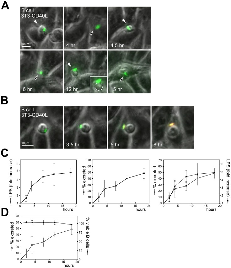
The observation that the number of primary B cells that had phagocytosed Salmonella dropped during prolonged culture of the Salmonella-containing B cells (Figure 2B, top right panel) suggested that Salmonella might be released from the B cells over time. To visualize the fate of phagocytosed Salmonella in B cells, primary human B cells infected with GFP-Salmonella were co-cultured on a monolayer of CD40L-expressing 3T3 cells and analyzed by time-lapse wide-field microscopy. Primary B cells that had phagocytosed GFP-Salmonella showed extensive invasive behaviour by continuously moving under and over the 3T3-CD40L monolayer (Figure 3A; see also Video S4). At later time points, a fraction of GFP-Salmonella appeared to be released from the B cell. To visualize this in more detail, GFP-Salmonella infected primary B cells were cultured in the presence of a low concentration of Texas-Red labeled anti-LPS mAb in the medium. GFP-Salmonella will attract and concentrate this antibody only upon exposure to the extracellular medium and are excluded from antibody recognition when confined to the B cell interior. Figure 3B shows a B cell with phagocytosed GFP-Salmonella that becomes accessible for anti-LPS antibodies in the medium after 5-8 hr of culture (Video S5). Salmonella excretion from primary B cells was quantified using FACS by detecting GFP-Salmonella and LPS-positive B cells. A strong increase in cell surface exposed LPS on cells that were initially GFP-Salmonella positive and LPS negative was observed (Figure 3C, left panel). This suggests that a large fraction of the phagocytosed Salmonella were exocytosed as in the example shown in Figure 3B. Accordingly, the population of GFP-Salmonella positive/LPS-negative B cells declined over time (Figure 3C, middle panel) with kinetics that were identical to the acquired LPS signal (Figure 3C, right panel), inferring an increased excretion. GFP-Salmonella infection of primary B cells did not affect B cell viability or induced apoptosis (Figure 3D). Note that during the first phase of excretion Salmonella was released, but remained associated to the B cells, hence the increased staining with the anti-LPS antibodies in the first 10h. The bacterium was later released from the B cell, leveling off further LPS labeling. Loss of the GFP-Salmonella signal from infected primary B cells increased over an 18h period in our experiments at which point more than 50% of the bacteria were released from B cells.
Figure 3. Salmonella is actively excreted by B cells.
(A) Primary B cells having phagocytosed anti-BCR coated GFP-Salmonella on a monolayer of 3T3-CD40L fibroblasts were imaged using widefield fluorescence microscopy. Depicted is the GFP signal projected on the transmission image with images taken every 30 min. Scalebar = 10 µm. Arrows indicate the B cell, white arrow: B cells moves op top of the monolayer, black arrow: B cells moves below the monolayer. Images are frames from Video S4. (B) Primary B cells having phagocytosed anti-BCR coated GFP-Salmonella on a monolayer of 3T3-CD40L fibroblasts were imaged using widefield fluorescence microscopy in the presence of TexasRed labeled anti-LPS mAbs. Depicted are GFP and Texas-Red signals projected on the transmission image. Scalebar = 10µm. Images are frames from Video S5. (C) Quantification of Salmonella secretion from B cells. Primary B cells were incubated with live uncoated GFP-Salmonella. Cells were stained with antibodies against LPS, fixed and analyzed using FACS. Left panel: increase in cell surface exposed LPS from bacteria exposed at the cell surface after initial uptake by B cells. Middle panel: percentage of B cells having excreted Salmonella as calculated from the percentage of B cells containing GFP-Salmonella followed in time. Right panel: left and middle panels are projected to illustrate that both processes show similar kinetics. Error bars represent SD from three independent experiments. (D) Primary B cells were incubated with live uncoated GFP-expressing Salmonella and followed for the time points indicated. The fraction of living B cells is plotted to demonstrate that loss of GFP-Salmonella positive B cells is not correlated with cell death.
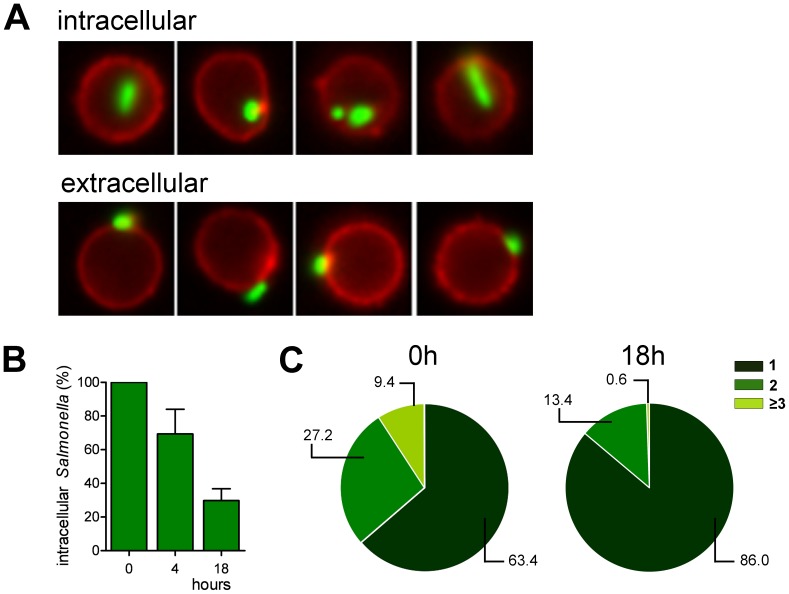
Bacterial excretion was further quantified by Imagestream analyses. This technique directly combines quantitative information obtained by flow cytometry with the visual information of subcellular bacterial localization by microscopy. Specific gating of B cells that had phagocytosed bacteria enabled exact follow-up of B cells with internalized bacteria over time (Figure 4A). Analysis showed a gradual decrease of intracellular Salmonella within 18 hrs of infection (Figure 4B). In addition, we tracked the numbers of internalized Salmonella per B cell over time, by separating the B cells that had phagocytosed Salmonella into different pools based on the GFP-signal per B cell (Figure S1). Immediately after infection, B cells had internalized between 1 and 4 bacteria per cell. Only 1 or 2 bacteria remained per B cell 18 hours later (Figure 4C). In conclusion, our data show that a substantial fraction of phagocytosed Salmonella is slowly secreted during the first 24 hrs. This mechanism is not tightly regulated as secretion occurs at an almost linear rate over the first 10 hours of culture post-infection.
Figure 4. Quantification of the fate of the GFP-expressing Salmonella in infected B cells.
(A) B cells were infected with anti-BCR coated GFP-expressing Salmonella (green).The plasma membrane of the B cells (red) was stained using an anti-CD20 mAb to discriminate between intracellular and extracellular Salmonella. (B) The relative amount of intracellular Salmonella were measured immediately after infection (0 h), or 4 h and 18 h post-infection. Error bars represent SD from two independent experiments. (C) The number of Salmonella per B cell was measured immediately after infection and 18h post-infection. A representative experiment of two individual experiments is shown.
Excretion of Salmonella is a B Cell Autonomous Process
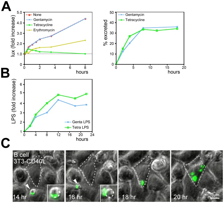
The excretion of Salmonella may be controlled by the pathogen or by the B cells. In our experiments we added antibiotic gentamicin at low concentrations to the medium to eliminate extracellular Salmonella (and prevent overgrowth of the cells by free Salmonella), as gentamicin does not affect intracellular Salmonella replication (see Figure 2A). Unlike gentamicin, the antibiotics tetracycline and erythromycin are able to enter phagosomes and eliminate intracellular Salmonella [27]. We validated this by testing growth of lux-Salmonella in Ramos cells. Tetracyclin and erythromycin inhibited intracellular Salmonella growth in Ramos cells (Figure 5A, left panel). In addition, no viable Salmonella bacteria were recovered in plating assays of infected B cells, while bacterial colonies were obtained from Salmonella-containing B cells exposed to gentamicin (data not shown). Salmonella secretion by primary B cells was measured in the presence of either gentamicin or tetracycline to test whether viable bacteria were required for B cell secretion. Tetracycline did not affect excretion of GFP-Salmonella from primary B cells. In fact, this occurred equally efficient as B cell excretion in the presence of gentamicin (Figure 5A, right panel), indicating that viability of Salmonella is not required for excretion. This was further confirmed by the observation that cell surface LPS levels increased at a similar rate when infected B cells were cultured in the presence of tetracycline as in the presence of gentamicin (Figure 5B). While Salmonella actively participates in uptake after capture by the BCR [20], excretion does not require viable Salmonella.
Figure 5. After excretion by B cells, Salmonella is capable of infecting secondary host cells.
(A) Left panel: the effect of antibiotics on the growth of lux-Salmonella in Ramos B cells. Right panel: the same FACS analysis with primary B cells as in 2C was performed in presence of either Gentamicin or Tetracycline to discriminate between host and bacteria mediated excretion. (B) Quantification of Salmonella secretion from B cells. Primary B cells were incubated with live uncoated GFP-Salmonella in presence of either Gentamicin or Tetracycline to discriminate between host versus bacterial-mediated excretion. Cells were stained with antibodies against LPS, fixed and analyzed using FACS. Increase in cell surface LPS levels is similar in the presence of Gentamicin and Tetracycline, indicating that viable Salmonella are not required for excretion. (C) Primary B cells having phagocytosed anti-IgM coated GFP-Salmonella on a monolayer of 3T3-CD40L fibroblasts were imaged using widefield fluorescence microscopy for the times indicated. Imaging conditions are similar as in 2A. GFP-Salmonella is excreted from a primary B cell (white arrowhead), followed by infection of the 3T3-CD40L monolayer (outline of infected cell marked by a dashed line). Inset shows zoom-in on primary B cell excreting GFP-Salmonella. Images are frames from Video S6.
Excretion of Salmonella from B Cells Allows Reproductive Reinfection of Other Cell Types
As Salmonella survives within antigen-specific B cells, Salmonella could also infect other cell types when released from B cells at distant sites. We co-cultured primary human B cells containing phagocytosed GFP-Salmonella on a monolayer of 3T3-CD40L and followed the behavior of Salmonella using time-lapse wide-field microscopy. Figure 5C shows an example of a phagocytosed GFP-Salmonella entering the field of imaging. The GFP-Salmonella is released from the B cells and infects the underlying fibroblast monolayer, followed by rapid expansion inside these fibroblasts (see Video S6). The released bacteria not only infected but also resumed replication in the fibroblast monolayer, demonstrating that its passage through primary B cells had not disabled bacterial replication in an irreversible manner. Collectively these data suggest that Salmonella can use primary Salmonella-specific B cells as a survival reservoir and transport vehicle allowing escape from immune attack and transfer to distant locations.
Salmonella-specific B Cells Mediate Spreading in Acute in vivo Infection
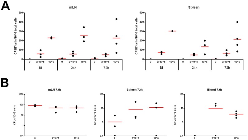
Our observations imply that the availability of B cells with pathogen-specific BCRs may support spreading of infection when used as carriers for Salmonella. In vivo, Salmonella first encounters B cells after crossing the intestinal epithelium via the M cells. The M cells are directly located over the gut-associated lymphoid tissue (GALT) sites where many B cells reside in Peyer’s patches. Among preferred distant sites of persistent infection for Salmonella are the spleen and mesenteric lymph nodes. Indeed, Salmonella has been isolated from splenic macrophages and splenic B cells of orogastrically infected mice [28]. How Salmonella spreads from the GALT to peripheral compartments is unclear. Transport of viable Salmonella by neutrophils [16] is probably inefficient given their efficient bactericidal capacity [5]. Also macrophages and DCs have been implicated, but their spreading beyond the mesenteric lymph nodes may be limited. Antigen-specific B cells as transport vehicles have not been considered. To test this option in vivo, different numbers of CD43- naive murine B cells (94% pure preparation) carrying a BCR specific for the HEL antigen were adoptively transferred into WT C57BL/6 mice. One day after transfer, mice were orally infected with surface HEL-expressing Salmonella. To directly establish if HEL-specific B cells circulate and mediate systemic dissemination of HEL-expressing Salmonella, we investigated both B cell and bacterial recovery from mesenteric lymph nodes, spleen and blood in the different experimental settings, 24 and 72 h after oral infection. Various organs or blood were collected and cells and Salmonella isolated. Transferred CFSE-labeled B cells were detected in the mesenteric lymph nodes and spleen and there were no signs of evident expansion (Figure 6A). Salmonella infection did not affect HEL+-B cell recovery from the organs. Lysis of organs recovered from infected mice followed by plating of the bacteria revealed the number of Colony Forming Units (CFU) in each compartment. Oral infection of mice with HEL-expressing Salmonella without adoptive B cell transfer resulted after 72 h after infection in prominent infiltration of the mesenteric lymph nodes and limited spreading to the spleen (Figure 6B). No bacteria were detected in circulating blood cells at the time of sampling, which points to low numbers of Salmonella infected cells in blood or a relative short transit time of the cells. Transfer of HEL-specific B cells mildly reduced bacterial dissemination to the mesenteric lymph nodes, but substantially enhanced spreading of Salmonella to the spleen (Figure 6B). In addition, Salmonella was now retrieved systemically from circulating cells in the blood compartment (Figure 6B).
Figure 6. Salmonella-specific B cells form a survival niche supporting in vivo Salmonella spreading to systemic sites.
C57BL/6 mice were adoptively transferred with 0, 2*105 or 106 HEL-specific CD43- naïve B cells labeled with CFSE, as indicated. Mice were orally infected with surface HEL-expressing Salmonella one day after B cell transfer. (A) Distribution of CFSE-labeled HEL-specific B cells in the mesenteric lymph nodes (mLN) and spleen before infection (BI), and 24 or 72 hours post-infection, as indicated. One representative example from 3 experiments with 4 mice for each experimental setting is shown. (B) Recovery of viable bacteria 72 hours post-infection from mesenteric lymph nodes (mLN) spleen (SP) and blood (BL) in infected mice transferred with 0, 2*105 or 106 HEL-specific B cells. Depicted are colony-forming units (CFU)/105 eukaryotic cells. One representative example from 3 experiments with 4 mice for each experimental setting is shown.
These data suggest that -in this oral-infection model- antigen-specific B cells do not only promote local spreading of Salmonella to the mesenteric lymph nodes, but strongly support systemic dissemination of Salmonella into the circulating blood pool and spleen. Pathogen-specific B cells thus form a novel example of immune cells that can be captured by bacteria for spreading and infection of distant sites.
Discussion
Recent data indicated that B cells from early vertebrates act as efficient phagocytes unlike mammalian B cells that did not show phagocytic behavior [26]. We demonstrated that human B cells have not lost this phagocytic capacity but require the BCR for phagocytosis of particles or pathogens [20]. Phagocytosed Salmonella infects and grows in many cell types, but can only be efficiently destroyed in specialized cells like macrophages and neutrophils by the NADPH-oxidase system [5]. A recent report showed also internalization of Salmonella in murine B cells, and demonstrated intracellular survival of Salmonella [29]. We previously noticed survival of Salmonella in B cells after BCR-mediated internalization and now studied the fate of these phagocytosed Salmonellae and the consequences for dissemination of infection in vivo.
We demonstrated that Salmonella can be excreted from primary B cells in time. The factors controlling Salmonella excretion are unclear. Our data suggest that the B cell rather than the bacterium controls this process, as both living and dead Salmonella can be excreted. Since many bacterial pathogens manipulate the host cell biology for survival, uptake by pathogen-specific B cells followed by release of the pathogen at distant sites may be a more general mechanism for pathogen spreading. Identification and manipulation of signaling pathways to prevent excretion would be a possible means to limit systemic spreading of Salmonella and potentially of other pathogens by B cells.
Does BCR-mediated immune escape and spreading by B cells play an important role during Salmonella infection? The bacteria may potentially encounter specific B cells very early during infection as they cross the intestinal epithelium and enter the GALT sites. Among preferred sites of persistent infection are localized mesenteric lymph nodes and the more distant spleen. Salmonella was thought to reach these locations after transport by neutrophils [16], a notion difficult to match with the efficient bactericidal capacity of neutrophils [5]. Our experiments indicate that Salmonella-specific B cells act as transport carriers for the in vivo spreading of Salmonella to the distant sites. In the event of excretion in B cell rich areas like the spleen and GALT, the bacterium might even go through multiple rounds of uptake (by other resident Salmonella-specific B cells), dissemination, and excretion. Salmonella thus abuses the specificity of the adaptive immune system to hide from the early innate immune defenses while hitch-hiking inside the B cell ensures systemic spreading of the infection. Dissemination of Salmonella to mesenteric lymph nodes does not require B cells, consistent with the concept that Salmonella-infected DCs can reach the mesenteric lymph nodes and may thus mediate this localized transport [3], [4], [30].
Salmonella survives in phagosomes in primary human B cells in a growth arrested manner. Salmonella replicates in other cell types via a pathway involving activation of the kinase Akt1 [10]. In B cells, Akt-signaling is negatively regulated after BCR-triggering by Rap1 and Rap2 GTPases [31]. Whether this explains the control of Salmonella replication in human primary B cells remains to be established. An effective CD8+ cytotoxic T cell response is essential to control Salmonella infection [32], [33], possibly to eliminate Salmonella hiding inside cells. Our data show that also systemic spreading of Salmonella will be limited by elimination of the intracellular bacteria in B cells. Thus, vaccination strategies that aim to induce both Salmonella-specific antibodies and Salmonella-restricted CD8+ T cells may yield strong synergistic effects.
In view of coevolution of pathogens and the eukaryotic immune system, it is striking that Salmonella abuses the specificity of the adaptive immune system. Previously, it was shown that cells of the innate immune system and erythrocytes can be hiked by pathogens for systemic spreading of infection within immunocompetent hosts. Examples include HIV (DCs) [34] and P. falciparum (erythrocytes) [35]. The adaptive immune system has evolved to clear infections, while simultaneously generating immunological memory to ensure rapid immunity against reinfection. We now show that pathogen-recognizing and antigen-specific immune cells from the adaptive immune system can be applied by bacteria to escape direct recognition by other immune cells and to mediate bacterial dissemination. Although the adaptive immune system responds to pathogens to limit infection, some pathogens, as illustrated here for Salmonella, have adapted to this and use the specific aspect in immune responses for survival and systemic spreading.
Supporting Information
GFP- Salmonella distribution per cell after 0 and 18 hours. B cells were infected with anti-BCR coated GFP-expressing Salmonella and the number of intracellular Salmonella was analyzed by ImagestreamX. Using IDEAS spot-count feature the numbers of intracellular Salmonella were discriminated by either one, two or three and more Salmonella per cell. Shown are examples of Salmonella count from one representative experiment of two independent experiments.
(TIF)
GFP- Salmonella growth in Ramos B cells. Widefield fluorescence microscopy of living cells at 37°C confirms the results from the FACS experiment in Fig. 2 B. Left panel: GFP signal, right panel: GFP signal projected on the transmission image, taken every 30 min. Bacterial growth is observed in a Ramos B cell that has phagocytosed anti-human BCR antibody coated GFP-expressing Salmonella. Total duration: 11.5 hours.
(MOV)
GFP- Salmonella does not grow in B cells. Widefield fluorescence microscopy of living primary human B cells at 37°C confirms the results from the FACS experiment in Fig. 2 B. Left panel: GFP signal, right panel: GFP signal projected on the transmission image, taken every 20 min. No growth of GFP-Salmonella is observed in primary B cells. Total duration: 14.5 hours.
(MOV)
GFP- Salmonella growth is actively suppressed in human primary B cells. Widefield fluorescence microscopy of living cells at 37°C as in Fig. 2 C. Left panel: GFP signal, right panel: GFP signal projected on the transmission image. Images taken every 30 min. Cells were treated with Edelfosine, an apoptosis-inducing alkyl-lysophospholipid. No bacterial growth is observed in primary B cells until the cell undergoes apoptosis after 2 h, showing clear apoptosis associated morphological changes of the nucleus and membrane blebbing. After the onset of apoptosis, Salmonella expansion is observed within the apoptosing cell. Total duration: 15 hours.
(MOV)
GFP- Salmonella containing human primary B cells show invasive behavior in a monolayer of CD40L-expressing 3T3 fibroblasts. A co-culture of living primary human B cells infected with GFP-expressing Salmonella and CD40L-expressing 3T3 cells was imaged using widefield fluorescence microscopy at 37°C. Depicted is the GFP signal projected on the transmission image with images taken every 30 min. The B cells repeatedly move under and over the monolayer. Arrows indicate the B cell, white: above the monolayer, black: under the monolayer. This Movie demonstrates the improved survival of B cells when co-cultured with CD40L expressing 3T3 cells. Also, excretion of the bacterium towards the end of the Movie (best visible at 12 h) is observed although the Salmonella is still contacting the B cell. Total duration: 15 hours.
(MOV)
GFP- Salmonella is excreted from primary human B cells. Living primary B cells infected with GFP-expressing Salmonella and co-cultured with CD40L-expressing 3T3 cells were imaged using widefield fluorescence microscopy at 37°C. Depicted are GFP and Texas-Red signals projected on the transmission image with images taken every 30 min. A single primary B cell that has phagocytosed anti-BCR coated GFP-expressing Salmonella is followed over time in medium containing anti-LPS antibodies labeled with Texas-Red. The bacterium is protected from staining by the extracellular antibodies as long as it resides intracellular. Double labeling is observed after 6.5 hr, showing access of the B cell-associated Salmonella to the antibody-containing medium. Total duration: 11.5 hours.
(MOV)
GFP- Salmonellae excreted from human primary B cells are capable of infecting 3T3-CD40L fibroblasts. Living primary human B cells infected with GFP-expressing Salmonella were co-cultured with CD40L-expressing 3T3 cells, and imaged by widefield fluorescence microscopy at 37°C. Depicted are GFP and Texas-Red signals projected on the transmission image. Images are collected every 30 min. 5 hours after the onset of the experiment a single human primary B cell infected with a GFP-expressing Salmonella moves from a distant location into the viewing plane from the upper right corner. The bacterium is excreted from the B cell and subsequently infects the 3T3-CD40L monolayer followed by rapid growth of Salmonella in the 3T3-CD40L cells. This further demonstrates that growth of the bacterium is actively suppressed inside the human primary B cell, while its viability is maintained. Primary B cells thus form a survival niche for Salmonella. Total duration: 15 hours.
(MOV)
Acknowledgments
We thank Marije Marsman and Coenraad Kuyl for discussions and help with the Salmonella experiments, Lauran Oomen and Lenny Brocks for support with CLSM imaging, Erik Mul, Floris van Alphen, Anita Pfauth and Frank van Diepen for flow cytometry and Nico Ong for photography.
Funding Statement
This work was supported by grants from the Dutch Cancer Society KWF (grant NKI 2001-2415), the Landsteiner Foundation for Blood Research (LSBR, grant 0533), Sanquin Blood Supply PPOC (PPOC 09-032) and the Netherlands Scientific Organization (N.W.O.). The funders had no role in study design, data collection and analysis, decision to publish, or preparation of the manuscript.
References
- 1. Jones BD, Falkow S (1996) Salmonellosis: host immune responses and bacterial virulence determinants. Annu Rev Immunol 14: 533–561. [DOI] [PubMed] [Google Scholar]
- 2. Jepson MA, Clark MA (2001) The role of M cells in Salmonella infection. Microbes Infect 3: 1183–1190. [DOI] [PubMed] [Google Scholar]
- 3. Rescigno M, Urbano M, Valzasina B, Francolini M, Rotta G, et al. (2001) Dendritic cells express tight junction proteins and penetrate gut epithelial monolayers to sample bacteria. Nat Immunol 2: 361–367. [DOI] [PubMed] [Google Scholar]
- 4. Vazquez-Torres A, Jones-Carson J, Baumler AJ, Falkow S, Valdivia R, et al. (1999) Extraintestinal dissemination of Salmonella by CD18-expressing phagocytes. Nature 401: 804–808. [DOI] [PubMed] [Google Scholar]
- 5. Fierer J (2001) Polymorphonuclear leukocytes and innate immunity to Salmonella infections in mice. Microbes Infect 3: 1233–1237. [DOI] [PubMed] [Google Scholar]
- 6. Wick MJ (2004) Living in the danger zone: innate immunity to Salmonella. Curr Opin Microbiol 7: 51–57. [DOI] [PubMed] [Google Scholar]
- 7. Patel JC, Galan JE (2005) Manipulation of the host actin cytoskeleton by Salmonella–all in the name of entry. Curr Opin Microbiol 8: 10–15. [DOI] [PubMed] [Google Scholar]
- 8. Gorvel JP, Meresse S (2001) Maturation steps of the Salmonella-containing vacuole. Microbes Infect 3: 1299–1303. [DOI] [PubMed] [Google Scholar]
- 9. Holden DW (2002) Trafficking of the Salmonella vacuole in macrophages. Traffic 3: 161–169. [DOI] [PubMed] [Google Scholar]
- 10. Kuijl C, Savage ND, Marsman M, Tuin AW, Janssen L, et al. (2007) Intracellular bacterial growth is controlled by a kinase network around PKB/AKT1. Nature 450: 725–730. [DOI] [PubMed] [Google Scholar]
- 11. Fields PI, Swanson RV, Haidaris CG, Heffron F (1986) Mutants of Salmonella typhimurium that cannot survive within the macrophage are avirulent. Proc Natl Acad Sci U S A 83: 5189–5193. [DOI] [PMC free article] [PubMed] [Google Scholar]
- 12. Meresse S, Unsworth KE, Habermann A, Griffiths G, Fang F, et al. (2001) Remodelling of the actin cytoskeleton is essential for replication of intravacuolar Salmonella. Cell Microbiol 3: 567–577. [DOI] [PubMed] [Google Scholar]
- 13. Hornef MW, Wick MJ, Rhen M, Normark S (2002) Bacterial strategies for overcoming host innate and adaptive immune responses. Nat Immunol 3: 1033–1040. [DOI] [PubMed] [Google Scholar]
- 14. Zwart W, Griekspoor A, Kuijl C, Marsman M, van Rheenen J, et al. (2005) Spatial separation of HLA-DM/HLA-DR interactions within MIIC and phagosome-induced immune escape. Immunity 22: 221–233. [DOI] [PubMed] [Google Scholar]
- 15. Gasem MH, Keuter M, Dolmans WMV, van der Ven-Jongekrijg J, Djokomoeljanto R, et al. (2003) Persistence of Salmonellae in blood and bone marrow: randomized controlled trial comparing ciprofloxacin and chloramphenicol treatments against enteric fever. Antimicrob Agents Chemother 47: 1727–1731. [DOI] [PMC free article] [PubMed] [Google Scholar]
- 16. Richter-Dahlfors A, Buchan AM, Finlay BB (1997) Murine salmonellosis studied by confocal microscopy: Salmonella typhimurium resides intracellularly inside macrophages and exerts a cytotoxic effect on phagocytes in vivo. J Exp Med 186: 569–580. [DOI] [PMC free article] [PubMed] [Google Scholar]
- 17. Mastroeni P (2002) Immunity to systemic Salmonella infections. Curr Mol Med 2: 393–406. [DOI] [PubMed] [Google Scholar]
- 18. Wu L, KewalRamani VN (2006) Dendritic-cell interactions with HIV: infection and viral dissemination. Nat Rev Immunol 6: 859–868. [DOI] [PMC free article] [PubMed] [Google Scholar]
- 19.Worley MJ, Nieman GS, Geddes K, Heffron F (2006) Salmonella typhimurium disseminates within its host by manipulating the motility of infected cells. Proc Natl Acad Sci U S A 103: 17915–17920. 0604054103 [pii];10.1073/pnas.0604054103 [doi]. [DOI] [PMC free article] [PubMed]
- 20. Souwer Y, Griekspoor A, Jorritsma T, de Wit J, Janssen H, et al. (2009) B cell receptor-mediated internalization of salmonella: a novel pathway for autonomous B cell activation and antibody production. J Immunol 182: 7473–7481. [DOI] [PubMed] [Google Scholar]
- 21. Marsman M, Jordens I, Kuijl C, Janssen L, Neefjes J (2004) Dynein-mediated vesicle transport controls intracellular Salmonella replication. Mol Biol Cell 15: 2954–2964. [DOI] [PMC free article] [PubMed] [Google Scholar]
- 22. Vesterlund S, Paltta J, Laukova A, Karp M, Ouwehand AC (2004) Rapid screening method for the detection of antimicrobial substances. J Microbiol Methods 57: 23–31. [DOI] [PubMed] [Google Scholar]
- 23. Cornelis P, Sierra JC, Lim A Jr, Malur A, Tungpradabkul S, et al. (1996) Development of new cloning vectors for the production of immunogenic outer membrane fusion proteins in Escherichia coli. Biotechnology (N Y ) 14: 203–208. [DOI] [PubMed] [Google Scholar]
- 24. Martinoli C, Chiavelli A, Rescigno M (2007) Entry route of Salmonella typhimurium directs the type of induced immune response. Immunity 27: 975–984. [DOI] [PubMed] [Google Scholar]
- 25. Ruiter GA, Zerp SF, Bartelink H, van Blitterswijk WJ, Verheij M (1999) Alkyl-lysophospholipids activate the SAPK/JNK pathway and enhance radiation-induced apoptosis. Cancer Res 59: 2457–2463. [PubMed] [Google Scholar]
- 26. Li J, Barreda DR, Zhang YA, Boshra H, Gelman AE, et al. (2006) B lymphocytes from early vertebrates have potent phagocytic and microbicidal abilities. Nat Immunol 7: 1116–1124. [DOI] [PubMed] [Google Scholar]
- 27. Kihlstrom E, Andaker L (1985) Inability of gentamicin and fosfomycin to eliminate intracellular Enterobacteriaceae. J Antimicrob Chemother 15: 723–728. [DOI] [PubMed] [Google Scholar]
- 28. Rosales-Reyes R, Alpuche-Aranda C, Ramirez-Aguilar ML, Castro-Eguiluz AD, Ortiz-Navarrete V (2005) Survival of Salmonella enterica serovar Typhimurium within late endosomal-lysosomal compartments of B lymphocytes is associated with the inability to use the vacuolar alternative major histocompatibility complex class I antigen-processing pathway. Infect Immun 73: 3937–3944. [DOI] [PMC free article] [PubMed] [Google Scholar]
- 29.Rosales-Reyes R, Perez-Lopez A, Sanchez-Gomez C, Hernandez-Mote RR, Castro-Eguiluz D, et al. (2012) Salmonella infects B cells by macropinocytosis and formation of spacious phagosomes but does not induce pyroptosis in favor of its survival. Microb Pathog 52: 367–374. S0882–4010(12)00073–3 [pii];10.1016/j.micpath.2012.03.007 [doi]. [DOI] [PubMed]
- 30. Bogunovic M, Ginhoux F, Helft J, Shang L, Hashimoto D, et al. (2009) Origin of the lamina propria dendritic cell network. Immunity 31: 513–525. [DOI] [PMC free article] [PubMed] [Google Scholar]
- 31. Christian SL, Lee RL, McLeod SJ, Burgess AE, Li AH, et al. (2003) Activation of the Rap GTPases in B lymphocytes modulates B cell antigen receptor-induced activation of Akt but has no effect on MAPK activation. J Biol Chem 278: 41756–41767. [DOI] [PubMed] [Google Scholar]
- 32. de Wit J, Souwer Y, Jorritsma T, Klaasse BH, ten Brinke A, et al. (2010) Antigen-specific B cells reactivate an effective cytotoxic T cell response against phagocytosed Salmonella through cross-presentation. PLoS One 5: e13016. [DOI] [PMC free article] [PubMed] [Google Scholar]
- 33. Salerno-Goncalves R, Sztein MB (2009) Priming of Salmonella enterica serovar typhi-specific CD8(+) T cells by suicide dendritic cell cross-presentation in humans. PLoS One 4: e5879. [DOI] [PMC free article] [PubMed] [Google Scholar]
- 34. Geijtenbeek TB, Kwon DS, Torensma R, van Vliet SJ, van Duijnhoven GC, et al. (2000) DC-SIGN, a dendritic cell-specific HIV-1-binding protein that enhances trans-infection of T cells. Cell 100: 587–597. [DOI] [PubMed] [Google Scholar]
- 35. Miller LH, Baruch DI, Marsh K, Doumbo OK (2002) The pathogenic basis of malaria. Nature 415: 673–679. [DOI] [PubMed] [Google Scholar]
Associated Data
This section collects any data citations, data availability statements, or supplementary materials included in this article.
Supplementary Materials
GFP- Salmonella distribution per cell after 0 and 18 hours. B cells were infected with anti-BCR coated GFP-expressing Salmonella and the number of intracellular Salmonella was analyzed by ImagestreamX. Using IDEAS spot-count feature the numbers of intracellular Salmonella were discriminated by either one, two or three and more Salmonella per cell. Shown are examples of Salmonella count from one representative experiment of two independent experiments.
(TIF)
GFP- Salmonella growth in Ramos B cells. Widefield fluorescence microscopy of living cells at 37°C confirms the results from the FACS experiment in Fig. 2 B. Left panel: GFP signal, right panel: GFP signal projected on the transmission image, taken every 30 min. Bacterial growth is observed in a Ramos B cell that has phagocytosed anti-human BCR antibody coated GFP-expressing Salmonella. Total duration: 11.5 hours.
(MOV)
GFP- Salmonella does not grow in B cells. Widefield fluorescence microscopy of living primary human B cells at 37°C confirms the results from the FACS experiment in Fig. 2 B. Left panel: GFP signal, right panel: GFP signal projected on the transmission image, taken every 20 min. No growth of GFP-Salmonella is observed in primary B cells. Total duration: 14.5 hours.
(MOV)
GFP- Salmonella growth is actively suppressed in human primary B cells. Widefield fluorescence microscopy of living cells at 37°C as in Fig. 2 C. Left panel: GFP signal, right panel: GFP signal projected on the transmission image. Images taken every 30 min. Cells were treated with Edelfosine, an apoptosis-inducing alkyl-lysophospholipid. No bacterial growth is observed in primary B cells until the cell undergoes apoptosis after 2 h, showing clear apoptosis associated morphological changes of the nucleus and membrane blebbing. After the onset of apoptosis, Salmonella expansion is observed within the apoptosing cell. Total duration: 15 hours.
(MOV)
GFP- Salmonella containing human primary B cells show invasive behavior in a monolayer of CD40L-expressing 3T3 fibroblasts. A co-culture of living primary human B cells infected with GFP-expressing Salmonella and CD40L-expressing 3T3 cells was imaged using widefield fluorescence microscopy at 37°C. Depicted is the GFP signal projected on the transmission image with images taken every 30 min. The B cells repeatedly move under and over the monolayer. Arrows indicate the B cell, white: above the monolayer, black: under the monolayer. This Movie demonstrates the improved survival of B cells when co-cultured with CD40L expressing 3T3 cells. Also, excretion of the bacterium towards the end of the Movie (best visible at 12 h) is observed although the Salmonella is still contacting the B cell. Total duration: 15 hours.
(MOV)
GFP- Salmonella is excreted from primary human B cells. Living primary B cells infected with GFP-expressing Salmonella and co-cultured with CD40L-expressing 3T3 cells were imaged using widefield fluorescence microscopy at 37°C. Depicted are GFP and Texas-Red signals projected on the transmission image with images taken every 30 min. A single primary B cell that has phagocytosed anti-BCR coated GFP-expressing Salmonella is followed over time in medium containing anti-LPS antibodies labeled with Texas-Red. The bacterium is protected from staining by the extracellular antibodies as long as it resides intracellular. Double labeling is observed after 6.5 hr, showing access of the B cell-associated Salmonella to the antibody-containing medium. Total duration: 11.5 hours.
(MOV)
GFP- Salmonellae excreted from human primary B cells are capable of infecting 3T3-CD40L fibroblasts. Living primary human B cells infected with GFP-expressing Salmonella were co-cultured with CD40L-expressing 3T3 cells, and imaged by widefield fluorescence microscopy at 37°C. Depicted are GFP and Texas-Red signals projected on the transmission image. Images are collected every 30 min. 5 hours after the onset of the experiment a single human primary B cell infected with a GFP-expressing Salmonella moves from a distant location into the viewing plane from the upper right corner. The bacterium is excreted from the B cell and subsequently infects the 3T3-CD40L monolayer followed by rapid growth of Salmonella in the 3T3-CD40L cells. This further demonstrates that growth of the bacterium is actively suppressed inside the human primary B cell, while its viability is maintained. Primary B cells thus form a survival niche for Salmonella. Total duration: 15 hours.
(MOV)


