

Abstract
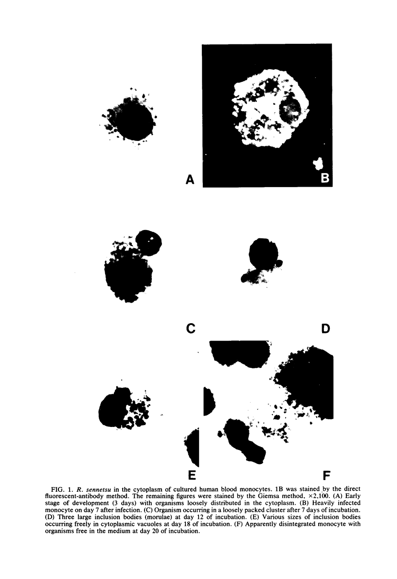
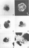
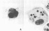
Microscopic examination of cultured human monocytes infected with Rickettsia sennetsu and stained by the Giemsa method revealed the presence of various organismal growth forms in the cytoplasm of the infected cells. The growth forms were loosely scattered individual organisms, clusters of organisms, various sizes of dense inclusion bodies in intact and vacuolated cytoplasm, and organisms in close proximity to disintegrated monocytes. The appearance and the morphology of these R. sennetsu growth forms were similar to those of Ehrlichia canis propagated in canine monocytes. Specific identification of R. sennetsu was made by staining cultured monocytes with fluorescein-conjugated globulins extracted from the pooled sera of patients recovering from sennetsu rickettsiosis. Mice infected with cultured R. sennetsu developed gross pathological changes indicative of infection, and the organism was demonstrated in their spleens, peritoneal macrophages, and mononuclear blood cells. Human monocyte culture appeared to be more sensitive than the previously used African green monkey kidney cell line (BSC-1) for the isolation of R. sennetsu from samples containing minute infectious quantities of this organism.
Full text
PDF

Images in this article
Selected References
These references are in PubMed. This may not be the complete list of references from this article.
- Anderson D. R., Hopps H. E., Barile M. F., Bernheim B. C. Comparison of the ultrastructure of several rickettsiae, ornithosis virus, and Mycoplasma in tissue culture. J Bacteriol. 1965 Nov;90(5):1387–1404. doi: 10.1128/jb.90.5.1387-1404.1965. [DOI] [PMC free article] [PubMed] [Google Scholar]
- Brown J. L., Huxsoll D. L., Ristic M., Hildebrandt P. K. In vitro cultivation of Neorickettsia helminthoeca, the causative agent of salmon poisoning disease. Am J Vet Res. 1972 Aug;33(8):1695–1700. [PubMed] [Google Scholar]
- Buhles W. C., Huxsoll D. L., Ruch G., Kenyon R. H., Elisberg B. L. Evaluation ofprimary blood monocyte and bone marrow cell culture for the isolation of Rickettsia rickettsii. Infect Immun. 1975 Dec;12(6):1457–1463. doi: 10.1128/iai.12.6.1457-1463.1975. [DOI] [PMC free article] [PubMed] [Google Scholar]
- DeShazo R. D., Boyce J. R., Osterman J. V., Stephenson E. H. Early diagnosis of Rocky Mountain spotted fever. Use of primary monocyte culture technique. JAMA. 1976 Mar 29;235(13):1353–1355. [PubMed] [Google Scholar]
- Huxsoll D. L., Hildebrandt P. K., Nims R. M., Walker J. S. Tropical canine pancytopenia. J Am Vet Med Assoc. 1970 Dec 1;157(11):1627–1632. [PubMed] [Google Scholar]
- Jones T. C. Macrophages and intracellular parasitism. J Reticuloendothel Soc. 1974 May;15(5):439–450. [PubMed] [Google Scholar]
- Nyindo M. B., Ristic M., Huxsoll D. L., Smith A. R. Tropical canine pancytopenia: in vitro cultivation of the causative agent--Ehrlichia canis. Am J Vet Res. 1971 Nov;32(11):1651–1658. [PubMed] [Google Scholar]
- Ristic M., Huxsoll D. L., Weisiger R. M., Hildebrandt P. K., Nyindo M. B. Serological diagnosis of tropical canine pancytopenia by indirect immunofluorescence. Infect Immun. 1972 Sep;6(3):226–231. doi: 10.1128/iai.6.3.226-231.1972. [DOI] [PMC free article] [PubMed] [Google Scholar]
- Shirai A., Sankaran V., Gan E., Huxsoll D. L. Early detection of Rickettsia tsutsugamushi in peripheral monocyte cultures derived from experimentally infected monkeys and dogs. Southeast Asian J Trop Med Public Health. 1978 Mar;9(1):11–14. [PubMed] [Google Scholar]
- Silverstein S. C. Endocytic uptake of particles by mononuclear phagocytes and the penetration of obligate intracellular parasites. Am J Trop Med Hyg. 1977 Nov;26(6 Pt 2):161–169. doi: 10.4269/ajtmh.1977.26.161. [DOI] [PubMed] [Google Scholar]
- Weiss E. Growth and physiology of rickettsiae. Bacteriol Rev. 1973 Sep;37(3):259–283. doi: 10.1128/br.37.3.259-283.1973. [DOI] [PMC free article] [PubMed] [Google Scholar]