Abstract
Gestational diabetes, occurring during the hyperglycemic period of pregnancy in maternal life, is a pathologic state that increases the incidence of complications in both mother and fetus. Offspring thus exposed to an adverse fetal and early postnatal environment may manifest increased susceptibility to a number of chronic diseases later in life. Compelling evidence for the role of epigenetic transmission in these complications has come from comparison of siblings born before and after the development of maternal diabetes, exposure to this intrauterine diabetic environment being shown to cause alterations in fetal growth patterns which predispose these infants to developing overweight and obesity later in life. Diabetes of the offspring is also mainly the consequence of exposure to the diabetic intrauterine environment, in addition to genetic susceptibility. Since obesity and diabetes are known to increase the risk of cardiovascular disease, cardiovascular sequelae in the offspring of diabetic mothers are virtually inevitable. Research data also suggest that exposure to a diabetic intrauterine environment during pregnancy is associated with an increase in dyslipidemia, subclinical vascular inflammation, and endothelial dysfunction processes in the offspring, all of which are linked with development of cardiovascular disease later in life. The main underlying mechanisms involve persistent hyperglycemia hyperinsulinemia and leptin resistance.
1. Introduction
The prevalence of obesity and type 2 diabetes amongst all age groups has, in the present time, reached alarming levels. As a result, more and more women of child-bearing age are either obese and/or diabetic during pregnancy. Pregnancy is a progressively hyperglycemic period, maternal hyperglycemia being necessary for the nutritional needs of the growing fetus [1]. However, maternal diabetes is a pathologic state that increases the incidence of complications in both the mother and the fetus [2], while it also exposes the women affected to higher risk for metabolic syndrome, subsequent type 2 diabetes, and cardiovascular disease later in life [3]. Long-term consequences for the offspring are also significant. Adipokines and inflammatory mediators are the main factors causing chronic subclinical inflammation which leads to insulin resistance and abnormality in glucose metabolism [1, 3], while there is additionally increasing evidence that exposure to an adverse fetal and/or early postnatal environment may increase susceptibility to a number of chronic diseases later in the life of the offspring. There is thus great interest in identifying the exact mechanisms by which maternal excess glucose may lead to diseases in the offspring later in life for the development of strategies to prevent this destructive cycle of metabolic dysfunction through generations.
Depending on the diagnostic and screening criteria, it has been observed that the prevalence of gestational diabetes ranges from 1.3% to 19.9% [4]. The recently established criteria use a 75 g oral glucose tolerance test (OGTT) without prior glucose challenge and diagnose gestational diabetes when the fasting glucose is ≥5.1 mmol/L and/or when the 1 h postload glucose is ≥10.0 mmol/L and/or when the 2 h postload glucose is ≥8.5 mmol/L [5]. Previousgestational diabetes, advanced maternal age, and obesity have the highest impact ongestational diabetesrisk. Racial/ethnic origin and family history of type 2diabeteshave a significant though moderate impact (except for type 2diabetesin siblings). Several nontraditionalfactors have recently been characterized, these being either physiological (low birthweight and short maternal height) or pathological (polycystic ovaries). The combination of the multiplicity ofrisk factorsand their interactions results in a low reliability ofriskprediction on an individual basis [6]. Traditional management includes diet, exercise, and short- and intermediate-acting insulin regimens. Meanwhile, use of metformin and glyburide is still controversial, but evidence substantiating their safety and efficacy is accumulating. Finally, postpartum screening with a glucose tolerance test rather than a fasting blood glucose level should be performed 6 weeks after delivery [7].
2. Offspring Diseases due to Hyperglycemic Intrauterine Environment
2.1. Offspring Growth and Adiposity
Offspring of diabetic mothers display excess fetal growth resulting in macrosomic and large for gestational age (LGA) infants [8], hence contributing to the increased risk for cesarean section or traumatic birth [9]. This excess fetal growth is caused by increased nutrients availability from the mother to the developing fetus through the placenta. Maternal serum glucose, which is the main excess nutrient in these circumstances, freely crosses the placenta, while maternal insulin does not. As a result of the thus induced fetal hyperglycemia, the fetal pancreas, although immature, is capable of producing increased levels of insulin, which in turn acts as a growth hormone and promotes growth and adiposity in the fetus [10]. The degree of hyperglycemia seems to determine the metabolic effect on the neonate [11]. Additionally to glucose excess, alterations in the delivery of amino acids and upregulation of placental transport systems also contribute to increased fetal growth [9, 12]. Notably, exposure to this intrauterine diabetic environment causes alterations in fetal growth patterns which predispose these infants to being overweight and obese also later in life, even in the absence of macrosomia at birth [13]. Insulin seems to play a key role, since amniotic fluid insulin levels during the third trimester correlate independently with infants' obesity [14].
2.2. Offspring Glucose Tolerance Disturbances
Many studies have shown that offspring of diabetic mothers have a statistically higher incidence of impaired glucose tolerance (IGT), this constituting a well known prediabetic state [15]. In the case of gestational diabetes, offspring have been shown to display reduced insulin secretion, while in the case of pre-existing diabetes, offspring have been shown to exhibit heightened insulin resistance, this possibly indicating a small difference in the underlying mechanisms [16]. Past studies also demonstrated that in all age groups of offspring who were exposed to hyperglycemia in utero, the incidence of diabetes was higher compared with the offspring of nondiabetic mothers, even though some of the latter would still develop diabetes in the future [17]. Therefore, offspring diabetes is mainly the consequence of exposure to a diabetic intrauterine environment, in conjunction with genetic susceptibility.
2.3. Offspring Cardiovascular Abnormalities
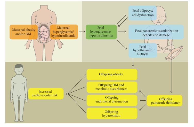
The main issue is whether these metabolic abnormalities of the offspring can augment their risk for cardiovascular diseases later in life, since such a finding would prompt closer glycemic control of their diabetic mothers and, possibly, closer health surveillance of the offspring themselves as a high risk population. However, few human studies have examined the effect of a diabetic intrauterine environment on cardiovascular risk factors of the offspring (Figure 1). Nevertheless, since obesity and diabetes are known to increase the risk of cardiovascular disease, the assumption is that cardiovascular consequences will arise in the offspring of diabetic mothers [18].
Figure 1.
Maternal hyperglycemia and hyperinsulinemia and, consequently, fetal hyperglycemia and hyperinsulinemia alter the function of every stage of fetal metabolism, including the hypothalamus, the pancreas, and adipose tissue. These metabolic disturbances pass through the offspring and, eventually, could increase cardiovascular risk for the maturing young adult.
Evidence of cardiovascular changes in pregnancies complicated by diabetes is already apparent during the third trimester of in utero life. The fetal heart shows reduced ventricular contractility compared with pregnancies not complicated by diabetes, even if the latter were complicated by hypertensive disease [19]. These findings suggest that the diabetic intrauterine environment induces biochemical alterations in the cardiovascular system that affect its function and that these changes are distinct from those caused by other poor intrauterine environments, such as those seen in hypertensive pregnancies.
In addition, systolic blood pressure, a well-known risk factor for cardiovascular diseases, of children born to diabetic mothers was significantly higher than that of those born to nondiabetic mothers [14, 20].
Research data also suggest that exposure to a diabetic intrauterine environment during pregnancy is associated with an increase in dyslipidemia, subclinical vascular inflammation, and endothelial dysfunction processes in the offspring, all of which are linked with development of cardiovascular disease later in life. Dyslipidemia is expressed by increased total and LDL cholesterol. The vascular inflammation and endothelial dysfunction markers examined were plasminogen activator inhibitor-1 (PAI-1), vascular adhesion molecule-1 (VCAM), intercellular adhesion molecule-1 (ICAM), E-selectin, insulin-like growth factor-1 (IGF-1), and others [21]. Thus, women with gestational diabetes and their fetuses demonstrate alterations in markers of eNOS uncoupling, oxidative stress, and endothelial dysfunction, and these changes correlate with the levels of hyperglycemia [22].
3. Epigenetic Modifications
Although diabetes type 2 is a disease with a known genetic component and is usually associated with a positive family history, this is not always the case. In fact, the current increasing incidence of the disease, which has risen to almost epidemic proportions, reveals that there must be a potent environmental component contributing to the disease as well. Many postnatal risk factors have been studied, like obesity, poor physical activity, and inappropriate diet. Nevertheless, prenatal exposure to a diabetic intrauterine environment seems to contribute to the development of the disease in the offspring later in life. Among females, it additionally contributes to the development of gestational diabetes, thus promoting a positive feedback loop for this disease. Animal models reveal that this metabolic imprinting can be transmitted across generations [23, 24].
Children who were exposed to a diabetic intrauterine environment during pregnancy are more likely to be obese [25], and there is a higher incidence of diabetes later in life among them than that is genetically expected, by almost 40% [26]. Adjustment for maternal weight does not explain the excess risk of the offspring, thereby supporting the hypothesis that nutrient-mediated developmental abnormalities in utero contribute independently to the development of obesity and diabetes in the offspring of diabetic mothers [13, 25].
Moreover, an excess of maternal to paternal transmission of diabetes has been widely reported, suggesting an epigenetic transmission [27, 28]. In other words, the extent to which maternal transmission exceeds paternal transmission can only be attributed to the intrauterine exposure to diabetes.
However, the strongest evidence for the role of epigenetic transmission has come from the comparison of siblings born before and after the development of maternal diabetes. Since siblings have the same risk of carrying the “diabetic” genes of their mothers, the excess risk that was observed in those born after the development of diabetes in their mothers can only be attributed to the hyperglycemic intrauterine environment [29].
Early epigenetic effects, such as those of an intrauterine hyperglycemic environment, play a significant role not only in the development of disease but also in its course. This is shown by studies in which offspring of mothers with diabetes before pregnancy developed diabetes at a younger age than those of mothers without diabetes who developed diabetes later after labor [30].
Finally, postnatal nutritional status is another epigenetic surfactant that influences the risk of diabetes in the offspring of diabetic mothers, with breastfeeding being protective [31]. We could thus reasonably suggest that the risk of diabetes is a combination of early prenatal and postnatal factors that act on a genetic basis and can trigger the disease sooner or even de novo.
4. Underlying Mechanisms
It is by now well established that insulin promotes storage of excess nutrition and fat mass, while at the same time leptin production from adipocytes acts as a safe mode negative loop mechanism that suppresses further insulin secretion. Interestingly, these two parameters, insulin and leptin, seem to play key roles in the metabolic disturbance [32].
There is evidence that hyperinsulinism can cause metabolic changes in the offspring of normoglycemic mothers. This evidence has come from animal experiments and reveals that hyperinsulinism rather than hyperglycemia could be the main surfactant acting on the offspring of diabetic mothers [33].
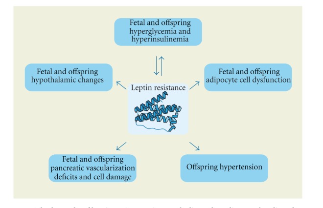
On the other hand, cord blood levels of leptin are also higher in the offspring of diabetic mothers than nondiabetic [34]. It cannot thus be hypothesized that a deficiency of leptin production in the case of diabetes has a role in the pathology of the offspring. Even more significantly, there is no connection between leptin and IGF-1 [35], though neither does the increase of leptin seem to act prophylactically for the offspring. What could hence be hypothesized is a type of leptin resistance in the case of neonates of diabetic mothers as a result of persistent hyperglycemia and hyperinsulinemia, just as insulin resistance is a progenitor of diabetic pathology. This leptin resistance might be the result of dysfunction of adipocytes or pancreatic cells and could be the cause of further hyperinsulinemia and a progressive positive loop mechanism (Figure 2).
Figure 2.
The key role of leptin resistance in metabolic and cardiovascular disturbances.
However, elevated leptin concentrations during diabetic pregnancy may be due to its secretion by adipocytes in the presence of elevated estrogen and also by placenta. In gestational diabetes, adipose tissue secretes low adiponectin (an anti-inflammatory and positive stimulator of insulin sensitizing) and high TNF-α and IL-6, which contribute to the inflammatory state and insulin resistance present in diabetic pregnancy as well as in macrosomia [36]. Leptin, being proinflammatory, is produced in abundance by adipose tissue during diabetic pregnancy and has been implicated in the pathogenesis of weight gain in macrosomic babies. Leptin may exert its effects by interacting with neuropeptide-Y in the hypothalamus, while the intrauterine hyperglycemia may act on the fetal hypothalamus and create what has been termed a “metabolic memory” which programs for development of obesity and metabolic syndrome in the offspring during adulthood [37].
As this condition goes on, further damage of pancreatic cells, as a result of severe hyperglycemia, is liable to cause defective insulin secretion and could be the starting point for initiation of glucose tolerance disturbances [38]. In addition, some data exist concerning intrauterine pancreatic vascularization deficits through alterations in factors like vascular endothelial growth factor (VEGF) and its receptors and intrauterine pancreatic innervation disturbances in diabetic pregnancies [38, 39].
5. Conclusions
The present-day diabetes epidemic incurs enormous costs, especially when one takes into account the adverse consequences wrought upon the future lives of both mother and offspring. Reducing obesity and type 2 diabetes should therefore be a primary goal of public health organizations and clinicians. Meanwhile, in the case of diabetic pregnancies, it is necessary to bear in mind that offspring metabolic dysfunction is a composite result of genetic and epigenetic factors, the latter also involving alterations in placental transport, endocrine molecules, like insulin and leptin, and inflammatory markers. Thus, with regard to the clinical management of women with diabetes during pregnancy, there may be the need to focus not only on achieving tight control of maternal blood glucose levels, but also on additional dietary and/or other changes, such as encouragement of breastfeeding. In summary, there is clearly an urgent need for the accumulation of specialized knowledge as to the most effective strategies to deal with metabolic disturbances and risk for chronic diseases in the offspring of diabetic mothers as a result of in utero exposure to diabetes.
References
- 1.Vrachnis N, Belitsos P, Sifakis S, et al. Role of adipokines and other inflammatory mediators in gestational diabetes mellitus and previous gestational diabetes mellitus. International Journal of Endocrinology. 2012;2012:12 pages. doi: 10.1155/2012/549748.549748 [DOI] [PMC free article] [PubMed] [Google Scholar]
- 2.Vitoratos N, Vrachnis N, Valsamakis G, Panoulis K, Creatsas G. Perinatal mortality in diabetic pregnancy. Annals of the New York Academy of Sciences. 2010;1205:94–98. doi: 10.1111/j.1749-6632.2010.05670.x. [DOI] [PubMed] [Google Scholar]
- 3.Vrachnis N, Augoulea A, Iliodromiti Z, Lambrinoudaki I, Sifakis S, Creatsas G. Previous gestational diabetes mellitus and markers of cardiovascular risk. International Journal of Endocrinology. 2012;2012:6 pages. doi: 10.1155/2012/458610.458610 [DOI] [PMC free article] [PubMed] [Google Scholar]
- 4.Simmons D. Epidemiology of diabetes in pregnancy. In: McCance D, Maresh M, editors. Practical Management of Diabetes in Pregnancy. London, UK: Blackwell Publishing.; 2010. [Google Scholar]
- 5.IADPSG Consensus Panel. International association of diabetes and pregnancy study groups (IADPSG) recommendations on the diagnosis and classification of hyperglycemia in pregnancy. Diabetes Care. 2010;33(7):676–682. doi: 10.2337/dc09-1848. [DOI] [PMC free article] [PubMed] [Google Scholar]
- 6.Galtier F. Definition, epidemiology, risk factors. Diabetes & Metabolism. 2010;36(6):628–651. doi: 10.1016/j.diabet.2010.11.014. [DOI] [PubMed] [Google Scholar]
- 7.Evensen AE. Update on gestational diabetes mellitus. Primary Care. 2012;39(1):83–94. doi: 10.1016/j.pop.2011.11.011. [DOI] [PubMed] [Google Scholar]
- 8.Lampl M, Jeanty P. Exposure to maternal diabetes is associated with altered fetal growth patterns: a hypothesis regarding metabolic allocation to growth under hyperglycemic-hypoxemic conditions. American Journal of Human Biology. 2004;16(3):237–263. doi: 10.1002/ajhb.20015. [DOI] [PubMed] [Google Scholar]
- 9.Jansson T, Cetin I, Powell TL, et al. Placental transport and metabolism in fetal overgrowth—a workshop report. Placenta. 2006;27:109–113. doi: 10.1016/j.placenta.2006.01.017. [DOI] [PubMed] [Google Scholar]
- 10.Ashworth MA, Leach FN, Milner RD. Development of insulin secretion in the human fetus. Archives of Disease in Childhood. 1973;48(2):151–152. doi: 10.1136/adc.48.2.151. [DOI] [PMC free article] [PubMed] [Google Scholar]
- 11.Catalano PM, Thomas A, Huston-Presley L, Amini SB. Increased fetal adiposity: a very sensitive marker of abnormal in utero development. American Journal of Obstetrics and Gynecology. 2003;189(6):1698–1704. doi: 10.1016/s0002-9378(03)00828-7. [DOI] [PubMed] [Google Scholar]
- 12.Ericsson A, Säljö K, Sjöstrand E, et al. Brief hyperglycaemia in the early pregnant rat increases fetal weight at term by stimulating placental growth and affecting placental nutrient transport. Journal of Physiology. 2007;581(3):1323–1332. doi: 10.1113/jphysiol.2007.131185. [DOI] [PMC free article] [PubMed] [Google Scholar]
- 13.Pettitt DJ, Knowler WC, Bennett PH. Obesity in offspring of diabetic Pima Indian women despite normal birth weight. Diabetes Care. 1987;10(1):76–80. doi: 10.2337/diacare.10.1.76. [DOI] [PubMed] [Google Scholar]
- 14.Silverman BL, Rizzo T, Green OC, et al. Long-term prospective evaluation of offspring of diabetic mothers. Diabetes. 1991;40(supplement 2):121–125. doi: 10.2337/diab.40.2.s121. [DOI] [PubMed] [Google Scholar]
- 15.Silverman BL, Metzger BE, Cho NH, Loeb CA. Impaired glucose tolerance in adolescent offspring of diabetic mothers: relationship to fetal hyperinsulinism. Diabetes Care. 1995;18(5):611–617. doi: 10.2337/diacare.18.5.611. [DOI] [PubMed] [Google Scholar]
- 16.Plagemann A, Harder T, Kohlhoff R, Rohde W, Dörner G. Glucose tolerance and insulin secretion in children of mothers with pregestational IDDM or gestational diabetes. Diabetologia. 1997;40(9):1094–1100. doi: 10.1007/s001250050792. [DOI] [PubMed] [Google Scholar]
- 17.Dabelea D, Pettitt DJ. Intrauterine diabetic environment confers risks for type 2 diabetes mellitus and obesity in the offspring, in addition to genetic susceptibility. Journal of Pediatric Endocrinology and Metabolism. 2001;14(8):1085–1091. doi: 10.1515/jpem-2001-0803. [DOI] [PubMed] [Google Scholar]
- 18.Halfon N, Verhoef PA, Kuo AA. Childhood antecedents to adult cardiovascular disease. Pediatrics in Review. 2012;33(2):51–60. doi: 10.1542/pir.33-2-51. [DOI] [PubMed] [Google Scholar]
- 19.Rasanen J, Kirkinen P. Growth and function of human fetal heart in normal, hypertensive and diabetic pregnancy. Acta Obstetricia et Gynecologica Scandinavica. 1987;66(4):349–353. doi: 10.3109/00016348709103651. [DOI] [PubMed] [Google Scholar]
- 20.Bunt JC, Antonio Tataranni P, Salbe AD. Intrauterine exposure to diabetes is a determinant of hemoglobin A1c and systolic blood pressure in pima indian children. Journal of Clinical Endocrinology and Metabolism. 2005;90(6):3225–3229. doi: 10.1210/jc.2005-0007. [DOI] [PMC free article] [PubMed] [Google Scholar]
- 21.Manderson J, Mullan B, Patterson C, Hadden D, Traub A, McCance D. Cardiovascular and metabolic abnormalities in the offspring of diabetic pregnancy. Diabetologia. 2002;45(7):991–996. doi: 10.1007/s00125-002-0865-y. [DOI] [PubMed] [Google Scholar]
- 22.Mordwinkin NM, Ouzounian JG, Yedigarova L, Montoro MN, Louie SG, Rodgers KE. Alteration of endothelial function markers in women with gestational diabetes and their fetuses. doi: 10.3109/14767058.2012.736564. Journal of Maternal-Fetal and Neonatal Medicine. In press. [DOI] [PMC free article] [PubMed] [Google Scholar]
- 23.Aerts L, Van Assche FA. Animal evidence for the transgenerational development of diabetes mellitus. International Journal of Biochemistry and Cell Biology. 2006;38(5-6):894–903. doi: 10.1016/j.biocel.2005.07.006. [DOI] [PubMed] [Google Scholar]
- 24.Gill-Randall RJ, Adams D, Ollerton RL, Alcolado JC. Is human Type 2 diabetes maternally inherited? Insights from an animal model. Diabetic Medicine. 2004;21(7):759–762. doi: 10.1111/j.1464-5491.2004.01225.x. [DOI] [PubMed] [Google Scholar]
- 25.Petitt DJ, Baird HB, Aleck KA. Excessive obesity in offspring of Pima Indian women with diabetes during pregnancy. The New England Journal of Medicine. 1983;308(5):242–245. doi: 10.1056/NEJM198302033080502. [DOI] [PubMed] [Google Scholar]
- 26.Dabelea D, Knowler WC, Pettitt DJ. Effect of diabetes in pregnancy on offspring: follow-up research in the Pima Indians. Journal of Maternal-Fetal and Neonatal Medicine. 2000;9(1):83–88. doi: 10.1002/(SICI)1520-6661(200001/02)9:1<83::AID-MFM17>3.0.CO;2-O. [DOI] [PubMed] [Google Scholar]
- 27.Harder T, Franke K, Kohlhoff R, Plagemann A. Maternal and paternal family history of diabetes in women with gestational diabetes or insulin-dependent diabetes mellitus type I. Gynecologic and Obstetric Investigation. 2001;51(3):160–164. doi: 10.1159/000052916. [DOI] [PubMed] [Google Scholar]
- 28.McLean M, Chipps D, Cheung NW. Mother to child transmission of diabetes mellitus: does gestational diabetes program Type 2 diabetes in the next generation? Diabetic Medicine. 2006;23(11):1213–1215. doi: 10.1111/j.1464-5491.2006.01979.x. [DOI] [PubMed] [Google Scholar]
- 29.Dabelea D, Hanson RL, Lindsay RS, et al. Intrauterine exposure to diabetes conveys risks for type 2 diabetes and obesity: a Study of Discordant Sibships. Diabetes. 2000;49(12):2208–2211. doi: 10.2337/diabetes.49.12.2208. [DOI] [PubMed] [Google Scholar]
- 30.Stride A, Shepherd M, Frayling TM, Bulman MP, Ellard S, Hattersley AT. Intrauterine hyperglycemia is associated with an earlier diagnosis of diabetes in HNF-1α gene mutation carriers. Diabetes Care. 2002;25(12):2287–2291. doi: 10.2337/diacare.25.12.2287. [DOI] [PubMed] [Google Scholar]
- 31.Pettitt DJ, Knowler WC. Long-term effects of the intrauterine environment, birth weight, and breast-feeding in Pima Indians. Diabetes Care. 1998;21(supplement 2):B138–B141. [PubMed] [Google Scholar]
- 32.Marroquí L, Gonzalez A, Ñeco P, et al. Role of leptin in the pancreatic β-cell: effects and signaling pathways. Journal of Molecular Endocrinology. 2012;49(1):9–17. doi: 10.1530/JME-12-0025. [DOI] [PubMed] [Google Scholar]
- 33.Susa JB, Boylan JM, Sehgal P, Schwartz R. Persistence of impaired insulin secretion in infant rhesus monkeys that had been hyperinsulinemic in utero. Journal of Clinical Endocrinology and Metabolism. 1992;75(1):265–269. doi: 10.1210/jcem.75.1.1619018. [DOI] [PubMed] [Google Scholar]
- 34.Persson B, Westgren M, Celsi G, Nord E, Örtqvist E. Leptin concentrations in cord blood in normal newborn infants and offspring of diabetic mothers. Hormone and Metabolic Research. 1999;31(8):467–471. doi: 10.1055/s-2007-978776. [DOI] [PubMed] [Google Scholar]
- 35.Christou H, Connors JM, Ziotopoulou M, et al. Cord blood leptin and insulin-like growth factor levels are independent predictors of fetal growth. Journal of Clinical Endocrinology and Metabolism. 2001;86(2):935–938. doi: 10.1210/jcem.86.2.7217. [DOI] [PubMed] [Google Scholar]
- 36.Atègbo JM, Grissa O, Yessoufou A, et al. Modulation of adipokines and cytokines in gestational diabetes and macrosomia. Journal of Clinical Endocrinology and Metabolism. 2006;91(10):4137–4143. doi: 10.1210/jc.2006-0980. [DOI] [PubMed] [Google Scholar]
- 37.Yessoufou A, Moutairou K. Maternal diabetes in pregnancy: early and long-term outcomes on the offspring and the concept of ‘metabolic memory’. Experimental Diabetes Research. 2011;2011:12 pages. doi: 10.1155/2011/218598.218598 [DOI] [PMC free article] [PubMed] [Google Scholar]
- 38.Fowden AL, Hill DJ. Intra-uterine programming of the endocrine pancreas. British Medical Bulletin. 2001;60:123–142. doi: 10.1093/bmb/60.1.123. [DOI] [PubMed] [Google Scholar]
- 39.Pinter E, Haigh J, Nagy A, Madri JA. Hyperglycemia-induced vasculopathy in the murine conceptus is mediated via reductions of VEGF-A expression and VEGF receptor activation. American Journal of Pathology. 2001;158(4):1199–1206. doi: 10.1016/S0002-9440(10)64069-2. [DOI] [PMC free article] [PubMed] [Google Scholar]


