Abstract
Background:
Orbital apex syndrome has been described previously as a syndrome involving damage to the oculomotor nerve (III), trochlear nerve (IV), abducens nerve (VI), and ophthalmic branch of the trigeminal nerve (V1), in association with optic nerve dysfunction. It may be caused by inflammatory, infectious, neoplastic, iatrogenic, or vascular processes.
Case Description:
A 73-year-old female having hypertension and rheumatoid arthritis stage 4 under long-term corticosteroid therapy presented to us with the right side orbital apex syndrome. Her magnetic resonance imaging (MRI) of orbit showed progression of a lesion at the right orbital apex and adjacent right superior orbital fissure with mild extension to the right posterior ethmoid sinus. She underwent endoscopic endonasal transethmoid approach with the removal of the lesion. The pathology showed a picture of fungal infection and the culture of the specimen proved Aspergillus fumigatus. Her postoperative course was smooth until 5 days after surgery, when she suffered a massive spontaneous subarachnoid hemorrhage resulting from a ruptured aneurysm, which was proven by computed tomography angiography (CTA) of brain. Unfortunately, she expired due to central failure.
Conclusion:
In cases of immunocompromised patients having orbital apex syndrome, fungal infection should be kept in mind. One of the most lethal but rare sequels of CNS fungal infection is intracranial aneurysms. Early diagnosis and radical resection, combined with antifungal medications is the key to save this particular group of patients.
Keywords: Aspergillosis, fungal aneurysm, orbital apex syndrome
INTRODUCTION
Orbital aspergillosis is usually seen in immunocompromised individuals. The paranasal sinuses are the usual portal of entry for the pathogen, with orbital extension when host defenses are impaired leading to orbital apex syndrome which is a paralysis of all three nerves supplying the external ocular muscles and a sensory deficit in the distribution of the first division of the trigeminal nerve, combined with an optic nerve lesion.[3,11] Orbital pain, proptosis, ophthalmoparesis, and optic neuropathy are the possible clinical features.[7,9,11] We report a case of 73-year-old female who got the right side orbital apex syndrome, but finally died of subarachnoid hemorrhage (SAH) probably due to ruptured fungal aneurysm.
CASE REPORT
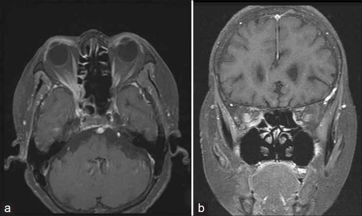
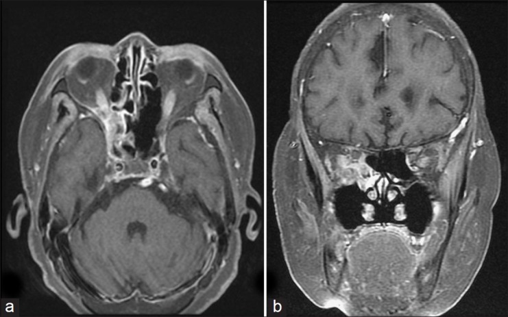
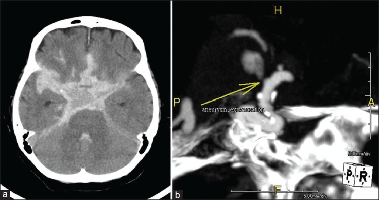
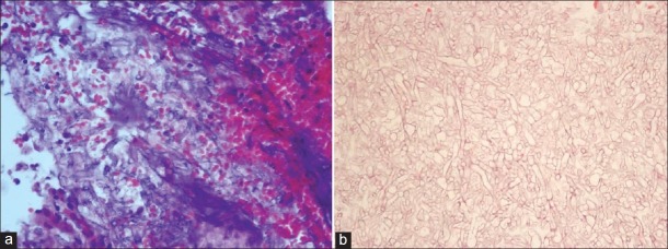
A 73-year-old woman with past medical history of hypertension and rheumatoid arthritis stage 4 under regular anti-hypertension medication and long-term corticosteroid therapy, presented to us in August 2010 with the chief complaint of decline of visual acuity of her right eye and right periorbital pain for 2 months. At the beginning of the clinical course, she had brain computed tomography (CT) scan and orbit magnetic resonance imaging (MRI) done in June 2010, which disclosed a small enhancing lesion, about 1.2 cm × 1.1 cm × 1 cm in size, near the right side orbital apex and adjacent right side superior orbital fissure with mild encasement of the right optic nerve, and this lesion showed mild extension to the adjacent right side posterior ethmoid sinus [Figure 1 a, b]. She had pulse steroid therapy in ophthalmology service, but it was ineffective. On admission, her neurological examination showed that she had right eye blindness, right ptosis, right ophthmaloplegia, and tingle in the territory of ophthalmic branch of right trigeminal nerve. Repeated MRI of orbit after admission in August 2010 showed the progression of the lesion which enlarged up to 1.5 cm × 1.3 cm × 1.2 cm [Figure 2 a, b]. She underwent endoscopic endonasal transethmoid approach with the removal of the lesion on 19 August 2010 under general anesthesia. After the surgery, she recovered well and her right periorbital pain was much released. However, 5 days after surgery, she experienced a severe headache followed by loss of consciousness. After endotracheal tube intubation and resuscitation, brain CT was checked which showed diffuse high-density acute SAH in the basal cistern, pre-pontine cistern, ambient cistern, quadrigeminal cistern, cerebellomedullary cistern, and right sylvian fissure, with acute hydrocephalus [Figure 3a]. Emergent external ventricular drainage was done followed by performing CT angiography which showed several bleb-like wide base aneurysms over right supraclinoid internal carotid artery (ICA), and one aneurysm, about 4 mm in size and located at the medial side of the right supraclinoid internal carotid artery, showed extravasation of contrast medium. The dome of the ruptured aneurysm projected medially and superiorly [Figure 3b]. On the same day, the histology examination reported that the lesion was composed of many fungal septate hyphae demonstrated on both HE stain and periodic acid-Schiff (PAS) stain [Figure 4a, b]. Fungal infection was diagnosed and the culture turned out to be Aspergillus fumigatus. Her intracranial aneurysms were probably fungal aneurysms, which are one of the sequels of central nervous system (CNS) fungal infection. Unfortunately, after the event, she remained in deep coma and finally she expired due to central failure.
Figure 1.

Orbit MRI T1-weighted post-Gadolinium enhancement. (a, axial view) A small enhancing lesion about 1.2 cm × 1.1 cm × 1 cm noted near the right side orbital apex and adjacent right side superior orbital fissure with mild extension to the adjacent right side posterior ethmoid sinus region. (b, coronal view) This lesion had mild encasement of the right optic nerve
Figure 2.

Repeated orbit MRI T1-weighted post-Gadolinium enhancement. (a) Axial view and (b) coronal view show the progression of the lesion
Figure 3.

(a) Non-contrast brain CT demonstrated diffuse high-density acute subarachnoid hemorrhage in the basal cistern, pre-pontine cistern, ambient cistern, quadrigeminal cistern, cerebellomedullary cistern, and right sylvian fissure. (b) computed tomography angiography showed the presence of several bleb-like wide base aneurysms over right supraclinoid internal carotid artery. One aneurysm of about 4 mm showed extravasation of contrast medium. The dome of the ruptured aneurysm projected medially and superiorly
Figure 4.
Histology of the lesion. (a) H and E, ×100. (b) PAS stain, ×200. Both showed many fungal septate hyphae
DISCUSSION
The orbital apex syndrome can be caused by tumors, trauma, aneurysm of internal carotid artery, inflammatory disorders, and infectious diseases, and aspergillosis is one of the causative pathologies.[3,7,11] Aspergillosis is the most commonly reported among all the CNS fungal infections.[1,2,7,11] A. fumigatus is the most frequent species isolated in human infections.[2,3,10] The route of infection is usually by inhalation of Aspergillus spores and conidia or airborne metabolites of Aspergillus.[3] The main routes of central nervous system contamination are hematogenous dissemination from a distant primary source, mainly lung, and contiguous spread from an adjacent focus such as orbit or paranasal sinuses.[3,4,10] Immunocompromised individuals such as those having acquired immunodeficiency syndrome (AIDS), patients with malignant tumors, diabetes, or on corticosteroids or immunosuppressive agents are at risk of developing aspergillosis.[3,5,7,9,11] One of the most lethal but rare sequels of CNS fungal infection is intracranial aneurysm.[1] Mukoyama et al. roughly classified the mode of development of aspergillosis of CNS into five types: 1) meningitis, 2) meningoencephalitis, 3) abscess, 4) granuloma, and 5) vasculitis.[4] Horten et al. described that Aspergillus infected vasculitis can follow three courses: 1) formation of thrombus, causing hemorrhagic infarction and abscess; 2) sudden massive hemorrhage; and 3) formation of an aneurysm.[4] Fungal aneurysms usually are found in the major vessels at the base of the brain, and they develop by direct invasion from the luminal surface and the adventitia rather than by involvement of vasa vasorum, which happens in bacterial aneurysms.[4,5,10] Rupture of fungal intracranial aneurysms usually leads to high mortality.[1,2,4–6,8,10] Fungal intracranial aneurysms are rare; however, with the widespread use of corticosteroids, broad-spectrum antibiotics, and immunosuppressive drugs, the frequency of opportunistic fungal infection is increasing; fungal aneurysms may become more common, and one should be aware of it in clinical practice.[2,6,8] It is learned from this painful clinical experience that in an immunocompromised patient having orbital apex syndrome with or without evidence of sinus disease on CT or MRI, fungal infection should be kept in mind. In those treated with steroid and whose symptoms and signs do not quickly improve, or the symptoms and signs relapse quickly, repeat imaging may reveal new findings. The ways to establish diagnosis include biopsy and culture of the excised specimen. Early diagnosis and radical resection, combined with antifungal medications is paramount to save this particular group of patients.
Footnotes
Available FREE in open access from: http://www.surgicalneurologyint.com/text.asp?2012/3/1/124/102349
Contributor Information
Chi-Man Yip, Email: yip_chiman@yahoo.com.
Shu-Shong Hsu, Email: sshsu@vghks.gov.tw.
Wei-Chuan Liao, Email: wcliao@vghks.gov.tw.
Jun-Yih Chen, Email: jychen@vghks.gov.tw.
Su-Hao Liu, Email: shliu@vghks.gov.tw.
Chih-Hao Chen, Email: chichchen@vghks.gov.tw.
REFERENCES
- 1.Ahsan H, Ajmal F, Saleem MF, Sonawala AB. Cerebral fungal infection with mycotic aneurysm of basilar artery and subarachnoid haemorrhage. Singapore Med J. 2009;50:22–5. [PubMed] [Google Scholar]
- 2.Ahuja GK, Jain N, Vijayaraghavan M, Roy S. Cerebral mycotic aneurysm of fungal origin Case report. J Neurosurg. 1978;49:107–10. doi: 10.3171/jns.1978.49.1.0107. [DOI] [PubMed] [Google Scholar]
- 3.Fernandes YB, Ramina R, Borges G, Queiroz LS, Maldaun MVC, Maciel JA., Jr Orbital Apex Syndrome due to Aspergillosis. Arq Neuropsiquiatr. 2001;59:806–8. doi: 10.1590/s0004-282x2001000500029. [DOI] [PubMed] [Google Scholar]
- 4.Iihara K, Makita Y, Nabeshima S, Tei T, Keyaki A, Nioka H. Aspergillosis of the Central Nervous System causing Subarachnoid Hemorrhage from Mycotic Aneurysm of the Basilar Artery- Case Report. Neurol Med Chir (Tokyo) 1990;30:618–23. doi: 10.2176/nmc.30.618. [DOI] [PubMed] [Google Scholar]
- 5.Ishikawa T, Kazumata K, Ni-iya Y, Kamiyama H, Andoh M. Subarachnoid hemorrhage as a result of fungal aneurysm at the posterior communicating artery associated with occlusion of the internal carotid artery Case Report. Surg Neurol. 2002;58:261–5. doi: 10.1016/s0090-3019(02)00839-x. [DOI] [PubMed] [Google Scholar]
- 6.Komatsu Y, Narushima K, Kobayashi E, Tomono Y, Nose T. Aspergillus Mycotic Aneurysm – Case Report. Neuro Med Chir (Tokyo) 1991;31:346–50. doi: 10.2176/nmc.31.346. [DOI] [PubMed] [Google Scholar]
- 7.Levin LA, Avery R, Shore JW, Woog JJ, Baker AS. The Spectrum of Orbital Aspergillosis: A Clinicopathological Review. Surv Ophthalmol. 1996;41:142–54. doi: 10.1016/s0039-6257(96)80004-x. [DOI] [PubMed] [Google Scholar]
- 8.Masago A, Fukuoka H, Yoshida T, Majima K, Tada T, Nagai H. Intracranial Mycotic Aneurysm caused by Aspergillus – Case Report. Neurol Med Chir (Tokyo) 1992;32:904–7. doi: 10.2176/nmc.32.904. [DOI] [PubMed] [Google Scholar]
- 9.O’Toole L’, Acheson JA, Kidd D. Orbital apex lesion due to Aspergillosis presenting in immunocompetent patients without apparent sinus disease. J Neurol. 2008;255:1798–801. doi: 10.1007/s00415-008-0977-5. [DOI] [PubMed] [Google Scholar]
- 10.Wilson WR, Hawrych A, Olan W. Rapid Development of Bilateral Internal Carotid Artery Aneurysm from Sphenoid Sinus Aspergillosis. Skull Base Surg. 1998;8:211–4. doi: 10.1055/s-2008-1058185. [DOI] [PMC free article] [PubMed] [Google Scholar]
- 11.Yeh S, Foroozan R. Orbital apex syndrome. Curr Opin Ophthalmol. 2004;15:490–8. doi: 10.1097/01.icu.0000144387.12739.9c. [DOI] [PubMed] [Google Scholar]


