Abstract
Background:
Angiosarcomas are high-grade endothelial tumors remarkable for their rarity and malignant behavior. Primary calvarial angiosarcoma is an extremely rare entity and its behavior usually sets it apart from other angiosarcoma types. We highlight the successful management of cranial angiosarcoma using a multidisciplinary approach.
Case Description:
We present a 16-year-old male who was first noted to have a right-sided parietal cranial mass that was biopsied in 2008. Pathology was initially thought to be Kaposiform hemangioendothelioma. The patient subsequently underwent chemotherapy with vincristine. The patient did well until early 2010, when he suffered a right-sided intraparenchymal intratumoral hemorrhage. At this time, the original pathologic diagnosis was revisited and the diagnosis was upgraded to an angiosarcoma. The patient underwent a second round of chemotherapy using vincristine, cyclophosphamide, and actinomycin. The tumor continued to progress despite this treatment and he developed extensive skull deformity. At this point more definitive surgical intervention was reconsidered. Preoperative embolization of the mass was performed followed by aggressive surgical resection of the bony disease. The patient tolerated the procedure well and was discharged 6 days postoperatively without any new deficits. The patient is currently in the process of completing radiation therapy to entire tumor bed. He has clinically done well with no neurologic deterioration and has demonstrated long-term survival (>3 years).
Conclusion:
With the combined efforts of pediatric oncology, radiation oncology, interventional neuroradiology, and neurosurgery, a survival of greater than 3 years is possible with this aggressive pathology.
Keywords: Angiosarcoma, cranium, hemangioendothelioma, pediatric
INTRODUCTION
Angiosarcomas are high-grade endothelial tumors remarkable for their rarity and malignant behavior. Their incidence is approximately 2–3 cases per 1,000,000.[6,22] Angiosarcoma can involve the central nervous system, arising from the mesenchymal elements or the bones of the cranium.[22]
Primary calvarial angiosarcoma (PCA) is an extremely rare entity and its aggressive behavior usually sets it apart from other angiosarcoma types. To our knowledge 5 histologically confirmed cases of primary skull angiosarcomas have been described in the pediatric population;[1,9,11,13,20] only 2 of these patients had a calvarial location.[1,13] We present a pediatric patient with PCA and review the pertinent literature.
CASE REPORT
A 14-year-old African-American male was admitted to our institution in October 2008 with a single episode of subjective left leg numbness and weakness. His past medical history was unremarkable and this was the first time he had experienced such an episode. General physical examination was normal and neurologic exam only revealed mild left arm numbness. Magnetic resonance imaging (MRI) scans revealed a 3 cm extra-axial, heterogeneously enhancing mass in the right parieto-occipital region with edema of the adjacent parenchyma [Figure 1]. Additional studies performed were negative for metastatic lesions. An uncomplicated biopsy of lesion was performed and the initial pathology was felt to be consistent with Kaposiform hemangioendothelioma.
Figure 1.
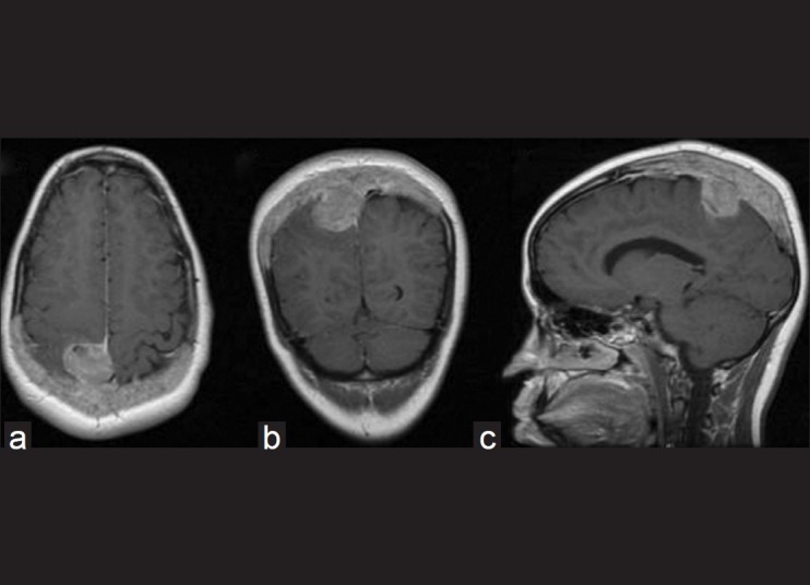
Preoperative (2008) axial (a), coronal (b), and sagittal (c) postcontrast T1-weighted magnetic resonance imaging scans show a 3 cm extra-axial, heterogeneously enhancing mass in the right parieto-occipital region
The patient was treated with monotherapy of vincristine. Initially, the tumor responded well to this treatment, but subsequently showed progression approximately 10 months after diagnosis. Due to the progression of the mass, the accuracy of the pathologic diagnosis was revisited and the initial diagnosis was changed to the more malignant angiosarcoma.
In January 2010, the patient had an acute onset of left-sided hemiparesis and headaches. Computed tomography (CT) scan revealed an intraparenchymal hematoma in the frontoparietal region, extending toward the lateral ventricle. An increase in the tumor size was also noticed [Figure 2]. The option of surgical intervention was presented to the patient and family, but due to the aggressive and vascular nature of the tumor, we advised that surgical resection would pose a high risk. The patient was subsequently started on a more aggressive multidrug regimen of vincristine, cyclophosphamide, and actinomycin.
Figure 2.
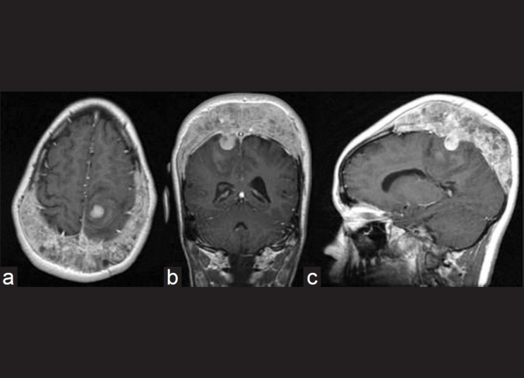
Posthemorrhage (2010) axial (a), coronal (b), and sagittal (c) T1-weighted postcontrast magnetic resonance imaging showing a posterior frontoparietal parenchymal hematoma, along with progression in tumor size
Tumor progression initially stabilized with multidrug therapy; however, after 8 months, a significant calvarial deformity was revealed on routine surveillance neuroimaging. In January 2011, repeat MRI brain again revealed progression of the tumor, which now encompassed both hemispheres and was accompanied by a marked calvarial deformity. On physical examination, it was noted that the patient's left-sided hemiparesis was worse. Given that there was significant tumor progression in spite of aggressive chemotherapy and clinical deterioration, surgical intervention was reconsidered. After considering the size, vascular nature, and lack of response to chemotherapy, the patient and family wished to proceed with surgery.
Surgery
Twenty-four hours prior to surgery, the patient underwent preoperative tumor embolization. Arterial supply was noted from the superficial temporal artery, middle meningeal artery, and branches of the occipital artery [Figure 3]. Ninety percent of the tumor was embolized with Gelfoam particles. The following day, the full extent of the extracranial subgaleal tumor along with the involved bone was removed. A bicoronal incision was made. A plane between the galea and tumor was easily identified. The interface between normal skull and tumor was identified circumferentially. We then performed a craniotomy around the tumor that would allow access to normal, intact dura around the entire mass. The tumor was then carefully dissected away from the underlying dura (except in the area where the mass traveled intradurally). As the tumor was debulked internally, defects in the calvarium were noted. An epidural plane was maintained throughout the resection except where the mass had already traveled intradurally. Any portions of the calvarium that were encountered were resected if tumor involvement was appreciated. Given the aggressive nature of the neoplasm, no attempt was made to reconstruct the bony defect at the time of initial surgery [Figure 4]. Elective cranioplasty was scheduled for after completion of the adjuvant treatment. Postoperatively, the patient had an uncomplicated hospital course and was discharged 6 days later. The patient has undergone a course of radiotherapy. On his latest follow-up, 8 months postsurgery, the patient has stable chronic paresis (3/5) in the right lower extremity (from the intratumoral hemorrhage), but no new deficits. He is able to ambulate without assistance and his scalp has healed well in spite of adjuvant radiotherapy. Postoperatively, MRI scans have not shown any recurrence thus far.
Figure 3.
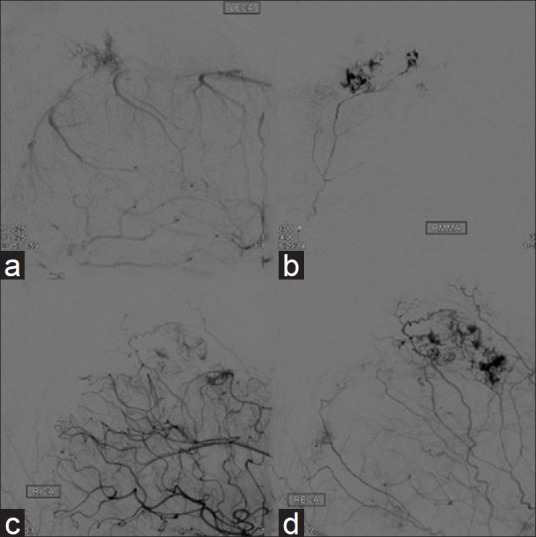
Pre-embolization angiography showing tumor blush from the branches of the left external carotid artery (a), right middle meningeal artery (b), right internal carotid artery (c), and the right external carotid artery (d)
Figure 4.
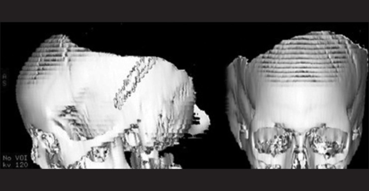
Postoperative computed tomography head (3D) scans showing the extent of calvarial resection
Pathology
Microscopic sections of the tumor demonstrated variable cellularity and growth pattern [Figure 5]. There were high-grade, neoplastic spindle cells with mitosis and rudimentary small vessel differentiation. The congested dilated vessels were lined by neoplastic endothelium with intraluminal budding. Immunohistochemically, the endothelial cells were positive for CD34, CD31, factor VIII; occasionally positive for PROX-1, but negative for CKAE1/AE3. The proliferative index (Ki-67) was increased and varied from area to area, with occasional foci demonstrating up to 30%. The final pathology was consistent with angiosarcoma.
Figure 5.
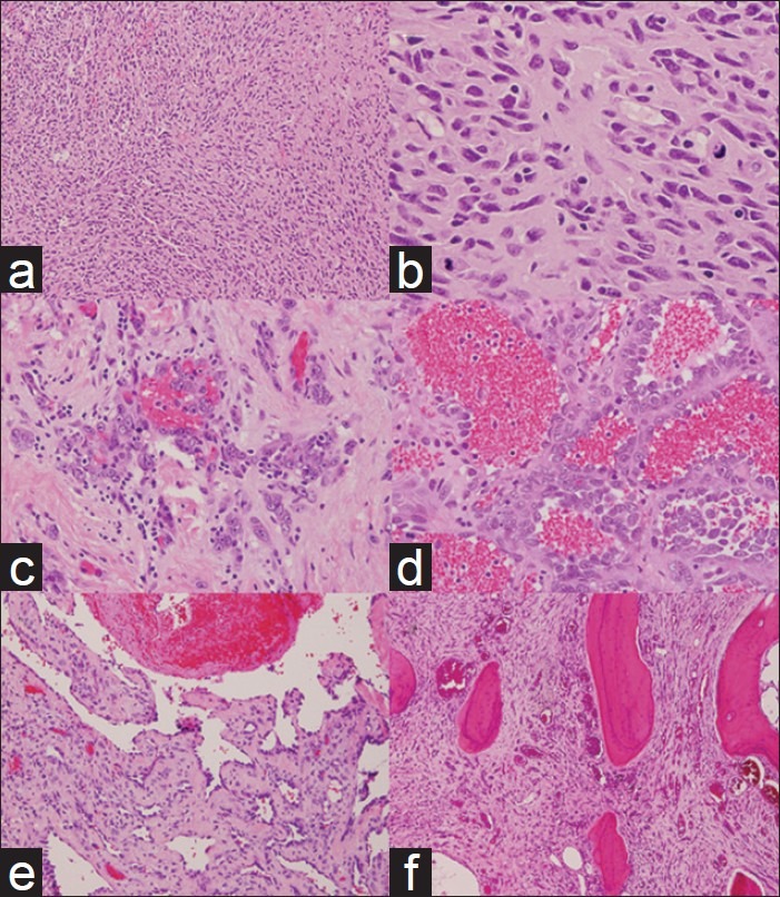
Microscopic sections of the tumor showing variable cellularity and growth pattern. (a) Cellular spindle cell area ×100 (b) High-grade, neoplastic spindle cells with mitosis showing rudimentary small vessel differentiation ×400. (c) Epithelioid foci showing round-to-oval cells with large, prominent nucleoli ×200. (d) Congested dilated vessels lined by neoplastic endothelium with intraluminal budding ×200. (e) Irregular, anastomosing vascular channels lined by flat neoplastic cells ×100. (f) Vascular tumor invading and destroying adjacent bone trabeculae ×100
DISCUSSION
Angiosarcomas are malignant neoplasms derived from the endothelial cells of arteries, veins, and lymphatics. They are classified as WHO grade 3 (or malignant) hemangioendotheliomas and belong to the larger class of soft tissue sarcomas that make up 1% of the overall malignancies in adults.[25] Only 2% of soft tissue sarcomas in adults are angiosarcomas.[14] In pediatric patients, they constitute an even lower percentage (0.3%) of all soft tissue sarcomas.[7] In children, most angiosarcomas are seen to arise in the mediastinum, pericardium, and heart.[20]
When bone is the source of origin, long bones of the extremities are most commonly affected.[22] Less commonly, this tumor can arise from the ribs, pelvis, or vertebrae.[22] The cranium remains an extremely unusual site of involvement for angiosarcoma in either children or adults. Cranial angiosarcomas have presented as a progressively noticeable lump on the head in the majority of the previously reported cases in all age groups.[4] If the tumor involves the temporal bone, symptoms such as tinnitus, hearing impairment, and otalgia may accompany the presence of an external deformity.[22] One reported patient presented with proptosis because the tumor was involving the roof of the orbit and the sphenoid and frontal bones.[23] Our patient's initial presentation of temporary left-sided numbness and weakness due to parenchymal compression was quite unusual. Also, there was no evidence of any cranial asymmetry or external deformity on initial presentation.
Diagnosis depends on imaging and pathologic findings. A plain skull X-ray typically shows a well-demarcated, expansile, lytic mass.[10,24] MRI shows a mass that is isointense with grey matter on T1-weighted images. T2-weighted images depict a hyperintense mass, possibly with lobulations or septations.[1] On gadolinum-enhanced images, avid contrast uptake is usually seen due to the rich vascularity of the lesion.[1] This enhancement might not always be present in the setting of immature vascular channels.[4]
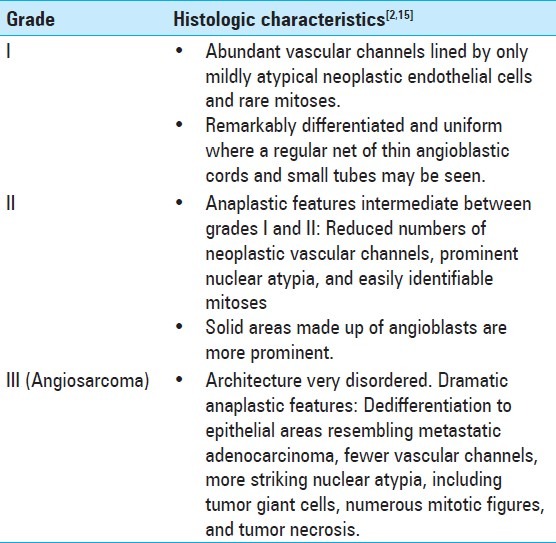
Histologic diagnosis relies on the architecture of the tumoral tissue and the features of the neoplastic cells. The hemangioendotheliomas family of tumors is summarized in Table 1. Histology generally shows networks of irregular vascular channels lined by plump endothelial cells with marked anaplasia.[14,22] The presence of vascular endothelial lineage of the cells can be confirmed with immunohistochemistry testing for the presence of factor VIII-related antigen, CD 34 and CD 31. These markers and the presence of pleomorphism help differentiate the lesion from meningiomas, intravascular papillary endothelial hyperplasia,[23] and epitheliod sarcomas.[1]
Table 1.
Grades of hemangioendothelioma depicting the histologic spectrum
The prognosis of angiosarcoma in general is very poor. For angiosarcoma involving the central nervous system, a mean overall survival of 8 months is reported.[15] The potential involvement of the brain in cranial angiosarcoma leads to a worse prognosis than other bony angiosarcomas.[23] Cranial angiosarcoma also has the tendency to encroach on the inner and outer tables of the skull and metastasize early in the course of the disease via the rich intradiploic blood supply of the skull.[2] The lungs and other bony structures seem to be the most common site of spread.[1] Given the propensity for early metastasis in cranial angiosarcoma, a full systemic evaluation should be performed at the time of presentation. Initial evaluation should include MRI (for the local lesion), chest X-rays, and bone scans (for metastatic evaluation). A follow-up assessment for metastasis is also imperative, which includes bone scans or positron emission tomography (PET) scans.[1]
Treatment of cranial angiosarcoma has not been standardized thus far due to the paucity of patients. Some authors suggest that these tumors should be treated similar to osteosarcomas, with wide surgical excision, adjuvant chemotherapy, and radiotherapy.[1,20] Head and neck angiosarcomas have also been treated with the same modalities, with 5-year disease-free survival reported to be between 4% and 33%.[4] Wide surgical resection is the first step in managing these patients, but might not be simple to undertake. The location of the tumor is always the primary determinant in the eventual success of a surgical procedure. Skull base lesions might be inaccessible for safe, total excision; sometimes, even obtaining a biopsy might pose a challenge.[20] Hypervascularity of the lesion is another determinant and presurgical embolization has been used to make the procedure safer.[22]
The role of adjuvant or neoadjuvant chemotherapy and radiotherapy is less well defined. The results of radiotherapy (combined with surgery in some instances) have been vague.[2,22] Larsson et al. describe a pediatric patient treated with high doses of radiation for tumor of the frontal bones and had been alive for 12 years.[13] Local control might be enhanced by adjuvant or stand-alone radiotherapy and may be a good option for palliation in patients where surgery is not a safe option.[1,2]
Lack of sufficient patient data on the efficacy of chemotherapy in patients with cranial angiosarcoma makes it difficult to establish standard guidelines.[16] Some investigators have successfully treated pediatric patients with alpha-interferon.[17] Paclitaxel has been shown to have good results in patients with scalp and face (soft tissue) angiosarcoma,[3,5,19] but it did not prevent pulmonary and bone metastases from a skull angiosarcoma as reported by Scholsem et al.[22]
Recently, there has been increased interest in treatment of angiosarcoma with monoclonal antibodies, in particular, bevacizumab. The inhibition of the vascular endothelial growth factor-vascular endothelial growth factor receptor (VEGF–VEGFR) pathway in angiosarcoma has shown some promise.[18] In one case report, a patient with cutaneous facial angiosarcoma showed partial response to bevacizumab alone.[21] Another case report demonstrated a dramatic response of cutaneous facial angiosarcoma when paclitaxel was combined with bevacizumab.[8] Preoperative radiotherapy and bevacizumab has also been used for angiosarcoma of the head and neck with decent results.[12]
CONCLUSION
Angiosarcomas are rare and highly aggressive tumors of the endothelial cells. Primary cranial involvement may be rarely encountered and usually portends a poor prognosis. Treatment is ill-defined due to the rarity of the pathology and generally consists of a combination of aggressive surgical excision, chemotherapy, and/or radiotherapy. Further studies need to be undertaken to clarify the best therapeutic route to take in these patients and to establish guidelines. Our case highlights the fact that with an aggressive multidisciplinary management protocol, survival greater than 3 years can be seen in pediatric patients with PCA.
Footnotes
Available FREE in open access from: http://www.surgicalneurologyint.com/text.asp?2012/3/1/134/102952
Contributor Information
Imad S. Khan, Email: imad.s.khan@Vanderbilt.edu.
Jai D. Thakur, Email: docjaideep@gmail.com.
Osama Ahmed, Email: oahmed@lsuhsc.edu.
Cedric D. Shorter, Email: cshorter@goodmancampbell.com.
Jaiyeola Thomas-Ogunniyl, Email: jtho19@lsuhsc.edu.
Mary T. Kim, Email: mkim@lsuhsc.edu.
Majed A. Jeroudi, Email: mjerou@lsuhsc.edu.
Bharat Guthikonda, Email: bguthi@lsuhsc.edu.
REFERENCES
- 1.Bourekas EC, Cohen ML, Kamen CS, Tarr RW, Lanzieri CF, Lewin JS. Malignant hemangioendothelioma (angiosarcoma) of the skull: Plain film, CT, and MR appearance. AJNR Am J Neuroradiol. 1996;17:1946–8. [PMC free article] [PubMed] [Google Scholar]
- 2.Campanacci M, Boriani S, Giunti A. Hemangioendothelioma of bone: A study of 29 cases. Cancer. 1980;46:804–14. doi: 10.1002/1097-0142(19800815)46:4<804::aid-cncr2820460427>3.0.co;2-1. [DOI] [PubMed] [Google Scholar]
- 3.Casper ES, Waltzman RJ, Schwartz GK, Sugarman A, Pfister D, Ilson D, et al. Phase II trial of paclitaxel in patients with soft-tissue sarcoma. Cancer Invest. 1980;16:442–6. doi: 10.3109/07357909809011697. [DOI] [PubMed] [Google Scholar]
- 4.Chou YC, Chang YL, Harnod T, Chen WF, Su CF, Lin SZ, et al. Primary angiosarcoma of the cranial vault: A case report and review of the literature. Surg Neurol. 2004;61:575–9. doi: 10.1016/j.surneu.2003.07.002. [DOI] [PubMed] [Google Scholar]
- 5.Fata F, O’Reilly E, Ilson D, Pfister D, Leffel D, Kelsen DP, et al. Paclitaxel in the treatment of patients with angiosarcoma of the scalp or face. Cancer. 1999;86:2034–7. [PubMed] [Google Scholar]
- 6.Fedok FG, Levin RJ, Maloney ME, Tipirneni K. Angiosarcoma: Current review. Am J Otolaryngol. 1999;20:223–31. doi: 10.1016/s0196-0709(99)90004-2. [DOI] [PubMed] [Google Scholar]
- 7.Ferrari A, Casanova M, Bisogno G, Cecchetto G, Meazza C, Gandola L, et al. Malignant vascular tumors in children and adolescents: A report from the Italian and German Soft Tissue Sarcoma Cooperative Group. Med Pediatr Oncol. 2002;39:109–14. doi: 10.1002/mpo.10078. [DOI] [PubMed] [Google Scholar]
- 8.Fuller CK, Charlson JA, Dankle SK, Russell TJ. Dramatic improvement of inoperable angiosarcoma with combination paclitaxel and bevacizumab chemotherapy. J Am Acad Dermatol. 63:e83–4. doi: 10.1016/j.jaad.2009.09.035. [DOI] [PubMed] [Google Scholar]
- 9.Henny FA. Angiosarcoma of the maxilla in a 3-month-old infant; report of case. J Oral Surg (Chic) 1949;7:250–2. [PubMed] [Google Scholar]
- 10.Ibarra RA, Kesava P, Hallet KK, Bogaev C. Hemangioendothelioma of the temporal bone with radiologic findings resembling hemangioma. AJNR Am J Neuroradiol. 2001;22:755–8. [PMC free article] [PubMed] [Google Scholar]
- 11.Kinkade JM. Angiosarcoma of the petrous portion of the temporal bone; Report of a case. Ann Otol Rhinol Laryngol. 1948;57:235–40. doi: 10.1177/000348944805700123. [DOI] [PubMed] [Google Scholar]
- 12.Koontz BF, Miles EF, Rubio MA, Madden JF, Fisher SR, Scher RL, et al. Preoperative radiotherapy and bevacizumab for angiosarcoma of the head and neck: Two case studies. Head Neck. 2008;30:262–6. doi: 10.1002/hed.20674. [DOI] [PubMed] [Google Scholar]
- 13.Larsson SE, Lorentzon R, Boquist L. Malignant hemangioendothelioma of bone. J Bone Joint Surg Am. 1975;57:84–9. [PubMed] [Google Scholar]
- 14.Lezama-del Valle P, Gerald WL, Tsai J, Meyers P, La Quaglia MP. Malignant vascular tumors in young patients. Cancer. 1998;83:1634–9. doi: 10.1002/(sici)1097-0142(19981015)83:8<1634::aid-cncr20>3.3.co;2-j. [DOI] [PubMed] [Google Scholar]
- 15.Lopes M, Duffau H, Fleuridas G. Primary spheno-orbital angiosarcoma: Case report and review of the literature. Neurosurgery. 1999;44:405–7. doi: 10.1097/00006123-199902000-00102. discussion 407-8. [DOI] [PubMed] [Google Scholar]
- 16.Mark RJ, Poen JC, Tran LM, Fu YS, Juillard GF. Angiosarcoma. A report of 67 patients and a review of the literature. Cancer. 1996;77:2400–6. doi: 10.1002/(SICI)1097-0142(19960601)77:11<2400::AID-CNCR32>3.0.CO;2-Z. [DOI] [PubMed] [Google Scholar]
- 17.Orchard PJ, Smith CM, 3rd, Woods WG, Day DL, Dehner LP, Shapiro R. Treatment of haemangioendotheliomas with alpha interferon. Lancet. 1989;2:565–7. doi: 10.1016/s0140-6736(89)90694-6. [DOI] [PubMed] [Google Scholar]
- 18.Park MS, Ravi V, Araujo DM. Inhibiting the VEGF-VEGFR pathway in angiosarcoma, epithelioid hemangioendothelioma, and hemangiopericytoma/solitary fibrous tumor. Curr Opin Oncol. 2010;22:351–5. doi: 10.1097/CCO.0b013e32833aaad4. [DOI] [PubMed] [Google Scholar]
- 19.Penel N, Bui BN, Bay JO, Cupissol D, Ray-Coquard I, Piperno-Neumann S, et al. Phase II trial of weekly paclitaxel for unresectable angiosarcoma: The ANGIOTAX Study. J Clin Oncol. 2008;26:5269–74. doi: 10.1200/JCO.2008.17.3146. [DOI] [PubMed] [Google Scholar]
- 20.Renukaswamy GM, Boardman SJ, Sebire NJ, Hartley BE. Angiosarcoma of skull base in a 1-year-old child: a case report. Int J Pediatr Otorhinolaryngol. 2009;73:1598–600. doi: 10.1016/j.ijporl.2009.07.025. [DOI] [PubMed] [Google Scholar]
- 21.Rosen A, Thimon S, Ternant D, Machet MC, Paintaud G, Machet L. Partial response to bevacizumab of an extensive cutaneous angiosarcoma of the face. Br J Dermatol. 2010;163:225–7. doi: 10.1111/j.1365-2133.2010.09803.x. [DOI] [PubMed] [Google Scholar]
- 22.Scholsem M, Raket D, Flandroy P, Sciot R, Deprez M. Primary temporal bone angiosarcoma: A case report. J Neurooncol. 2005;75:121–5. doi: 10.1007/s11060-005-0375-0. [DOI] [PubMed] [Google Scholar]
- 23.Shuangshoti S, Chayapum P, Suwanwela N, Suwanwela C. Unilateral proptosis as a clinical presentation in primary angiosarcoma of skull. Br J Ophthalmol. 1988;72:713–9. doi: 10.1136/bjo.72.9.713. [DOI] [PMC free article] [PubMed] [Google Scholar]
- 24.Thananopavarn P, Smith JK, Castillo M. MRI of angiosarcoma of the calvaria. AJR Am J Roentgenol. 2003;181:1432–3. doi: 10.2214/ajr.181.5.1811432. [DOI] [PubMed] [Google Scholar]
- 25.Wold LE, Unni KK, Beabout JW, Ivins JC, Bruckman JE, Dahlin DC. Hemangioendothelial sarcoma of bone. Am J Surg Pathol. 1982;6:59–70. doi: 10.1097/00000478-198201000-00006. [DOI] [PubMed] [Google Scholar]


