Abstract
The piggyBac transposon system is naturally active, originally derived from the cabbage looper moth1,2. This non-viral system is plasmid based, most commonly utilizing two plasmids with one expressing the piggyBac transposase enzyme and a transposon plasmid harboring the gene(s) of interest between inverted repeat elements which are required for gene transfer activity. PiggyBac mediates gene transfer through a "cut and paste" mechanism whereby the transposase integrates the transposon segment into the genome of the target cell(s) of interest. PiggyBac has demonstrated efficient gene delivery activity in a wide variety of insect1,2, mammalian3-5, and human cells6 including primary human T cells7,8. Recently, a hyperactive piggyBac transposase was generated improving gene transfer efficiency9,10.
Human T lymphocytes are of clinical interest for adoptive immunotherapy of cancer11. Of note, the first clinical trial involving transposon modification of human T cells using the Sleeping beauty transposon system has been approved12. We have previously evaluated the utility of piggyBac as a non-viral methodology for genetic modification of human T cells. We found piggyBac to be efficient in genetic modification of human T cells with a reporter gene and a non-immunogenic inducible suicide gene7. Analysis of genomic integration sites revealed a lack of preference for integration into or near known proto-oncogenes13. We used piggyBac to gene-modify cytotoxic T lymphocytes to carry a chimeric antigen receptor directed against the tumor antigen HER2, and found that gene-modified T cells mediated targeted killing of HER2-positive tumor cells in vitro and in vivo in an orthotopic mouse model14. We have also used piggyBac to generate human T cells resistant to rapamycin, which should be useful in cancer therapies where rapamycin is utilized15.
Herein, we describe a method for using piggyBac to genetically modify primary human T cells. This includes isolation of peripheral blood mononuclear cells (PBMCs) from human blood followed by culture, gene modification, and activation of T cells. For the purpose of this report, T cells were modified with a reporter gene (eGFP) for analysis and quantification of gene expression by flow cytometry.
PiggyBac can be used to modify human T cells with a variety of genes of interest. Although we have used piggyBac to direct T cells to tumor antigens14, we have also used piggyBac to add an inducible safety switch in order to eliminate gene modified cells if needed7. The large cargo capacity of piggyBac has also enabled gene transfer of a large rapamycin resistant mTOR molecule (15 kb)15. Therefore, we present a non-viral methodology for stable gene-modification of primary human T cells for a wide variety of purposes.
Keywords: Immunology, Issue 69, Molecular Biology, Medicine, Genetics, Cellular Biology, Virology, Human T cells, Transposons, piggyBac, transgene
Protocol
Day 0
1. Isolation of PBMCs from Human Blood
Collect 20 ml of fresh human blood using venipuncture into Na-heparin vacutainer tubes.
Mix blood and Advanced RPMI 1,640 in 1:1 (v/v) ratio.
Add 20 ml lymphoprep medium to a 50 ml centrifuge tube (25 °C). Slowly layer 25-30 ml of blood-RPMI 1,640 mix on top of the lymphoprep.
Centrifuge at 400 x g for 40 min without brakes.
Collect both distinct and fuzzy layers using a disposable pipette into 10 ml of 1x PBS (25 °C) and bring the volume up to 50 ml with 1x PBS.
Centrifuge at 450 x g for 10 min.
Aspirate the supernatant completely and add 20 ml of Advanced RPMI 1,640.
Centrifuge at 400 x g for 5 min.
Aspirate the supernatant. Add 10 ml of complete T cell media supplemented with 5 ng/ml rhIL-15 prewarmed at 37 °C.
Count the number of cells and plate in a 24 well tissue culture coated plate at 2 x 10⁶ cells/well in complete T cell media supplemented with 5 ng/ml rhIL-15 Add sterile water to surrounding wells. Incubate overnight in a humidified incubator at 37 °C, 5% CO2.
Day 1
2. Coating Plates with anti-CD28 and anti-CD3 Antibody for Stimulating T cells
Dilute anti-CD28 and anti-CD3 antibodies in sterile water at a concentration of 1 μg/ml each.
Add 500 μl each antibody solution to 5 marked wells of a non-coated tissue culture 24 well plate.
Add sterile water to rest of the wells. Wrap the plate in shrink wrap and place in a 4 °C refrigerator.
3. Nucleofection of Unstimulated T Cells
Prewarm T cell media at 37 °C and supplement with 5 ng/ml of rhIL-15. Prepare complete nucleofector solution by adding 500 μl of Nucleofector Supplement 1 to 2.25 ml of Nucleofector solution.
Aliquot 5 μg each of transposon (Zeo-pT-CMV-eGFP) and transposase (pCMV-piggyBac) in a 1.5 ml microfuge tube. Note: it may be necessary to optimize the transposase and transposon DNA amount for optimal gene delivery and to minimize cellular toxicity. The plasmids can be obtained from the authors by request.
Harvest PBMCs from the 24 well tissue culture plate into a 50 ml tube. Count the number of cells. Save 2 x 10⁶ cells for use as control during flow cytometry.
Add 7-10 x 106 cells to a 15 ml tube and centrifuge at 400 x g, 5 min, aspirate supernatant and finger-flick the pellet.
Add 100 μl of T cell complete nucleofection solution to loosened cell pellet.
Add the solution-cell mixture to the tube containing the plasmids.
Add the solution-cell-plasmid mixture to the bottom of the nucleofection cuvette. Be careful not to introduce any bubbles.
Nucleofect the cells using program U-014 (Unstimulated T cells, human, http://bio.lonza.com/resources/product-instructions/protocols/).
Immediately add 500 μl of prewarmed media with rhIL-15 to the cuvette. Transfer the cells to a well of a 24 well plate with 1.5 ml of prewarmed media with rhIL15.
Incubate overnight in a humidified incubator at 37 °C, 5% CO2.
Day 2
4. Nonspecific Stimulation of T Cells
Harvest cells and determine cell numbers. Set aside 0.5 x 10⁶ cells for flow cytometry to determine the frequency of GFP-positive cells. Use the non-transfected PBMCs as controls.
Aspirate the CD3/28 antibody solution from the non tissue culture coated plate and rinse each well with T cell media.
Resuspend the nucleofected cells at 0.5 x 10⁶ cells per ml in complete CTL media supplemented with 5 ng/ml rhIL-15. Add 2.0 ml each of the nucleofected cells in 4 wells of the non tissue culture plate. Add 0.5 x 10⁶ non nucleofected cells to the 5th well.
Incubate for 3 days in a humidified incubator at 37 °C, 5% CO2.
Day 5
Harvest the stimulated T cells from the non-coated tissue culture plate.
Count and replate the cells in a 24 well coated tissue culture plate at 0.7 x 10⁶ cells/ml in T cell media with 5 ng/ml IL-15.
Day 7
7. Analysis of Gene Expression
Harvest cells and stain with anti-human CD8 antibody (could also use anti-CD3 and anti-CD4) and analyze with flow cytometry for %GFP expression.
Day 8 (Optional)
8. Expansion of T cells
On day 8 after transfection, T cells can be replated in T-cell media with IL-15 at a density of 0.7 x 10⁶ cells per well for further expansion16.
Representative Results
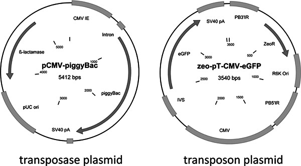
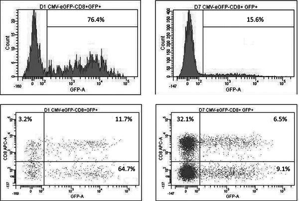
A schematic demonstrating the steps in genetically modifying human T lymphocytes with a reporter gene (eGFP) is shown in Figure 1. These plasmids are available upon request from the authors. A schemtic demonstrating the steps in genetically modified human T lymphocytes with a reporter gene (eGFP) is showin in Figure 2. It is necessary to activate T cells in order to get them to divide, expand, and propagate in culture. Modified human T cells were then cultured and analyzed using flow cytometry for gene expression on Day 1 and Day 7. Shown are results from one donor in Figure 3. Cells were stained with allophycocyanin (APC)-conjugated anti-CD8, analyzed for eGFP (the transgene) and APC fluorescence by a FACSCalibur equipped with the filter set for 4 fluorescence signals using Cell Quest software (Becton Dickinson). We have previously observed gene modification of both CD4 and CD8 positive T cells and herein demonstrate CD8 positive cells as an example7. Although we analyzed for single transgene expression herein, piggyBac has also been used for multi-gene (or multiplexed) gene transfer in human cells17. The decrease in eGFP expression between day 1 and day 7 is likely due to the fact that not all transfected cells undergo stable integration of transposon DNA.
 Figure 1. Schematic of plasmids used for piggyBac mediated gene modification of human T cells. CMV, cytomegalovirus promoter; intron, SV40 intron for mRNA stabilization; piggyBac, transposase cDNA; SV40 pA, polyadenylation site; pUC, origin of replication; b-lactamase, ampicillin resistance gene; PB3'IR, piggyBac 3' inverted repeat; PB5'IR, piggyBac 5' inverted repeat; ZeoR, zeocin resistance gene; R6K Ori, origin of replication; IVS, intervening sequence; eGFP, flourescent reporter gene. Note: antibiotics were used for bacterial selection and growth but were not used in T cell cultures. Click here to view larger figure.
 Figure 2. Schematic describing the modification of primary human T cells using the piggyBac transposon system.
 Figure 3. Stable transgene expression in T cells modified with the piggyBac transposon system. Left panels: eGFP expression on Day 1. Right panels: eGFP expression on day 7. Click here to view larger figure.
Discussion
The method described herein enables stable transgene modification of primary human T lymphocytes. We have previously tested the use of the piggyBac transposon system to modify T cells to express a reporter gene (for more than 4 weeks), a non-immunogenic suicide gene, a chimeric antigen receptor for adoptive immunotherapy (for more than 100 days), and to engineer resistance to immunosuppressive medications7,13-15. Non-viral modification of T cells for adoptive immunotherapy and other applications should be much less expensive and therefore more widely utilized than retroviral transduction. The use of new hyperactive piggyBac elements should increase the feasibility of manufacturing of stable transgene modified human T cells9. Although not described herein, one can achieve necessary numbers of stably transfected T cells for potential clinical application. Using a combination of piggyBac-mediated gene transfer and aK562 (artificial antigen presenting) feeder cells, an initial yield of about 2 x 10⁶ stably transfected T cells can be expanded by 4 to 5 logs to over 1010 transduced T cells in 4 to 5 weeks and to 1012 in 6 to 7 weeks7. Analysis of piggyBac integration sites in human T cells showed no bias towards proto-oncogenes, however, it did show a predilection for integrating into highly expressed genes in activated T cells when using the nucleofection technology outlined above13. PiggyBac represents a promising methodology for stable genetic modification of human T cells for a wide variety of applications.
Disclosures
No conflicts of interest declared.
Acknowledgments
SS is supported in part by the HHMI Med into Grad Training Grant through the TBMM Program. MHW is supported in part by a career development award from the Department of Veterans Affairs and the generous support of Dr. and Mrs. Harold M. Selzman. This work was also supported in part by NIH lymphoma SPORE grant P50CA126752 and NIH R01 DK093660.
References
- Cary LC. Transposon mutagenesis of baculoviruses: analysis of Trichoplusia ni transposon IFP2 insertions within the FP-locus of nuclear polyhedrosis viruses. Virology. 1989;172(1):156. doi: 10.1016/0042-6822(89)90117-7. [DOI] [PubMed] [Google Scholar]
- Fraser MJ. Assay for movement of Lepidopteran transposon IFP2 in insect cells using a baculovirus genome as a target DNA. Virology. 1995;211(2):397. doi: 10.1006/viro.1995.1422. [DOI] [PubMed] [Google Scholar]
- Ding S. Efficient transposition of the piggyBac (PB) transposon in mammalian cells and mice. Cell. 2005;122(3):473. doi: 10.1016/j.cell.2005.07.013. [DOI] [PubMed] [Google Scholar]
- Saridey SK. PiggyBac transposon-based inducible gene expression in vivo after somatic cell gene transfer. Mol. Ther. 2009;17(12):2115. doi: 10.1038/mt.2009.234. [DOI] [PMC free article] [PubMed] [Google Scholar]
- Nakanishi H. piggyBac transposon-mediated long-term gene expression in mice. Mol. Ther. 2010;18(4):707. doi: 10.1038/mt.2009.302. [DOI] [PMC free article] [PubMed] [Google Scholar]
- Wilson MH, Coates CJ, George AL., Jr PiggyBac Transposon-mediated Gene Transfer in Human Cells. Mol. Ther. 2007;15(1):139. doi: 10.1038/sj.mt.6300028. [DOI] [PubMed] [Google Scholar]
- Nakazawa Y. Optimization of the PiggyBac transposon system for the sustained genetic modification of human T lymphocytes. J. Immunother. 2009;32(8):826. doi: 10.1097/CJI.0b013e3181ad762b. [DOI] [PMC free article] [PubMed] [Google Scholar]
- Raja Manuri PV. piggyBac transposon/transposase system to generate CD19-specific T cells for treatment of B-lineage malignancies. Hum. Gene Ther. 2010;21(4):427. doi: 10.1089/hum.2009.114. [DOI] [PMC free article] [PubMed] [Google Scholar]
- Doherty JE. Hyperactive piggyBac gene transfer in human cells and in vivo. Hum. Gene Ther. 2011. In Press. [DOI] [PMC free article] [PubMed]
- Yusa K. A hyperactive piggyBac transposase for mammalian applications. Proc. Natl. Acad. Sci. U.S.A. 2011;108(4):1531. doi: 10.1073/pnas.1008322108. [DOI] [PMC free article] [PubMed] [Google Scholar]
- Bonini C. Genetic modification of T cells. Biol. Blood Marrow Transplant. 2011;17(1):S15–S20. doi: 10.1016/j.bbmt.2010.09.019. [DOI] [PMC free article] [PubMed] [Google Scholar]
- Hackett PB, Largaespada DA, Cooper LJ. A transposon and transposase system for human application. Mol. Ther. 2010;18(4):1531. doi: 10.1038/mt.2010.2. [DOI] [PMC free article] [PubMed] [Google Scholar]
- Galvan DL. Genome-wide mapping of PiggyBac transposon integrations in primary human T cells. J. Immunother. 2009;32(8):837. doi: 10.1097/CJI.0b013e3181b2914c. [DOI] [PMC free article] [PubMed] [Google Scholar]
- Nakazawa Y. PiggyBac-Mediated Cancer Immunotherapy Using EBV-Specific Cytotoxic T-Cells Expressing HER2-Specific Chimeric Antigen Receptor. Mol. Ther. 2011;19(12):2133. doi: 10.1038/mt.2011.131. [DOI] [PMC free article] [PubMed] [Google Scholar]
- Huye LE. Combining mTor Inhibitors With Rapamycin-resistant T Cells: A Two-pronged Approach to Tumor Elimination. Mol. Ther. 2011;19(12):2239. doi: 10.1038/mt.2011.179. [DOI] [PMC free article] [PubMed] [Google Scholar]
- Vera JF. Accelerated production of antigen-specific T cells for preclinical and clinical applications using gas-permeable rapid expansion cultureware (G-Rex) J. Immunother. 2010;33(3):305. doi: 10.1097/CJI.0b013e3181c0c3cb. [DOI] [PMC free article] [PubMed] [Google Scholar]
- Kahlig KM. Multiplexed transposon-mediated stable gene transfer in human cells. Proc. Natl. Acad. Sci. U.S.A. 2010;107(4):1343. doi: 10.1073/pnas.0910383107. [DOI] [PMC free article] [PubMed] [Google Scholar]


