Keratinocytes and dermal fibroblasts express adhesive proteins that ensure the epidermis remains attached to the skin basement membrane and to the papillary dermis. Congenital deficiency of any of at least 15 such proteins results in a blistering condition, termed epidermolysis bullosa (EB)(Fine et al., 2008). The most severe form of EB is the Herlitz variant of junctional EB (JEB-H) caused by loss-of-function mutations in one of the three genes (LAMA3, LAMB3, and LAMC2) encoding one of three chains of the heterotrimeric protein laminin 332 (LM-332)(Kiritsi et al., 2011). LM-332 is secreted by keratinocytes and interacts with integrin receptors α3β1 and α6β4 to form focal adhesions and stable anchoring contacts in the dermalepidermal junction (DEJ). Children with this autosomal recessive genodermatosis develop generalized skin blistering; extensive mucosal erosions in the upper respiratory, gastrointestinal, and genitourinary tracts; infections; and, despite supportive measures, typically die within the first year of life.
Even though it has been largely accepted that JEB-H is untreatable(Yuen et al., 2012), evidence from a gene therapy trial for the less severe (non-Herlitz) form of JEB(Mavilio et al., 2006), and from LAMB3 gene correction of human JEB-H cells –(Robbins et al., 2001; Sakai et al., 2010), suggests novel treatment options.
Prominent among these, the novel technology of reprogramming skin cells into pluripotent stem cells (iPSCs), already applied to EB (Bilousova et al., 2011; Itoh et al., 2011; Tolar et al., 2010) by a combination of specific transcription factors has the dual potential of generating patient-specific, highly proliferative cells for gene-correction strategies and of providing a tool for better understanding the biology of JEB-H. We hypothesized that such iPSCs can be derived from JEB-H individuals. Thus, in principle, an inexhaustible supply of patient-specific stem cells can be generated for local wound therapy and for systemic administration aimed at reaching both skin and internal mucosal membranes.
To investigate this, we obtained skin biopsies from two individuals with JEB-H, who carried mutations in the LAMB3 gene: patient 1 P1; c.1365_1366del (p.Asn456ArgfsX7); c.2207C>A (p.Ser736X(Varki et al., 2006)) and patient 2 (P2; c.1903 C>T (p.Arg635X); c.1117C>T (p.Gln373X). Samples were obtained with written informed consent and following a protocol approved by the University of Minnesota Institutional Review Board and with adherence to the Helsinki Guidelines. All mutations create premature stop codons in the open reading frame, with expected nonsense-mediated decay of the mRNA or truncation of the protein product. The children experienced extensive areas of mucocutaneous lesions from birth, hoarseness and stridor, numerous infections, and progressive severe malnutrition. Examination of skin sections revealed the absence of laminin β3 chain at the DEJ. Electron microscopy examination showed infrequent and underdeveloped hemidesmosomes. Collectively these molecular, clinical, biochemical, and ultrastructural features were consistent with the diagnosis of JEB-H.
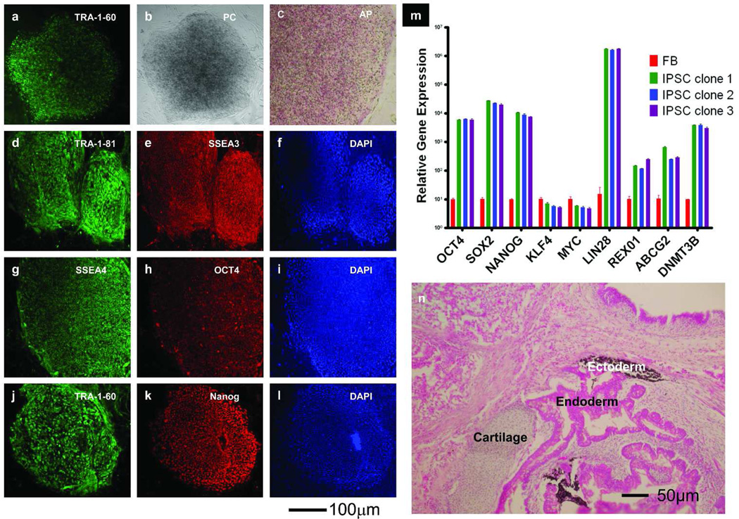
To derive JEB-iPSCs, we transduced skin fibroblasts with the four transcription factors OCT4, SOX2, KLF4, and c-MYC, which are known to induce pluripotency in somatic cells(Tolar et al., 2010). Within three weeks of culture, the patient-specific JEB-iPSCs emerged as raised clusters of cells (Figure 1 a–c). To document the embryonic stem cell-like cellular state, we examined their mRNA and protein expression patterns. When compared with the parental fibroblasts, the JEB-iPSCs expressed the genes coding for nuclear, cytoplasmic, and cell surface proteins (e.g., TRA-1-60, TRA-1-81, stage-specific embryonic antigens 3 and 4, Lin28, Rex1, ABCG2 and DNMT3b, OCT4, and NANOG) in a pattern consistent with embryonic stem cell and iPSC phenotype (Figure 1 d–m). In support of the known activation of endogenous expression of stem cell genes by exogenous reprogramming factors, the maintenance of pluripotency became independent of the original exogenous reprogramming factors (data not shown). As expected in fully reprogrammed iPSCs, the epigenetic profiles showed that endogenous OCT4 and NANOG promoters were largely demethylated (Supplementary Figure 1). The JEB-iPSC lines were maintained for more than 20 passages, and they showed no evidence of genomic instability as evidenced by cytogenetic analysis (Supplementary Figure 2). To exclude the possibilities of cell contamination or the mosaicism observed in JEB(Pasmooij et al., 2007), we verified the authenticity of the JEB-iPSCs by genomic finger-typing with competitive polymerase chain reaction of a variable number of tandem repeat polymorphisms and by sequencing of LAMB3 gene mutations in the JEB-iPSCs (data not shown). To show that JEB-iPSCs are capable of differentiating into cells of endodermal, mesodermal, and ectodermal origin, we injected them into immune-deficient mice lacking T cells, B cells, and natural killer cells, and having a macrophage defect that makes them reliable recipients of human cells. In 6–8 weeks, cystic teratomas formed and cells derived from all three embryonic layers were seen (Figure 1 n). In aggregate, these data show that fully reprogrammed iPSCs can be derived from skin cells of JEB-H individuals.
Figure 1. Expression profile of JEB-iPSCs.
(a–l) To assess the ability of expressing embryonic stem cell proteins, JEB-iPSCs were live stained with TRA-1-60, viewed in phase contrast (PC), immunostained with TRA-1-81, SSEA3, SSEA4, OCT4, TRA-1-60, and NANOG. Corresponding images stained with 4,6-diamidino-2-phenylindole (DAPI) show nuclei of individual cells in the colonies. Scale bar = 100 µm. (m) Quantitative reverse transcription PCR analysis of OCT4, SOX2, NANOG, KLF4, c-MYC, LIN28, REX01, ABCG2, and DNMT3b in parental JEB-H fibroblasts (FB, red bars) and three independent JEB-H iPSC lines (iPSC clones 1, 2, and 3 denoted with green, blue, and violet bars, respectively). All values were normalized against endogenous GAPDH expression. (n) JEB-H iPSC-derived teratoma differentiated into cells of all three germ lines. Same histologic section of mature teratoma from an immunodeficient mouse shows melanocytes of ectodermal origin, cartilage of mesodermal origin, and columnar epithelium with goblet cells of endodermal origin. Hematoxylin-eosin. The findings were analogous for both P1 and P2, therefore data for P1 are shown here as a representative example. Detailed methods are included as supplementary information.
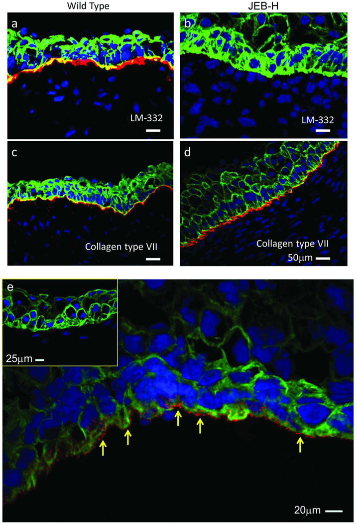
JEB-iPSCs can provide means for drug screening and to model cellular interactions among various mucocutaneous cell types derived from the same individual. To our knowledge, this is a previously unreported use of iPSCs as a cellular tool to study the skin pathology in JEB. We showed first that skin-like structures formed in the process of in vivo JEB-iPSC differentiation (Supplementary Figure 3). In contrast to wild-type iPSCs, the skin-like structures arising from the JEB-iPSCs expressed no detectable laminin β3, but expressed collagen type VII, the DEJ protein deficient in distinct, dystrophic forms of EB (Figure 2 a–d). Next, to substantiate the proof-of-concept that skin cell cultures can be derived from JEB-iPSCs, we differentiated JEB-iPSCs into keratinocytes (Supplementary Figures 4 and 5). Lastly, to demonstrate that this operating procedure can serve as a platform for gene correction of these highly proliferative cells, we transduced the JEB-iPSCs with LAMB3 gene. After transduction, the JEB-iPSC-derived cells expressed and secreted laminin β3 protein (Figure 2 e).
Figure 2. Skin cells derived from JEB-iPSCs.
(a–d) Immunofluorescent staining of JEB-H iPSC-derived skin-like structures showed that both wild-type and JEB-H iPSC-derived cells stained for collagen type VII (red, bottom panels), but only wild-type skin-like structures were positive for LM-332 (red, top panels). Scale bar = 50 µm (e) In contrast to JEB-H skin-like structures (inset), skin-like structures derived in vivo from LAMB3-corrected JEB-H iPSCs expressed extracellular laminin 332 (arrows). Nuclei are stained with DAPI (blue) and keratinocytes are stained with cytokeratin 5 antibody in all the images shown. Detailed methods are included as supplementary information. Scale bar = 20 µm
In summary, we have shown that the LAMB3 defect does not preclude reprogramming into pluripotency, as has been observed in other genetic diseases(Raya et al., 2009). We have also shown that the JEB-iPSCs—in addition to establishing a reliable stem cell source for gene therapy interventions in JEB-H—can be used in the study of early human skin formation and compared to LM-332 in early development. With the ultimate clinical application of iPSC technology in mind, it is worth noting that strategies exist for genome-nonintegrating reprogramming, for depletion of tumor-inducing cells from differentiated iPSC cultures, and—as an JEB-H individual can develop anti-LM-332 antibody—for induction of immunological tolerance to disease-correcting transgenes(Vailly et al., 1998; Wu and Hochedlinger, 2011). Thus, gene-corrected JEB-iPSCs can inform medical advances in this severe and lethal blistering disease, as well as additional extracellular matrix disorders of the skin and other tissues(McGowan and Marinkovich, 2000).
Supplementary Material
ACKNOWLEDGEMENTS
This study was supported in part by grants from DebRA International, Epidermolysis Bullosa Research Fund, University of Minnesota Department of Pediatrics and Academic Health Center, Jackson Gabriel Silver Foundation, Pioneering Unique Cures for Kids Foundation, Minnesota Medical Foundation, and the Children’s Cancer Research Fund, Minneapolis, Minnesota. We would like to acknowledge the use of confocal microscope made available through an NCRR Shared Instrumentation Grant (#1 S10 RR16851), and to thank Paul Khavari for the LAMB3 cDNA. We gratefully acknowledge the US Veterans Affairs Office of Research and Development (MPM), and the National Institutes of Health grant R01 AR047223 (MPM), which supported this work.
Footnotes
CONFLICT OF INTEREST
The authors state no conflict of interest.
REFERENCES
- Bilousova G, Chen J, Roop DR. Differentiation of mouse induced pluripotent stem cells into a multipotent keratinocyte lineage. J Invest Dermatol. 2011;131:857–864. doi: 10.1038/jid.2010.364. [DOI] [PubMed] [Google Scholar]
- Fine JD, Eady RA, Bauer EA, Bauer JW, Bruckner-Tuderman L, Heagerty A, et al. The classification of inherited epidermolysis bullosa (EB): Report of the Third International Consensus Meeting on Diagnosis and Classification of EB. J Am Acad Dermatol. 2008;58:931–950. doi: 10.1016/j.jaad.2008.02.004. [DOI] [PubMed] [Google Scholar]
- Itoh M, Kiuru M, Cairo MS, Christiano AM. Generation of keratinocytes from normal and recessive dystrophic epidermolysis bullosa-indluced pluripotent stem cells. Proc Natl Acad Sci U S A. 2011;108:8797–8802. doi: 10.1073/pnas.1100332108. [DOI] [PMC free article] [PubMed] [Google Scholar]
- Kiritsi D, Kern JS, Schumann H, Kohlhase J, Has C, Bruckner-Tuderman L. Molecular mechanisms of phenotypic variability in junctional epidermolysis bullosa. J Med Genet. 2011;48:450–457. doi: 10.1136/jmg.2010.086751. [DOI] [PubMed] [Google Scholar]
- Mavilio F, Pellegrini G, Ferrari S, Di Nunzio F, Di Iorio E, Recchia A, et al. Correction of junctional epidermolysis bullosa by transplantation of genetically modified epidermal stem cells. Nat Med. 2006;12:1397–1402. doi: 10.1038/nm1504. [DOI] [PubMed] [Google Scholar]
- McGowan KA, Marinkovich MP. Laminins and human disease. Microsc Res Tech. 2000;51:262–279. doi: 10.1002/1097-0029(20001101)51:3<262::AID-JEMT6>3.0.CO;2-V. [DOI] [PubMed] [Google Scholar]
- Pasmooij AM, Pas HH, Bolling MC, Jonkman MF. Revertant mosaicism in junctional epidermolysis bullosa due to multiple correcting second-site mutations in LAMB3. J Clin Invest. 2007;117:1240–1248. doi: 10.1172/JCI30465. [DOI] [PMC free article] [PubMed] [Google Scholar]
- Raya A, Rodriguez-Piza I, Guenechea G, Vassena R, Navarro S, Barrero MJ, et al. Disease-corrected haematopoietic progenitors from Fanconi anaemia induced pluripotent stem cells. Nature. 2009;460:53–59. doi: 10.1038/nature08129. [DOI] [PMC free article] [PubMed] [Google Scholar]
- Robbins PB, Lin Q, Goodnough JB, Tian H, Chen X, Khavari PA. In vivo restoration of laminin 5 beta 3 expression and function in junctional epidermolysis bullosa. Proc Natl Acad Sci U S A. 2001;98:5193–5198. doi: 10.1073/pnas.091484998. [DOI] [PMC free article] [PubMed] [Google Scholar]
- Sakai N, Waterman EA, Nguyen NT, Keene DR, Marinkovich MP. Observations of skin grafts derived from keratinocytes expressing selectively engineered mutant laminin-332 molecules. J Invest Dermatol. 2010;130:2147–2150. doi: 10.1038/jid.2010.85. [DOI] [PMC free article] [PubMed] [Google Scholar]
- Tolar J, Xia L, Riddle MJ, Lees CJ, Eide CR, McElmurry RT, et al. Induced Pluripotent Stem Cells from Individuals with Recessive Dystrophic Epidermolysis Bullosa. J Invest Dermatol. 2010;131:848–856. doi: 10.1038/jid.2010.346. [DOI] [PMC free article] [PubMed] [Google Scholar]
- Vailly J, Gagnoux-Palacios L, Dell'Ambra E, Roméro C, Pinola M, Zambruno G, et al. Corrective gene transfer of keratinocytes from patients with junctional epidermolysis bullosa restores assembly of hemidesmosomes in reconstructed epithelia. Gene Ther. 1998;5:1322–1332. doi: 10.1038/sj.gt.3300730. [DOI] [PubMed] [Google Scholar]
- Varki R, Sadowski S, Pfendner E, Uitto J. Epidermolysis bullosa. I. Molecular genetics of the junctional and hemidesmosomal variants. J Med Genet. 2006;43:641–652. doi: 10.1136/jmg.2005.039685. [DOI] [PMC free article] [PubMed] [Google Scholar]
- Wu SM, Hochedlinger K. Harnessing the potential of induced pluripotent stem cells for regenerative medicine. Nat Cell Biol. 2011;13:497–505. doi: 10.1038/ncb0511-497. [DOI] [PMC free article] [PubMed] [Google Scholar]
- Yuen WY, Duipmans JC, Molenbuur B, Herpertz I, Mandema JM, Jonkman MF. Long-term follow-up of patients with Herlitz type junctional epidermolysis bullosa. Br J Dermatol. 2012 Apr 18; doi: 10.1111/j.1365-2133.2012.10997.x. advance online publication. [DOI] [PubMed] [Google Scholar]
Associated Data
This section collects any data citations, data availability statements, or supplementary materials included in this article.


