Abstract
Inherited dentin defects are classified into three types of dentinogenesis imperfecta (DGI) and two types of dentin dysplasia (DD). The genetic etiology of DD-I is unknown. Defects in dentin sialophosphoprotein (DSPP) cause DD type II and DGI types II and III. DGI type I is the oral manifestation of osteogenesis imperfecta (OI), a systemic disease typically caused by defects in COL1A1 or COL1A2. Mutations in MSX1, PAX9, AXIN2, EDA and WNT10A can cause non-syndromic familial tooth agenesis. In this study a simplex pattern of clinical dentinogenesis imperfecta juxtaposed with a dominant pattern of hypodontia (mild tooth agenesis) was evaluated, and available family members were recruited. Mutational analyses of the candidate genes for DGI and hypodontia were performed and the results validated. A spontaneous novel mutation in COL1A2 (c.1171G>A; p.Gly391Ser) causing only dentin defects and a novel mutation in PAX9 (c.43T>A; p.Phe15Ile) causing hypodontia were identified and correlated with the phenotypic presentations in the family. Bone radiographs of the proband’s dominant leg and foot were within normal limits. We conclude that when no DSPP mutation is identified in clinically determined isolated DGI cases, COL1A1 and COL1A2 should be considered as candidate genes. PAX9 mutation p.Phe15Ile within the N-terminal β-hairpin structure of the PAX9 paired domain causes tooth agenesis.
Introduction
Osteogenesis imperfecta (OI) is a hereditary, bone fragility disease that varies in severity from mild bone defects to neonatal lethality. Mutations in the type-I collagen genes, COL1A1 and COL1A2, have been identified in approximately 90% of individuals with OI [1]. Dentinogenesis imperfecta type I (DGI-I) is a common phenotypic feature of OI [2]. Non-syndromic dentin defects categorized as dentin dysplasia type II (DD-II), dentinogenesis imperfecta type II (DGI-II) and type III (DGI-III) are generally caused by dominant mutations in dentin sialophosphoprotein (DSPP), which encodes the most abundant non-collagenous matrix component of dentin [3]. DSPP is critical for predentin formation and dentin mineralization [4], although the dentin malformations associated with DSPP mutations are likely due to odontoblast pathology caused by dominant negative or gain of function effects [5], [6].
Familial tooth agenesis is distinguished from conditions with multiple missing teeth occurring in syndromes, such as ectodermal dysplasia. MSX1, PAX9, AXIN2, WNT10A, and EDA are proven candidate genes for familial tooth agenesis [7], [8]. In this study we identified a Caucasian family with familial tooth agenesis going back at least three generations, but isolated dentin defects occurred only in the proband. Using a target gene approach, we identified a novel missense mutation in PAX9 (p.Phe15Ile) that is responsible for the tooth agenesis. Characterization of the DSPP 5-prime regulatory region, intron/exon borders, and coding region identified no potential disease-causing mutations. Despite the absence of skeletal abnormalities, mutational analyses of the COL1A1 and COL1A2 genes identified a novel COL1A2 mutation (p.Gly391Ser) only in the proband that explains his dentin phenotype.
Materials and Methods
Human subjects
The human study protocol and subject consents were reviewed and approved by the Institutional Review Board at the University of Michigan. Study participants signed appropriate written consents after an explanation of their contents and after their questions about the study were answered. Any minors age 8 or older signed a written assent form after their parent completed a written parental consent for participation of the minor.
A five-year-old boy of European descent was referred by a genetics clinic for evaluation of dentin defects. The proband’s pediatrician and geneticist considered OI, but decided that DGI-II was a better diagnosis due to the lack of systemic signs and history of bone fractures. The proband’s growth and developmental parameters were within normal limits. He was the only family member with dentin defects in the known history of the extended family. Oral photographs and radiographs were obtained from the family’s dental care provider. The dental radiographs revealed the tooth agenesis in the proband and his mother. The mother reported that this trait came from her father and that her siblings were unaffected. Blood samples (5 cc) were collected from the proband, his brother, and their biological parents. Radiographic evaluation of weight-bearing bones on the proband’s dominant side was conducted and a report compiled by the Radiology Department, Spectrum Health, Grand Rapids, Michigan.
Mutational analysis
The MSX1 and PAX9 coding exons and intron junctions were amplified by PCR as previously reported [9]. The amplification products were purified and characterized by direct DNA sequencing at the University of Michigan DNA Sequencing Core. Because the dentin defects were observed only in the proband, fifteen polymorphic markers were amplified using maternal, paternal and proband DNA for paternity testing. In assessing the DSPP sequence, the putative promoter region (1–2000 bp upstream to the transcription start site) and the first four exons were analyzed by PCR amplification and direct DNA sequencing. DSPP exon 5 was characterized by cloning and sequencing due to the highly variable repeat region of DPP. The PCR primers for DPP (DSPP-FRF 5′-AGTCCATGCAAGGAGATGATCC-3′ and DSPP-FRR 5′-CTAATCATCACTGGTTGAGTGG-3′) annealed at 57°C and generated a 2534-bp amplification product that was subcloned (TOPO cloning kit, Invitrogen, Calsbad, CA). Twenty clones from two separate PCR amplifications were sequenced using forward and reverse M13 primers and two custom primers (5′-CAGACAGCAGCAAATCAGAG-3′, 5′-GATAGCGACAGCAGCAATAGA-3′). The COL1A1 and COL1A2 coding sequences were characterized by Athena Diagnostics (Worcester, MA, USA). The putative PAX9 and COL1A2 disease causing mutations were analyzed using PolyPhen-2 (Polymorphism Phenotyping version 2.2.2) [10].
Results
Pedigree analyses
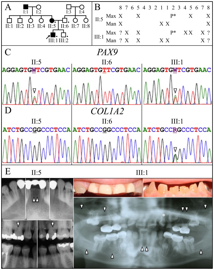
A 3-generation pedigree was constructed (Fig 1A) based upon family histories provided by the mother and maternal grandmother, and was consistent with an autosomal dominant pattern of inheritance for tooth agenesis. Dentin defects occurred only in the proband.
Figure 1. Family pedigree, missing teeth and disease causing mutations. A:
The family pedigree follows the tooth agenesis trait for 3 generations and is consistent with an autosomal-dominant pattern of inheritance. Key: A filled icon indicates tooth agenesis. A dot indicates individual who donated samples. B: Chart of missing teeth in mother (II:5) and the proband (III:1). C: DNA sequencing chromatograms show that the affected mother (II:5) and proband (III:1) had a T or A (W) (arrowhead) at position g.5368 (NCBI Ref. Seq. NC_000014.8). This PAX9 mutation (g.5368T>A; c.43T>A; p.Phe15Ile) caused the tooth agenesis. D: DNA sequencing chromatograms show both parents (II:5; II:6) had the wild-type G, while the proband had a G or A (R) (arrowhead) at position g.15941 (NCBI Ref. Seq. NG_007405.1). This spontaneous COL1A2 mutation (g.15941G>A; c.1171G>A; p.Gly391Ser) caused the dentin defects in the proband. E: Radiographs of the mother (II:5) and proband (III:1) document the missing teeth (arrowheads) and the peg lateral (*) in the proband. Oral photos show the proband’s primary anterior teeth show the brownish discoloration and attrition. His maxillary incisors were removed because of severe attrition and a pediatric partial denture was placed. The proband’s radiographs show the bulbous crowns with cervical constrictions and thin, narrow roots.
Clinical and radiographic findings
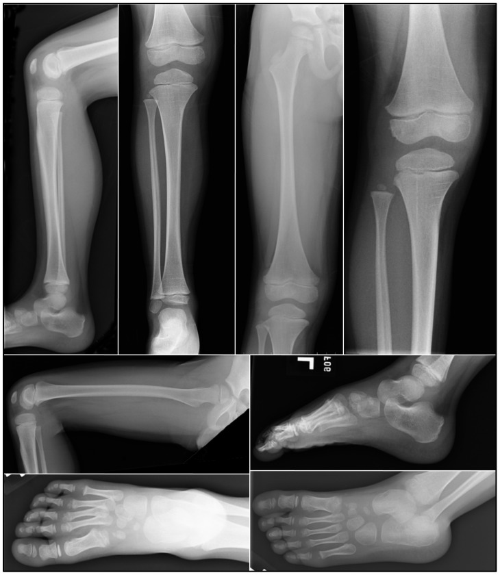
The maternal grandfather, mother and the proband exhibited tooth agenesis. The mother was missing tooth numbers 4, 13, 24, 25, and all third molars (Fig 1B, 1E). In both the mother and the proband, tooth 10 was a peg lateral incisor. The proband at age 8.0 showed no radiographic evidence of developing tooth germs for tooth numbers 2, 5, 12, 13, 15, 18, 24, 25, 29, 31 and was too young to determine the status of his third molars (Fig 1B, 1E). His primary teeth had bulbous crowns, enlarged pulp chambers, and opalescent dentin consistent with the phenotypic features of dentinogenesis imperfecta, while none of those features were observed in the radiographs of his brother, mother and father (data not shown). Radiographs of the dominant (left) leg of the proband (Fig 2), including his femur, tibia, fibula, foot, and knee, revealed no significant osteopenia, bony destructive process, periosteal reactions, or evidence of any acute fractures, dislocations, injuries, or remote traumatic changes. Thus there was no evidence supporting a diagnosis of osteogenesis imperfecta.
Figure 2. Radiographs of the proband’s left.
(dominant) leg at age 6. Radiographs show the femur, tibia, fibula, knee, and foot. No significant osteopenia, bony destructive process, periosteal reactions, or evidence of any acute fractures, dislocations, injuries, or remote traumatic changes were observed.
Identification of a PAX9 and a COL1A2 missense mutation
The proband and his mother had a novel missense mutation (c.43T>A; p.Phe15Ile) in PAX9 (Fig 1C) that was not in unaffected family members, the dbSNP database or in 1000 Genomes Project Pilot Data [11]. No potential disease-causing mutations were identified in MSX1. The mutated amino acid is invariant in vertebrates and the p.Phe15Ile substitution was predicted to be probably damaging by PolyPhen-2 analyses. Supported by the finding of other PAX9 mutations causing familial tooth agenesis (Table 1), we conclude that this mutation causes the tooth agenesis in this family.
Table 1. PAX9 mutations causing familial tooth agenesis.
| cDNA | Protein | Reference | |
| 1) | PAX9 deletion | p.0 | [21] |
| 2) | c.1A>G; | p.M1V | [22] |
| 3) | c.16G>A | p.G6R | [19] |
| 4) | c.43T>A | p.F15I | This Report |
| 5) | c.62T>C | p.L21P | [23] |
| 6) | c.76C>T | p.R26W | [24] |
| 7) | c.83>C | p.R28P | [25] |
| 8) | c.109_110insG | p.I37Sfs*41 | [26] |
| 9) | c.128G>A | p.S43K | [19] |
| 10) | c.139C>T | p.R47W | [27] |
| 11) | c.151G>C | p.G51S | [28] |
| 12) | c.175C>T | p.R59* | [29] |
| 13) | c.175_176ins288 | p.R59Zfs*177 | [23] |
| 14) | c.218_219insG | p.S74Qfs*317 | [30] |
| 15) | c.259A>T | p.I87F | [31] |
| 16) | c.271A>G | p.K91E | [23] |
| 17) | c.340A>T | p.K114* | [32] |
| 18) | c.433C>T | p.Q145* | [33] |
| 19) | c.503C>G | p.A168G | [34] |
| 20) | c.619_621delATCins24bp | p.I207Yfs*211 | [28] |
| 21) | c.793insC | p.V265RfsX315 | [35] |
No potential disease-causing mutations were identified in the promoter, coding regions, or intron/exon boundaries in DSPP. Analyses covering all intron/exon junctions of COL1A1 identified only known polymorphisms (Table 2). A novel missense mutation (c.1171G>A; p.Gly391Ser) in one COL1A2 allele only in the proband was identified. This sequence variation is not reported in the SNP database. As paternity was confirmed, this was likely a spontaneous germ-line mutation. The mutated amino acid is invariant in vertebrates and the p.Gly391Ser substitution was predicted to be probably damaging by PolyPhen-2 analyses. Supported by Pallos et al, documenting a family with dentinogenesis imperfecta associated with a minimal bone phenotype caused by a COL1A1 mutation [12], we conclude that the spontaneous COL1A2 mutation caused the proband’s dentin defects.
Table 2. Collagen (COL1A1 and COL1A2) sequence variations identified in the proband.
Discussion
The objective of this study was to identify the causative gene mutation (s) for the dentin defects and familial tooth agenesis in this family. The dentin defects were caused by a novel spontaneous missense mutation that substituted serine for a conserved glycine in the triple helical region of COL1A2. The familial tooth agenesis segregated as an autosomal dominant trait and resulted from a novel missense mutation affecting the conserved N-terminal DNA binding domain of PAX9.
COL1A2 p.G391S mutation and OI/DGI
Type-I collagen, the major extracellular matrix protein of bone, dentin, and skin, is comprised of two pro-á1 chains and one pro-á2 chain encoded by COL1A1 and COL1A2, respectively. Mutations in these two genes generally cause dominant forms of OI, characterized by bone fragility, altered sclera hue, hearing loss, dentin defects, and soft tissue dysplasia. However, the clinical presentation of OI is highly heterogeneous. Since 85% of organic component in tooth dentin is type-I collagen, dentin defects are observed in many OI cases. Pallos et al reported a family with autosomal-dominant OI in which affected members exhibited dentin defects without obvious bone abnormalities, except hyperextensible joints and joint pain. After checking DSPP, they identified the disease-causing mutation in COL1A1 [12]. Our investigation followed a similar course. After finding no potential disease-causing mutations in DSPP, we analyzed COL1A1 and COL1A2 and identified the missense mutation (p.Gly391Ser) in COL1A2. We then searched more carefully for bone defects. Radiographs of the weight-bearing bones on the proband’s dominant side revealed no pathologic bone defects. Based upon Pallos’ report and our present study, we propose that COL1A1 and COL1A2 should be regarded as strong candidate genes for isolated dentin defects when no mutation can be identified in DSPP.
The repeating Gly-X-Y sequence motif in collagen is critical for its triple helical conformation. Glycine must occupy every third position as its side chain is the only one small enough to fit into the interior position of the helix. Substitutions for invariant glycines are the most common cause of clinically significant OI [13]. The clinical severity of different glycine mutations varies significantly from lethality to a mild connective tissue disorder, depending upon the location in the chain where the glycine substitution occurs. Several models have been developed to correlate glycine mutations with the severity of OI phenotypes. Bodian et al proposed a model to predict the clinical lethality of collagen glycine mutations [14] and Rauch et al provided a detailed picture of genotype–phenotype correlations in OI patients with glycine mutations [2]. Besides affecting the helix, crucial regions may represent certain specific ligand-binding sites [15]. Finding that the p.Gly391Ser substitution in the pro-á2 chain, leads to dentin defects without a clinically-detectable skeletal phenotype suggests that this glycine may reside in a region that interacts with DSPP.
PAX9 p.F15I mutation and familial tooth agenesis
Tooth formation is a sequential process of epithelial-mesenchymal interactions controlled by numerous molecules and signaling pathways [16]. Mutations in genes critical for the early stages of tooth formation lead to familial tooth agenesis. At present, mutations causing non-syndromic tooth agenesis have been identified in MSX1, PAX9, AXIN2, EDA, and WNT10A [7]. In general, the pattern of missing teeth correlates with the causative gene. Second bicuspids and third molars are frequently involved in MSX1-associated tooth agenesis. PAX9 mutations lead to tooth agenesis of second bicuspids, second molars, and some central incisors [9]. AXIN2 aberrations cause multiple missing teeth, intestinal polyposis, and predispose to colorectal cancer [17]. EDA-associated tooth agenesis is more likely to miss multiple anterior teeth [18]. In the present study, agenesis of third molars, second bicuspids, and mandibular central incisors in the mother, suggested MSX1-associated tooth agenesis. No MSX1 mutation could be identified, but a PAX9 missense mutation substituting isoleucine for a highly conserved phenylalanine (p.Phe15Ile) was identified in the paired box domain crucial for DNA binding. Wang et al identified a PAX9 mutation (p.Gly6Arg) causing a mild hypodontia phenotype almost identical to that of our proband’s mother [19]. Interestingly, our p.Phe15Ile substitution is structurally close to the p.Gly6Arg mutation. Both of these amino acids reside in the N-terminal beta hairpin of the paired domain [20]. Initially this suggested to us that substitutions within the N-terminal beta hairpin of PAX9 cause a relatively minor functional deficit. However, our proband had substantially more missing teeth than his mother, highlighting the variable expressivity of the tooth agenesis phenotype. Characterizing additional PAX9 mutations affecting the N-terminal beta hairpin of the paired domain and documenting their associated dental phenotypes will be necessary before we can conclude that such substitutions generally cause a minor functional deficit.
Acknowledgments
We thank the family for their enthusiastic participation in this investigation. The authors declare there are no competing interests.
Funding Statement
This study was supported by NIDCR National Institute of Dental and Craniofacial Research – /NIH research grant DE015846. The funders had no role in study design, data collection and analysis, decision to publish, or preparation of the manuscript.
References
- 1. Basel D, Steiner RD (2009) Osteogenesis imperfecta: recent findings shed new light on this once well-understood condition. Genet Med 11: 375–385. [DOI] [PubMed] [Google Scholar]
- 2. Rauch F, Lalic L, Roughley P, Glorieux FH (2010) Genotype-phenotype correlations in nonlethal osteogenesis imperfecta caused by mutations in the helical domain of collagen type I. Eur J Hum Genet. 18: 642–647. [DOI] [PMC free article] [PubMed] [Google Scholar]
- 3. Lee KE, Kang HY, Lee SK, Yoo SH, Lee JC, et al. (2011) Novel dentin phosphoprotein frameshift mutations in dentinogenesis imperfecta type II. Clin Genet 79: 378–384. [DOI] [PubMed] [Google Scholar]
- 4. Suzuki S, Sreenath T, Haruyama N, Honeycutt C, Terse A, et al. (2009) Dentin sialoprotein and dentin phosphoprotein have distinct roles in dentin mineralization. Matrix Biol 28: 221–229. [DOI] [PMC free article] [PubMed] [Google Scholar]
- 5. Wang SK, Chan HC, Rajderkar S, Milkovich RN, Uston KA, et al. (2011) Enamel malformations associated with a defined dentin sialophosphoprotein mutation in two families. Eur J Oral Sci 119: 158–167. [DOI] [PMC free article] [PubMed] [Google Scholar]
- 6. McKnight DA, Suzanne Hart P, Hart TC, Hartsfield JK, Wilson A, et al. (2008) A comprehensive analysis of normal variation and disease-causing mutations in the human DSPP gene. Hum Mutat 29: 1392–1404. [DOI] [PMC free article] [PubMed] [Google Scholar]
- 7. Nieminen P (2009) Genetic basis of tooth agenesis. J Exp Zool B Mol Dev Evol 312B: 320–342. [DOI] [PubMed] [Google Scholar]
- 8. van den Boogaard MJ, Creton M, Bronkhorst Y, van der Hout A, Hennekam E, et al. (2012) Mutations in WNT10A are present in more than half of isolated hypodontia cases. J Med Genet 49: 327–331. [DOI] [PubMed] [Google Scholar]
- 9. Kim JW, Simmer JP, Lin BP, Hu JC (2006) Novel MSX1 Frameshift Causes Autosomal-dominant Oligodontia. J Dent Res 85: 267–271. [DOI] [PMC free article] [PubMed] [Google Scholar]
- 10. Adzhubei IA, Schmidt S, Peshkin L, Ramensky VE, Gerasimova A, et al. (2010) A method and server for predicting damaging missense mutations. Nat Methods 7: 248–249. [DOI] [PMC free article] [PubMed] [Google Scholar]
- 11. 1000 Genomes Project Consortium (2010) A map of human genome variation from population-scale sequencing. Nature 467: 1061–1073. [DOI] [PMC free article] [PubMed] [Google Scholar]
- 12. Pallos D, Hart PS, Cortelli JR, Vian S, Wright JT, et al. (2001) Novel COL1A1 mutation (G559C) [correction of G599C] associated with mild osteogenesis imperfecta and dentinogenesis imperfecta. Arch Oral Biol 46: 459–470. [DOI] [PubMed] [Google Scholar]
- 13. Marini JC, Forlino A, Cabral WA, Barnes AM, San Antonio JD, et al. (2007) Consortium for osteogenesis imperfecta mutations in the helical domain of type I collagen: regions rich in lethal mutations align with collagen binding sites for integrins and proteoglycans. Hum Mutat 28: 209–221. [DOI] [PMC free article] [PubMed] [Google Scholar]
- 14. Bodian DL, Madhan B, Brodsky B, Klein TE (2008) Predicting the clinical lethality of osteogenesis imperfecta from collagen glycine mutations. Biochemistry 47: 5424–5432. [DOI] [PubMed] [Google Scholar]
- 15. Sweeney SM, Orgel JP, Fertala A, McAuliffe JD, Turner KR, et al. (2008) Candidate cell and matrix interaction domains on the collagen fibril, the predominant protein of vertebrates. J Biol Chem 283: 21187–21197. [DOI] [PMC free article] [PubMed] [Google Scholar]
- 16. Tummers M, Thesleff I (2009) The importance of signal pathway modulation in all aspects of tooth development. J Exp Zool B Mol Dev Evol 312B: 309–319. [DOI] [PubMed] [Google Scholar]
- 17. Lammi L, Arte S, Somer M, Jarvinen H, Lahermo P, et al. (2004) Mutations in AXIN2 cause familial tooth agenesis and predispose to colorectal cancer. Am J Hum Genet 74: 1043–1050. [DOI] [PMC free article] [PubMed] [Google Scholar]
- 18. Han D, Gong Y, Wu H, Zhang X, Yan M, et al. (2008) Novel EDA mutation resulting in X-linked non-syndromic hypodontia and the pattern of EDA-associated isolated tooth agenesis. Eur J Med Genet 51: 536–546. [DOI] [PubMed] [Google Scholar]
- 19. Wang Y, Wu H, Wu J, Zhao H, Zhang X, et al. (2009) Identification and functional analysis of two novel PAX9 mutations. Cells Tissues Organs 189: 80–87. [DOI] [PMC free article] [PubMed] [Google Scholar]
- 20. Wang Y, Groppe JC, Wu J, Ogawa T, Mues G, et al. (2009) Pathogenic mechanisms of tooth agenesis linked to paired domain mutations in human PAX9. Hum Mol Genet 18: 2863–2874. [DOI] [PMC free article] [PubMed] [Google Scholar]
- 21. Das P, Stockton DW, Bauer C, Shaffer LG, D’Souza RN, et al. (2002) Haploinsufficiency of PAX9 is associated with autosomal dominant hypodontia. Hum Genet 110: 371–376. [DOI] [PubMed] [Google Scholar]
- 22. Klein ML, Nieminen P, Lammi L, Niebuhr E, Kreiborg S (2005) Novel mutation of the initiation codon of PAX9 causes oligodontia. J Dent Res 84: 43–47. [DOI] [PubMed] [Google Scholar]
- 23. Das P, Hai M, Elcock C, Leal SM, Brown DT, et al. (2003) Novel missense mutations and a 288-bp exonic insertion in PAX9 in families with autosomal dominant hypodontia. Am J Med Genet A 118: 35–42. [DOI] [PMC free article] [PubMed] [Google Scholar]
- 24. Lammi L, Halonen K, Pirinen S, Thesleff I, Arte S, et al. (2003) A missense mutation in PAX9 in a family with distinct phenotype of oligodontia. Eur J Hum Genet 11: 866–871. [DOI] [PubMed] [Google Scholar]
- 25. Jumlongras D, Lin JY, Chapra A, Seidman CE, Seidman JG, et al. (2004) A novel missense mutation in the paired domain of PAX9 causes non-syndromic oligodontia. Hum Genet 114: 242–249. [DOI] [PubMed] [Google Scholar]
- 26. Zhao JL, Chen YX, Bao L, Xia QJ, Wu TJ, et al. (2005) [Novel mutations of PAX9 gene in Chinese patients with oligodontia]. Zhonghua Kou Qiang Yi Xue Za Zhi 40: 266–270. [PubMed] [Google Scholar]
- 27. Zhao J, Hu Q, Chen Y, Luo S, Bao L, et al. (2007) A novel missense mutation in the paired domain of human PAX9 causes oligodontia. Am J Med Genet A 143A: 2592–2597. [DOI] [PubMed] [Google Scholar]
- 28. Mostowska A, Kobielak A, Biedziak B, Trzeciak WH (2003) Novel mutation in the paired box sequence of PAX9 gene in a sporadic form of oligodontia. Eur J Oral Sci 111: 272–276. [DOI] [PubMed] [Google Scholar]
- 29. Tallon-Walton V, Manzanares-Cespedes MC, Arte S, Carvalho-Lobato P, Valdivia-Gandur I, et al. (2007) Identification of a novel mutation in the PAX9 gene in a family affected by oligodontia and other dental anomalies. Eur J Oral Sci 115: 427–432. [DOI] [PubMed] [Google Scholar]
- 30. Stockton DW, Das P, Goldenberg M, D’Souza RN, Patel PI (2000) Mutation of PAX9 is associated with oligodontia. Nature Genet 24: 18–19. [DOI] [PubMed] [Google Scholar]
- 31. Kapadia H, Frazier-Bowers S, Ogawa T, D’Souza RN (2006) Molecular characterization of a novel PAX9 missense mutation causing posterior tooth agenesis. Eur J Hum Genet 14: 403–409. [DOI] [PubMed] [Google Scholar]
- 32. Nieminen P, Arte S, Tanner D, Paulin L, Alaluusua S, et al. (2001) Identification of a nonsense mutation in the PAX9 gene in molar oligodontia. Eur J Hum Genet 9: 743–746. [DOI] [PubMed] [Google Scholar]
- 33. Hansen L, Kreiborg S, Jarlov H, Niebuhr E, Eiberg H (2007) A novel nonsense mutation in PAX9 is associated with marked variability in number of missing teeth. Eur J Oral Sci 115: 330–333. [DOI] [PubMed] [Google Scholar]
- 34. Boeira Junior BR, Echeverrigaray S (2012) Novel missense mutation in PAX9 gene associated with familial tooth agenesis. J Oral Pathol Med 2: 1600–0714. [DOI] [PubMed] [Google Scholar]
- 35. Frazier-Bowers SA, Guo DC, Cavender A, Xue L, Evans B, et al. (2002) A novel mutation in human PAX9 causes molar oligodontia. J Dent Res 81: 129–133. [PubMed] [Google Scholar]
- 36. Chan TF, Poon A, Basu A, Addleman NR, Chen J, et al. (2008) Natural variation in four human collagen genes across an ethnically diverse population. Genomics 91: 307–314. [DOI] [PMC free article] [PubMed] [Google Scholar]
- 37. Liang CL, Hung KS, Tsai YY, Chang W, Wang HS, et al. (2007) Systematic assessment of the tagging polymorphisms of the COL1A1 gene for high myopia. J Hum Genet 52: 374–377. [DOI] [PubMed] [Google Scholar]
- 38. Bodian DL, Chan TF, Poon A, Schwarze U, Yang K, et al. (2009) Mutation and polymorphism spectrum in osteogenesis imperfecta type II: implications for genotype-phenotype relationships. Hum Mol Genet 18: 463–471. [DOI] [PMC free article] [PubMed] [Google Scholar]


