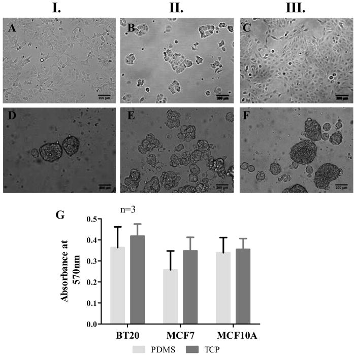
Fig. 1.
Morphology of BT20 (I.), MCF7 (II.) and MCF10A (III.) cell lines propagating as a monolayer on TCP (AC) and spheroids on PDMS (D–F) 48h after seeding [seeding density=50,000 cells/well of a 24-well plate, scale bars=200 μm]. MTT assay for determining the metabolic activity of cells indicate that there is no statistically significant difference in the metabolic activity of cells on PDMS and TCP (G).

