Abstract
Aim
Detection of the possible impact of surgical management on the occurrence of minimal residual disease (MRD) in patients with colorectal carcinoma (CRC) in bone marrow samples, portal and peripheral blood samples. Comparison of MRD prevalence in patients with laparoscopic resection of CRC and those with open colorectal resection. Assessment of the potential impact of MRD on the relapse of the disease and overall survival of patients.
Material and methods
The study included 124 patients with primary CRC without proven distant metastases indicated for elective laparoscopic resection and operated on between September 21, 2006 and December 31, 2008 at the Department of Surgery, Hospital and J.G. Mendel Oncological Centre Novy Jicin. 6 samples were collected from each patient to determine MRD (system venous blood and bone marrow at the beginning of surgery, venous blood from mesenteric bloodstream, system venous blood after the resection, system venous blood and bone marrow 1 month after the surgery). Detection of MRD on the basis of CEA expression was performed by real-time RT-PCR technique. The results were compared with those obtained within the similar research using the same methodology at the 2nd Department of Surgery, University Hospital in Olomouc (the group included 230 patients treated with open resection of CRC).
Results
In the group of patients treated with laparoscopic resection, a correlation between positive MRD in the sample of bone marrow collected before the surgery and the stage of the disease was found (p < 0.035). We also recorded the impact of surgical management on MRD occurrence in system venous blood in primary negative patients (p < 0.025). However, in the course of the short period of time we have not found a statistically significant correlation between the finding in patients with stage I-III, and the period prior to the relapse of the disease (p < 0.59). With regard to the results obtained, we can expect a potential direct correlation between a positive MRD finding in system venous blood taken prior to surgery in patients with stage I-III CRC and shorter time of survival (p < 0.075). In the group of patients treated with open resection of CRC, no statistically significant relationship between the stage of the disease and MRD occurrence was found. Incidence of circulating tumour cells (CTC) in the samples of venous blood taken prior to surgery is a prognostically important factor (p < 0.05) from the viewpoint of disease-free survival (DFS). Another prognostically important factor with regard to DFS seems to be the occurrence of disseminated tumour cells (DTC) in the bone marrow taken 1 month after the surgery.
Conclusions
The data recorded suggest a relationship between MRD finding and the disease prognosis. Collection of samples as well as evaluation of results continue as we strive to include more patients in our study and to obtain more data within 5-10 years of the follow-up. The comparison between the data obtained in the laparoscopic approach and the data obtained in open resection performed from the viewpoint of molecular biology did not show a significant difference in MRD detection in the samples collected after the surgery.
Keywords: minimal residual disease, colorectal carcinoma, laparoscopic resection
Introduction
Minimal residual disease (MRD) in patients with solid tumours is characterised by the occurrence of cancer cells in patients who underwent resection of the primary tumour and are currently without clinical manifestation of the disease. These may include circulating tumour cells (CTC), disseminated tumour cells in bone marrow (DTC) or cells within the lymphatic system [1]. These isolated cells are considered precursors of micro-metastases. The idea of MRD detection in solid tumours is to use it for the identification of epithelial cells in mesenchymal compartments, i.e. in bone marrow, blood, and the lymphatic system [2].
Isolated tumour cells are usually present in concentrations which cannot be detected with standard methods. These cells are detected with techniques of molecular biology, quantitative real-time RT-PCR is the most sensitive method. The principle of real-time RT-PCR is the detection of a specific marker of tumour epithelial cells at the level of the RNA sequence (information RNA-mRNA). The real-time RT-PCR method makes it possible – on the basis of quantification of the expression of tumour markers – to detect the presence of a single tumour cell in a population of 10 million non-tumour cells. This method is approximately 3 times more sensitive than immunohistochemical determination, and – with regard to the absolute quantification in an oncological patient – it is able to determine the difference in the number of tumour cells present in peripheral blood and in bone marrow [2–6].
Material and methods
Between September 21, 2006 and December 31, 2008, 124 patients diagnosed with colorectal carcinoma were operated on with the laparoscopic approach at the Department of Surgery, Hospital and J.G. Mendel Oncological Centre Novy Jicin [7]. The results were compared with 147 patients with colorectal carcinoma, stage I-III and R0 resection performed with open surgery at the 1st and 2nd Departments of Surgery, University Hospital in Olomouc, between 2004 and 2008 [8, 9]. Patients with treatment other than resection (i.e. stoma, bypass, exploration), those with duplicate tumour disease, and those who underwent any type of oncological treatment in the past, were not included in the study. In patients included in the study there were not (at the time of surgery) diagnosed synchronous distant metastases of colorectal carcinoma, and in all the patients radical (R0) resection was performed. Details on stage of the disease are given in Table I.
Table I.
Description of the group of 124 patients with laparoscopic resection (LS) and 147 patients with open surgery (OS) based on disease staging [10]
| TNM classification | LS, n (%) | OS, n (%) |
|---|---|---|
| Stage I | 23 (18.6) | 41 (27.9) |
| Stage II | 53 (42.7) | 59 (40.1) |
| Stage III | 48 (38.7) | 47 (32.0) |
In the patients included in the study 6 samples were taken to detect isolated tumour cells:
system venous blood sample collected at the beginning of surgery (pSVB),
bone marrow sample collected at the beginning of surgery (pBM),
venous blood of mesenteric bloodstream (v. mes. inf., v. ileocolica, v. colica media) (iPVB),
system venous blood sample collected immediately after the resection (aSVB),
system venous blood sample collected one month after the resection and prior to oncological treatment (mSVB),
bone marrow sample collected one month after the resection and prior to oncological treatment (mBM).
After the patients were anaesthetized, before surgical management pSVB and pBM samples were collected immediately. To avoid contamination of the sample with skin epithelium, a second portion of peripheral blood was sampled. Bone marrow puncture was performed from the sternum (with a minimum skin incision). At the beginning of the resection the iPVB sample was collected. The place of sampling was chosen according to the location of the primary carcinoma, according to the assumed subregion/ catchment area. The sample was taken after a selected vein was clipped, from the spot located immediately under and going peripheral to the clip. After the resection, the second sample of system venous blood was collected (aSVB). One month after the surgery and prior to oncological therapy the control samples of system venous blood (mSVB) and bone marrow (mBM) were collected. Blood and bone marrow samples were stored in PAXgene Blood RNA Tubes (Qiagen, Valencia, CA, USA). The tubes contain stabilization solution. 2.5 ml of blood or bone marrow was injected into each tube, and the contents were properly mixed. Tubes were stored for 2 h at room temperature to fix core cells and to lyse erythrocytes. Afterwards the tubes were stored at –20°C.
As the tumour marker for the detection of MRD, carcinoembryonic antigen (CEA) was used. The real-time RT-PCR was performed using 100 ng cDNA, overall RNA entering into reverse transcription, with the reaction volume of 25 µl. MasterMix was prepared from 2.5 µl 10× PCR buffer, 0.5 µl 10 mM dNTP, optimized amount of primers, probe, and MgCl2, DEPC water, and 1U Taq polymerase. After real-time RT-PCR the analysis itself was performed. The obtained results of individual marker expression were standardised in the amount of isolated RNA entering into reverse transcription. After the complete quantification of CEA expression with the real-time RT-PCR technique, we had to create a standard curve. The standards were 10× diluted PCR amplicons containing the sequence of the marker tested. The calculation of the number of copies was based on the known molecular weight (length of an amplicon), spectrophotometric given concentration, and Avogadro constant.
Cut-off values of the positivity in the sample of venous blood from mesenteric bloodstream were determined at the value ≥ 100 copies in µg of RNA, in the sample of venous blood from peripheral vein at the value ≥ 100 copies in µg of RNA, and in the bone marrow sample at the value ≥ 200 copies in µg of RNA. Cut-off values were defined on the basis of the values in the period before the relapse of the disease, and on the basis of the overall survival of patients in the group (disease-free survival – DFS, overall survival – OS), which were related to the value of MRD. The cut-off value is the value that makes the maximum difference between DFS and OS curves.
Statistical analysis
Statistical analyses of the data obtained were performed with the software Statistica 8 (StatSoft, Inc.). The level of significance was α = 0.05 (5%). Oncological data (DFS, OS) were processed with the Kaplan-Meier approach. With contingency tables the mutual dependency/independency of positivity in individual markers was verified, as well as dependency/independency of positivity related to the stage of colorectal carcinoma. The Wilcoxon pair test was used to compare pairs of two MRD values in different samples.
Results
The group of patients with laparoscopic management
Comparison of the finding in system venous blood collected at the beginning of surgery (pSVB) and that in system venous blood collected immediately after carcinoma resection (aSVB) in patients with laparoscopic surgery shows a statistically significant impact of surgical treatment on recording a positive finding of CTC in system venous blood in patients who are primarily negative (p < 0.025). A statistically significant relationship (p < 0.015) was recorded in comparison of CTC presence in system venous blood drawn immediately after the resection (aSVB), and in system venous blood collected one month after the surgical management (mSVB).
The follow-up period of the patients with laparoscopic resection of colorectal carcinoma (CRC) varies between 1.08 and 36.6 months (median = 21.4 months). One hundred percent of patients in this subgroup were monitored.
The evaluation of the disease-free survival (DFS) brought the following results: all patients with stage I were without relapse during the follow-up. One patient (1.9%) with stage II was diagnosed with metastases in both lung lobes 10 months after the primary surgical management. In stage III no locoregional relapse was found. In 13 patients with stage III (27.1%) generalization of the disease was recorded. In 7 patients metachronous metastases of the liver were diagnosed; in 1 patient the metastases were accompanied with the finding in the lungs. Lung metastases were found in 1 patient, metastases in distant lymph nodes in 1 patient, and carcinosis of parietal and visceral peritoneum was recorded in 4 patients.
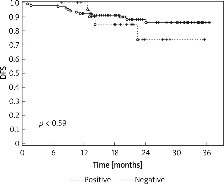
Within the short follow-up period there was not found a statistically significant correlation between MRD finding and relapse of the disease (p < 0.59). However, Kaplan-Meier analysis suggests an association that might, in the following period, show a relationship between MRD and the period of time before the relapse of the disease (Figure 1).
Figure 1.
Results of DFS follow-up related to the detection of MRD in the subgroup of patients with laparoscopic surgery
When detecting a possible correlation between MRD findings in different samples, we are only able to record an indication of some trends. Due to the fact that only a small subgroup of patients experienced relapse of the disease or death due to the disease progression, and that a large group of patients with positive MRD findings is currently being censored, the preliminary results regarding DFS can be made in selected samples only. Within the short follow-up period, there has not been recorded a relationship between CTC finding in pSVB (p < 0.33), aSVB (p < 0.14), mBM (p < 0.24) and DFS. However, in all analyses we can see a certain trace of prediction of a potential relationship with a positive MRD finding and higher risk of early relapse of the disease.
To evaluate the overall survival related to MRD finding in individual samples, and thus related to potential prediction of the eventual progression of the disease, we have to evaluate results after a longer period of time since the resection. However, even in the follow-up with a median of 21.4 months, it is possible to see some correlations. In the group with stage I no morbidity was recorded. One patient with stage II (1.9%) died on the 33rd day after the surgery due to multiple organ failure. The death was not due to progression of the malignant process. In stage III 9 patients died (18.8%). One patient with the anamnesis of chronic renal insufficiency died on the 53rd day after the surgery due to renal failure, and the death of 8 patients (16.7%) was related to generalization of colorectal carcinoma.
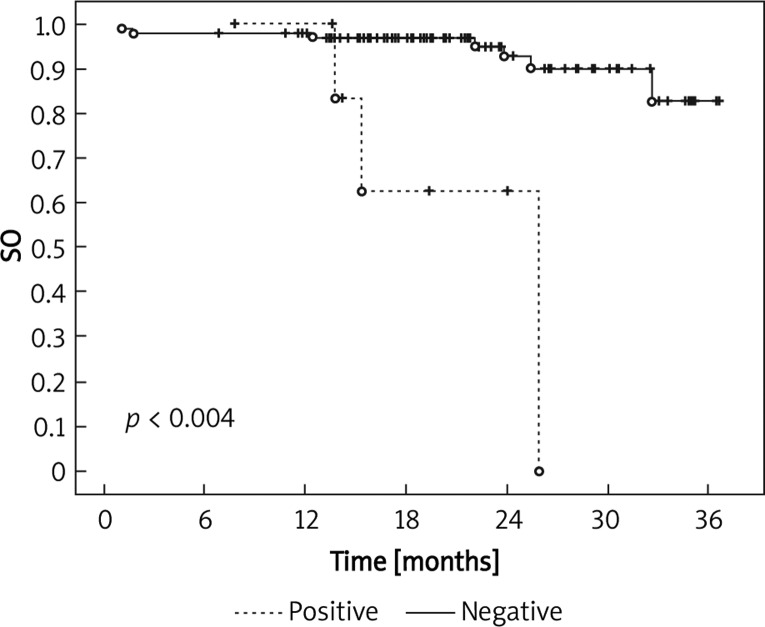
The evaluation of the finding in pSVB showed no statistically significant correlation (p < 0.075) between CTC presence and cancer-specific survival (CSS) of patients. Nevertheless, we may expect a potential direct correlation between positive CTC finding in pSVB and shorter survival of these patients. A statistically significant relationship (p < 0.52) was not found between CTC finding in iPVB and CSS within the period of the follow-up either. However, the evaluation of CSS related to CTC presence in system venous blood collected immediately after the resection (aSVB) showed a statistically significant correlation (p < 0.004). In comparison with negative patients those with a positive finding in this sample had significantly shorter CSS during the follow-up period (Figure 2).
Figure 2.
Results of CSS follow-up related to CTC finding in the sample of system venous blood collected immediately after the resection of carcinoma (aSVB) in the subgroup of patients with laparoscopic surgery
The group of patients with open surgery
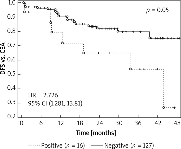
The median follow-up period in patients with open surgery was 21.2 months. The prognostically important factor (p < 0.05) suggesting higher risk of relapse in this group of patients was CTC finding in the sample of system venous blood taken prior to surgery (Figure 3).
Figure 3.
Results of DFS follow-up related to CTC finding in the sample of system venous blood collected prior to surgery (pSVB) in the subgroup of patients with open surgery
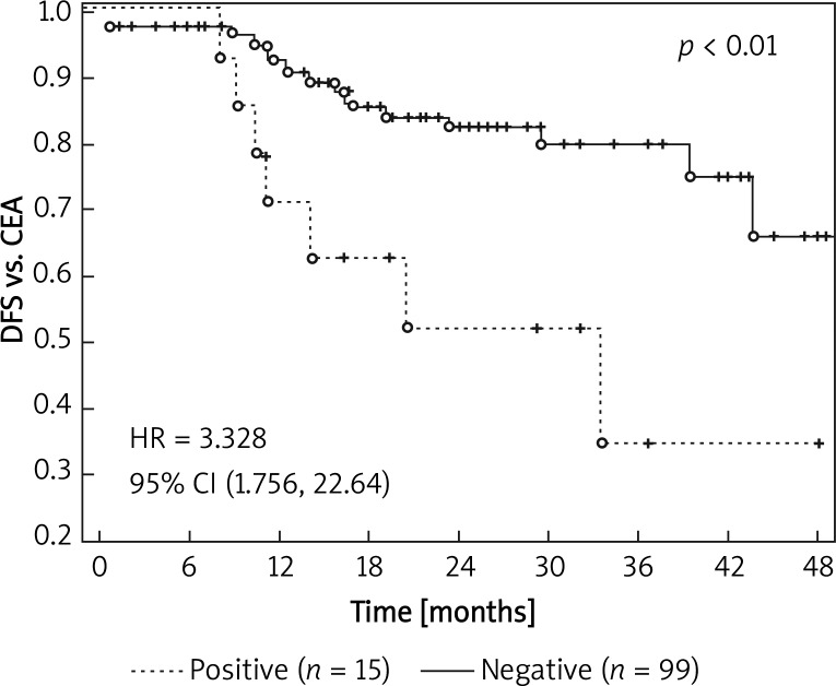
Even more significant factor (p < 0.01) – from the viewpoint of prognosis – seems to be a DTC positive finding in bone marrow taken a month after the resection (Figure 4).
Figure 4.
Results of DFS follow-up related to DTC finding in the sample of bone marrow collected 1 month after the surgery (mBM) in the subgroup of patients with open surgery
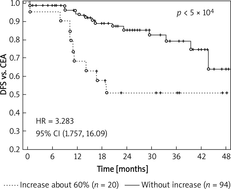
Furthermore, we identified a specific risk group of patients with a negative DTC finding in bone marrow sampled prior to surgery (pBM) and with the minimum 60% increase in the expression of CEA (mBM) after the surgery. In this subgroup of patients the risk of relapse of the malignant disease was significantly higher (p < 5 × 10⁴) (Figure 5).
Figure 5.
Results of DFS follow-up related to positive DTC finding in patients with negative finding of DTC in bone marrow prior to surgery (pBM) and minimum 60% increase of CEA expression after the surgery (mBM)
The risk of relapse is almost four times higher (HR = 3.729) in the group of MRD positive patients compared with the group of MRD negative patients. In the whole group of patients with open surgery the DFS median was 19.7 months [8, 9].
Discussion
So far, the evaluations of laparoscopic resections for colorectal carcinoma have been focused on clinical aspects [10–13]. There are only a few published studies about MRD detection in patients with laparoscopic resection of colorectal carcinoma (CRC). Chen et al. worked with a set of 42 patients with laparoscopic resection of CRC. They discuss the impact of mini-invasive surgery on circulating tumour cells (CTC). The CTC was detected with real-time RT-PCR by determination of guanylyl cyclase C (GCC) mRNA in the peripheral blood sampled prior to surgery, during the surgical management, and two weeks after the surgery. Despite the increasing interest in CTC detection related to the progression of the disease, they did not record any significant difference in the level of CTC determined prior to surgery in individual stages of the disease. Compared with values obtained prior to surgery, laparoscopic management did not result in elevation of the CTC level. With regard to relapse and overall survival, patients with a persistently high CTC level had a bad prognosis even 14 days after the surgery [14].
Bessa et al. compared the results of the CEA mRNA detection with RT-PCR in venous peripheral blood in a group of 50 patients – 26 patients with laparoscopically assisted resection of CRC, and 24 patients with open surgery. With regard to CTC detection in peripheral venous blood sampled prior to surgery, immediately after the management, and 24 h after the resection, the authors did not find any difference between the two subgroups of patients [15]. Our results for pSVB and aSVB show that there is statistically significant impact of laparoscopic surgery on positive CTC finding in system venous blood in primary negative patients (p < 0.025).
Hardingham et al. point out the important relationship between cancer cells present in peripheral blood detected with the PCR method and DFS in patients with CRC [16]. However, this opinion is not accepted generally. Individual circulating cancer cells are commonly destroyed by protective mechanisms of the body. Fidler proved in an animal model that only less than 0.1% of circulating cancer cells may become the basis for the occurrence of distant metastases [17]. Nevertheless, some authors maintain the position that the presence of circulating cancer cells is prognostically important. Patel et al. report the relationship between MRD, the stage of the disease, and surgical management. Prior to surgery they examined samples of peripheral blood in 116 patients with CRC. In 81 patients (70%) they found with the RT-PCR method positive CEA or CK20 in peripheral blood. Moreover, the number of RT-PCR positive patients decreased significantly over 24 h after the surgery. However, the dependence was recorded in the subset of patients with Dukes’ A or B stage and the decrease was statistically significant [18]. Similar conclusions were stated by Ito et al. and Wang et al. The authors, independently from each other, demonstrated a relationship between CEA mRNA expression detected with the RT-PCR approach with poor prognosis of the disease and the occurrence of metachronous metastases in the liver in patients after resection of CRC [19, 20]. The opinions on the importance of MRD detection in peripheral blood and the impact on the clinical development of the oncological disease vary. There still remain questions about technical problems of the examination. Another problem is represented by the potential impact of manipulation with the tissue affected with tumour during the surgical treatment, when cancer cells may penetrate the blood stream. Though this theory is not generally accepted, it is supported by the increased concentration of mRNA after the surgery [21, 22]. In our research no statistically significant correlation between the stage of the disease and CTC present in system venous blood sampled in laparoscopic patients at the beginning of surgery (p < 0.49), or in system venous blood sampled immediately after the resection (p < 0.61) was found. However, results for the sample of system venous blood taken a month after the surgery suggest a potential relationship between the stage of the disease and CTC finding (p < 0.075). Evaluations of DFS at stage I-III of the disease did not show a correlation between DFS and detected CTC in pSVB (p < 0.33) or in aSVB (p < 0.14) in patients treated with laparoscopic resection. In comparison with negative patients (p < 0.004), patients with a positive finding in system venous blood sampled after the surgery (aSVB) showed significantly shorter overall survival during the follow-up. In patients with open surgery the finding of CTC in the sample of system venous blood collected prior to surgery (pSVB) became a prognostically important factor (p < 0.05) referring to higher risk of disease relapse.
The liver is considered the primary extranodular location for the origin of CRC distant metastases. Therefore, we have been focusing on the detection of cancer cells from portal blood stream. Sadahiro et al. did not report a statistically significant relationship between positive finding in peripheral blood and in portal blood. On the other hand, they found an unexpectedly high incidence of positive findings in portal blood also in patients with early detected CRC [23]. Taniguchi et al. describe a significant relationship between CEA mRNA expression and depth of the invasion of tumour in an organ, vascular and lymphatic invasion and histological type of the carcinoma. We recorded a decrease of two-year DFS in patients with CEA mRNA detected in the portal circulation, compared to those with a negative finding. However, an identical difference was also recorded in examination of the peripheral blood [24]. Iinuma et al. used real-time RT-PCR and demonstrated a significant correlation between positive finding of CEA/CK20 mRNA in portal blood and the depth of cancer invasion, vascular invasion, and presence of metastases in lymphatic nodes and liver. They observed significantly shorter DFS in patients with CEA/CK20 mRNA expression in peripheral and portal blood, compared to those with negative findings [25]. In our research we did not record a direct relationship between positive finding of CTC in iPVB and the stage of the disease (p < 0.80) in patients treated with laparoscopic resection. The short time evaluation of the overall survival with regard to CTC finding in iPVB has not shown any statistically significant correlation (p < 0.52).
Practical clinical implications of DTC incidence in bone marrow are still unclear. Lindemann et al. informed about the shorter DFS in patients with positive findings of DTC in the bone marrow aspirate [26]. Flatmark et al. focused on the detection of DTC in bone marrow of patients with CRC using an immuno-magnetic approach. The positive finding of DTC in bone marrow was recorded in 17% of patients who underwent surgery. The proportion is lower in comparison with studies using the RT-PCR technique. Nevertheless, they report a direct relationship between the increased number of positive findings and advanced stage of the disease [27]. In our set of patients treated with laparoscopic resection, no statistically significant relationship between the stage of the disease and DTC finding in bone marrow sampled prior to surgery (p < 0.035) was found. The analysis of bone marrow sampled one month after laparoscopic resection (mBM) proved no statistically significant relationship (p < 0.24) between DTC finding and DFS during the short follow-up period. A positive finding of DTC in bone marrow sampled 1 month after resection (mBM) seems to be an important prognostic factor (p < 0.01) in the subset of patients with open surgery. Patients with negative DTC detection in bone marrow sampled prior to surgery (pBM) and with the minimum of 60% increase of CEA expression after the surgery (mBM) represented a risk group. In this subgroup of patients we found a statistically significant (p < 5 × 10⁴) increase in relapse of the disease.
Conclusions
The results obtained for patients with primary colorectal carcinoma treated with laparoscopic resection and open surgery are not completely clear. However, they suggest a direct correlation between MRD presence and relapse of the disease in patients after R0 resection of colorectal carcinoma, and overall survival of the patients. The comparison between the data obtained in the laparoscopic approach and the data obtained in open resection performed from the viewpoint of molecular biology did not show a significant difference in MRD detection in the samples collected after the surgery.
The presented results should be considered a preliminary report assessing the potential impact of the new prognostic marker in patients with CRC.
A more precise evaluation of the relationship between MRD detection and prognosis for patients with CRC may be given only on the basis of further collection of samples and evaluation of results within the ongoing research of at least 5 to 10-year follow-up period.
References
- 1.Tsavellas G, Huang A, McCullough T, et al. Flow cytometry correlates with RT-PCR for detection of spiked but not circulating colorectal cancer cells. Clin Exp Metastas. 2002;19:495–502. doi: 10.1023/a:1020350117292. [DOI] [PubMed] [Google Scholar]
- 2.Tsavellas G, Patel H, Allen-Mersh TG. Detection and clinical significance of occult tumour cells in colorectal cancer. Br J Surg. 2001;88:1307–20. doi: 10.1046/j.0007-1323.2001.01863.x. [DOI] [PubMed] [Google Scholar]
- 3.Smith BM, Slade MJ, English J, et al. Response of circulating tumor cells to systematic therapy in patients with metastatic breast cancer: comparison of quantitave polymerase chain reaction and immunocytochemical techniques. J Clin Oncol. 2000;18:1432–9. doi: 10.1200/JCO.2000.18.7.1432. [DOI] [PubMed] [Google Scholar]
- 4.Gerhard M, Juhl H, Kalthoff H, et al. Specific detection of carcinoembryonic antigen-expressing tumor cells in bone marrow aspirates by polymerase chain reaction. J Clin Oncol. 2001;12:725–9. doi: 10.1200/JCO.1994.12.4.725. [DOI] [PubMed] [Google Scholar]
- 5.Wong IHN, Yeo W, Chan AT, Johnson PJ. Quantitaive relationship of the circulating tumor burden assessed by reverse transcription-polymerase chain reaction for cytokeratin 19 mRNA in peripheral blood of colorectal cancer patients with Dukes′ stage, serum carcinoembryonic antigen level and tumor progression. Cancer Lett. 2001;162:65–73. doi: 10.1016/s0304-3835(00)00630-3. [DOI] [PubMed] [Google Scholar]
- 6.Vlems FA, Diepstra JHS, Cornelissen IMHA, et al. Limitations of cytokeratin 20 RT-PCR to detect disseminated tumour cells in blood and bone marrow of patients with colorectal cancer: expression in controls and downregulation in tumour tissue. J Clin Pathol Mol Pathol. 2002;55:156–63. doi: 10.1136/mp.55.3.156. [DOI] [PMC free article] [PubMed] [Google Scholar]
- 7.Skrovina M, Duda M, Srovnal J, et al. Detection of minimal residual disease and its significance for establishing prognose in patients with laparoscopic resection of colorectal carcinomas. Rozhl Chir. 2010;89:362–9. [PubMed] [Google Scholar]
- 8.Duda M, Vyslouzil K, Skalicky P, et al. Minimal residual disease in colorectal cancer – the new prognostic marker in surgical oncology. Slovenska Chirurgie. 2006;3:16–22. [Google Scholar]
- 9.Duda M, Vyslouzil K, Srovnal J. 2008. Detection and clinical significance of minimal residual disease and micrometastases in patients after colorectal cancer surgery. Záverečná zpráva o řešení programového projektu (NR 7804) podpořeného IGA MZ ČR. [Google Scholar]
- 10.International Union Against Cancer (UICC) TNM klasifikace zhoubných novotvaru, Šesté vydání 2002; 2004. pp. 13–14. Ústav zdravotníckých informací a statistiky České republiky, Praha. [Google Scholar]
- 11.Skrovina M, Czudek S, Bartos J, et al. Colorectal cancer complications of laparoscopic resection. Videosurgery and Other Miniinvasive Techniques. 2006;1:142–9. [Google Scholar]
- 12.Dostalik J, Martinek L, Vavra P, et al. Laparoscopic colorectal surgery for carcinomas – assessment of the autor′s patient group. Rozhl Chir. 2006;85:35–40. [PubMed] [Google Scholar]
- 13.Piatkowski J, Jackowski M. Laparoscopic colon resection – own experience report. Videosurgery and Other Miniinvasive Techniques. 2009;4:135–7. [Google Scholar]
- 14.Chen WS, Chung MY, Liu JH, et al. Impact of circulating free tumor cells in the peripheral blood of colorectal cancer patients during laparoscopic surgery. World J Surg. 2004;28:552–7. doi: 10.1007/s00268-004-7276-9. [DOI] [PubMed] [Google Scholar]
- 15.Bessa X, Castells A, Lacy AM, et al. Laparoscopic-assisted vs. open colectomy for colorectal cancer: influence on neoplastic cell mobilization. J Gastrointest Surg. 2001;5:66–73. doi: 10.1016/s1091-255x(01)80015-9. [DOI] [PubMed] [Google Scholar]
- 16.Hardingham JE, Kotasek D, Farmer B, et al. Imunobead-PCR: a technique for the detection of circulating tumor cells using immunomagnetic beads and the polymerase chain reaction. Cancer Res. 1993;53:3455–8. [PubMed] [Google Scholar]
- 17.Fidler IJ. Critical factors in the biology of human cancer metastasis: twenty-eighth G.H.A. Clowes memorial award lecture. Cancer Res. 1990;50:6130–8. [PubMed] [Google Scholar]
- 18.Patel H, Le Marer N, Wharton RQ, et al. Clearance of circulating tumor cells after excision of primary colorectal cancer. Ann Surg. 2002;235:226–31. doi: 10.1097/00000658-200202000-00010. [DOI] [PMC free article] [PubMed] [Google Scholar]
- 19.Ito S, Nakanishi H, Hirai T, et al. Quanatitative detection of CEA expressing free tumor cells in the peripheral blood of colorectal cancer patients during surgery with real-time RT-PCR on a LightCycler. Cancer Lett. 2002;183:195–203. doi: 10.1016/s0304-3835(02)00157-x. [DOI] [PubMed] [Google Scholar]
- 20.Wang JY, Wu CH, Lu CY, et al. Molecular detection of circulating tumor cells in the peripheral blood of patients with colorectal cancer using RT-PCR: significance of the prediction of postoperative metastasis. World J Surg. 2006;30:1007–13. doi: 10.1007/s00268-005-0485-z. [DOI] [PubMed] [Google Scholar]
- 21.Feezor RJ, Copeland EM, Hochwald SN. Significance of micrometastases in colorectal cancer. Ann Surg Oncol. 2002;9:944–53. doi: 10.1007/BF02574511. [DOI] [PubMed] [Google Scholar]
- 22.Yamaguchi K, Takagi Y, Aoki S, et al. Significant detection of circulating cancer cells in the blood by reverse transcriptase-polymerase chain reaction during colorectal cancer resection. Ann Surg. 2000;232:58–65. doi: 10.1097/00000658-200007000-00009. [DOI] [PMC free article] [PubMed] [Google Scholar]
- 23.Sadahiro S, Suzuki T, Tokunaga N, et al. Detection of tumor cells in the portal and peripheral blood of patients with colorectal carcinoma using competetive reverse transcriptase-polymerase chain reaction. Cancer. 2001;92:1251–8. doi: 10.1002/1097-0142(20010901)92:5<1251::aid-cncr1445>3.0.co;2-o. [DOI] [PubMed] [Google Scholar]
- 24.Taniguchi T, Makino M, Suzuki K, Kaibara N. Prognostic significance of reverse transcriptase-polymerase chain reaction measurement of carcinoembryonic antigen mRNA levels in tumor drainage blood and peripheral blood of patients with colorectal carcinoma. Cancer. 2000;89:970–6. [PubMed] [Google Scholar]
- 25.Iinuma H, Okinaga K, Egami H, et al. Usefulness and clinical significance of quantitative real-time RT-PCR to detect isolated tumor cells in the peripheral blood and tumor drainage blood of patients with colorectal cancer. Int J Oncol. 2006;28:297–306. [PubMed] [Google Scholar]
- 26.Lindemann F, Schlimok G, Dirschedl P, et al. Prognostic significance of micrometastatic tumor cells in bone marrow of colorectal cancer patients. Lancet. 1992;340:685–9. doi: 10.1016/0140-6736(92)92230-d. [DOI] [PubMed] [Google Scholar]
- 27.Flatmark K, Bjornland K, Johannessen HO, et al. Study Group for Micrometastases in BM in Colorectal Cancer. Immunomagnetic detection of micrometastatic cells in bone marrow of colorectal cancer patients. Clin Cancer Res. 2002;8:444–9. [PubMed] [Google Scholar]


