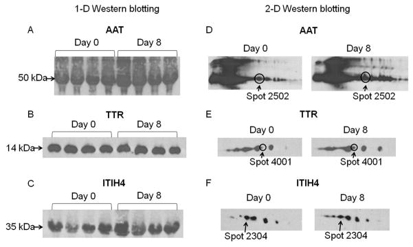
Figure 3.
Western blotting of AAT, TTR and ITIH4. A–C, 1-D Western blotting of AAT, TTR and ITIH4. Serum samples from the same four subjects at day 0 and day 8 of GH treatment phase were loaded and immunoblotted with antibodies against AAT, TTR and ITIH4. D–F, 2-D Western blotting of AAT, TTR and ITIH4. The isoform corresponding to the one identified by 2-DE was circled and/or indicated by an arrow.

