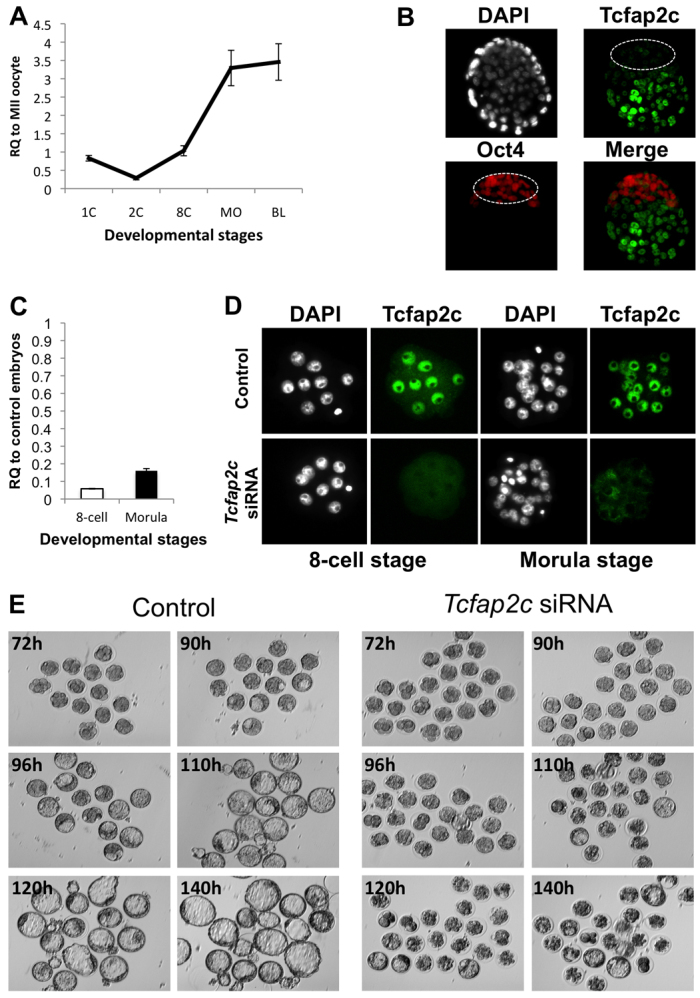
Fig. 1.

Developmental expression and RNAi-mediated ablation of Tcfap2c in mouse preimplantation embryos. (A) qRT-PCR analysis of Tcfap2c transcripts in mouse MII oocytes and preimplantation embryos. Expression data from each stage were normalized to exogenous GFP and are relative to MII oocytes. 1C, one-cell zygote; 2C, two-cell; 8C, eight-cell; MO, morula; BL, blastocyst. (B) Immunocytochemistry (ICC) analysis revealed that Tcfap2c protein is enriched in the blastocyst mural trophectoderm (TE). Blastocysts were double stained for Oct4 to label the inner cell mass (ICM; outlined). Nuclei were counterstained with DAPI. (C) Validation of siRNA-mediated knockdown (KD) of Tcfap2c transcripts in eight-cell and morula stage embryos by qRT-PCR. (D) Confirmation of Tcfap2c protein ablation in eight-cell and morula stage embryos by ICC. (E) Depletion of Tcfap2c blocks blastocyst formation. Representative images of Tcfap2c KD and control embryos cultured for 72 to 140 hph. Error bars indicate mean ± s.e.m. RQ, relative quantification.
