Abstract
The development of functionally equivalent fibrocartilage remains elusive despite efforts to engineer tissues such as the knee menisci, intervertebral disc, and TMJ disc. Attempts to engineer these structures often fail to create tissues with mechanical properties on par with native tissue, resulting in constructs unsuitable for clinical applications. The objective of this study was to engineer a spectrum of biomimetic fibrocartilages representative of the distinct functional properties found in native tissues. Using the self-assembly process, different co-cultures of meniscus cells (MCs) and articular chondrocytes (ACs) were seeded into agarose wells and treated with the catabolic agent chondroitinase-ABC (C-ABC) and the anabolic agent transforming growth factor-β1 (TGF-β1) via a two-factor (cell ratio and bioactive treatment), full factorial study design. Application of both C-ABC and TGF-β1 resulted in a beneficial or positive increase in the collagen content of treated constructs compared to controls. Significant increases in both the collagen density and fiber diameter were also seen with this treatment, increasing these values 32% and 15%, respectively, over control values. Mechanical testing found the combined bioactive treatment to synergistically increase the Young’s modulus and ultimate tensile strength of the engineered fibrocartilages compared to controls, with values reaching the lower spectrum of those found in native tissues. Together, these data demonstrate that C-ABC and TGF-β1 interact to develop a denser collagen matrix better able to withstand tensile loading. This study highlights a way to optimize the tensile properties of engineered fibrocartilage using a biochemical and biophysical agent together to create distinct fibrocartilages with functional properties mimicking those of native tissue.
1. Introduction
Fibrocartilage is uniquely situated within specific joint spaces of the body to facilitate the distribution of peak loads. Viscoelastic in nature, this tissue is capable of deforming its shape to effectively spread loads across a greater surface area. Found, for example, in the knee, intervertebral, and temporomandibular joints, fibrocartilage is distinctively situated to allow for congruent meshing of the articulating structures within these joint spaces, ensuring smooth, stable articulation and stress absorption [1–3]. However, fibrocartilage lacks an innate ability to self-repair following disease or injury. With a lack of clinically available treatment options for patients suffering from injured or diseased fibrocartilage, the need for an effective remedy is crucial. Fibrocartilages engineered in vitro offer new hope as a long-term treatment option to repair or replace injured or degenerated fibrocartilaginous tissues.
Collagens make up the majority of the extracellular matrix (ECM) of a fibrocartilaginous tissue. Within a specific fibrocartilage, these collagen fibers align with the peak tensile loads in a given joint space, situating the tissue to best distribute complex forces. While fibrocartilages are primarily composed of collagen type I, they also contain lower amounts of collagen type II as well as glycosaminoglycans (GAGs). The levels at which these main ECM components are found are characteristic of the anatomical location and function. The heterogeneous distribution and organization of ECM result in anisotropic biomechanical properties specific to each fibrocartilage type. Efforts to tissue engineer fibrocartilages able to withstand the mechanical complexity of a joint space must therefore focus on creating a well-developed, collagen-rich matrix.
Capitalizing on chondrocytes’ unique ability to form dense, ECM-rich clusters when seeded at high densities, a scaffold-free, self-assembly approach to cartilage tissue engineering has been developed [4]. With primary TMJ disc and intervertebral disc cells presenting as an implausible cell source due to limited cell numbers in the tissue as well as low levels of matrix production in vitro, this technology has recently been extended to co-cultures of varying ratios of meniscus cells (MCs) and articular chondrocytes (ACs), allowing for the formation of a spectrum of fibrocartilages [5–7]. While current efforts to tissue engineer fibrocartilage have succeeded in creating constructs with compressive properties on par with native tissue values, tensile properties have lagged greatly behind [7–11]. This is a significant problem for fibrocartilages, as they function under both compression and tension. For instance, resistance to compression in the knee menisci occurs via tensile, hoop stresses developed in the wedged-shape of this tissue. Likewise, tension-compression is also observed in the TMJ disc [1]. Here, ligaments around the periphery of the disc secure the tissue within in the joint space, and as the condyle translates during jaw opening, it compresses the disc, causing inherent tension much like that of a trampoline [12]. Exogenous stimuli such as TGF-β1 and chondroitinase-ABC (C-ABC) have therefore been investigated to enhance the tensile properties of self-assembled, engineered fibrocartilages [7, 13–15].
Previous work with C-ABC has shown that this enzyme’s temporary depletion of GAG enhances the tensile properties of treated articular cartilage explants [16]. It has also been shown that there is a window of effectiveness for C-ABC’s application; applying C-ABC at both t = 2 wk and t = 4 wk of culture is superior to a single treatment at either time point for enhancing the tensile properties of tissue-engineered articular cartilage [15]. In another study, tissue-engineered meniscus cartilage was treated with C-ABC at an earlier time point to determine if treating a more immature matrix would have a greater effect on tissue development, as chondrocytes produce the majority of their matrix during the first week of self-assembly [17]. Results found a single, early application of C-ABC at t = 1 wk to be beneficial in enhancing the tensile properties of the tissue [13].
Recent work has shown concurrent application of C-ABC and TGF-β1 results in further enhancement of the mechanical properties of tissue-engineered cartilage compared to treatment with either agent alone [13, 18]. Application of these two agents on tissue-engineered neocartilage was found to synergistically increase collagen content and additively increase the tissue’s tensile strength [18]. In a separate study, engineered meniscus fibrocartilage experienced a 5-fold increase in the radial tensile modulus as well as a 196% increase in collagen content after being treated with both C-ABC and TGF-β1 [13]. Through depleting GAG, C-ABC releases GAG fragments, such as hyaluronic acid fragments, into the tissue, and such fragments have been reported to have an anabolic effect on surrounding cells [19]. Microarray analysis of self-assembled articular cartilage treated with C-ABC, however, has found no changes in gene expression as a result of this treatment, suggesting the GAG fragments are being effectively removed from the tissue [18]. Further, inspection of C-ABC treated engineered articular cartilage via scanning electron microscopy (SEM) has found C-ABC to work via a biophysical mechanism, through altering the physical structure of a collagen network. Microarray analysis work has also found that TGF-β1 promotes chondrocytes to synthesize more ECM through a MAPK biochemical pathway [18]. It was concluded that these bioactive agents are able to collaboratively increase the functional properties of tissue-engineered cartilage because they act via different mechanisms: one being biophysical, the other biochemical. With previous work on enhancing the functional properties of a spectrum of engineered fibrocartilage focused on TGF-β1 alone, it is important to institute both TGF-β1 and C-ABC with the goal of further enhancing the functional properties of such tissues.
The overall objective of this study was to generate biomimetic fibrocartilages representative of the vast array of distinct fibrocartilages found in the body. Given the previous success of engineering such tissues with compressive properties on par with native tissue values, this study emphasized enhancing the tensile properties of engineered fibrocartilage. To capture the heterogeneous nature of the different types of fibrocartilage, a spectrum of fibrocartilaginous tissues was generated using the self-assembly process and optimized using C-ABC and TGF-β1 alone or in combination. Tissues having different ratios of MCs and ACs were created to recapitulate the diversity of collagen type I to collagen type II found in fibrocartilages. After optimizing a C-ABC treatment regimen, tissues were treated with the bioactive agents C-ABC and TGF-β1 using a two-factor, full factorial study design with cell ratio and bioactive treatment as the factors. Overall, it was hypothesized that the combination of C-ABC and TGF-β1 would significantly increase the tensile properties of each of the co-culture constructs without compromising their compressive properties. It was further hypothesized that by increasing the tensile properties, the combined bioactive treatment regimen would generate co-culture ratio-dependent fibrocartilages having distinct functional properties within the range of native tissue.
2. Materials and Methods
2.1 Cell Isolation and Self-Assembly into Fibrocartilage Constructs
Bovine articular chondrocytes and meniscus cells were harvested from juvenile bovine knee joints (Research 87, Boston, MA). Minced articular cartilage was digested in base medium of Dulbecco’s modified Eagle’s medium (DMEM) with 1% penicillin/streptomycin/fungizone (PSF), containing 0.2% collagenase (Worthington, Lakewood, NJ) for 18 hours. Minced menisci were digested in base medium containing 0.25% Pronase E (Sigma, St. Louis, MO) for 1 hour prior to 18 hours in 0.2% collagenase-containing base medium. Isolated cells were frozen in base medium containing 20% fetal bovine serum and 10% dimethyl sulfoxide until needed for seeding.
Cells were self-assembled at 50:50 and 75:25 MC:AC co-culture ratios as previously described [4, 7]. Based on preliminary work, a 100:0 MC:AC cell ratio was not used in this study, as neither C-ABC nor TGF-β1 alone or in combination was found to have significant effects on this cell ratio, resulting in constructs with subpar functional properties compared to constructs containing both MCs and ACs. Briefly, MC and AC cell suspensions were seeded into 5 mm diameter, 2% agarose wells at t = 0 hr. By t = 4 hr, the cells had assembled into constructs. Constructs were fed 500 μl of chondrogenic medium consisting of DMEM, 1% PSF, 1% non-essential amino acids, 100 nM dexamethasone (Sigma, St. Louis, MO), 1% ITS+ (BD Scientific, Franklin Lakes, NJ), 40 μg/mL L-proline, 50 μg/mL ascorbate-2-phosphate, and 100 μg/mL sodium pyruvate (Fischer Scientific, Pittsburgh, PA), every day. At t = 1 wk, constructs were removed from the agarose molds and placed into 48 well plates for the duration of the 5 wk culture period.
2.2 Bioactive Treatment Regimens
Two bioactive agents were investigated in this study: C-ABC (Sigma-Aldrich, St. Louis, MO) and TGF-β1 (PeproTech, Rocky Hill, NJ). Constructs treated with C-ABC received 2 U/mL C-ABC with a 0.05 M acetate activator in chondrogenic medium for 4 hr at 37°C once at t = 7 d and again at t = 21 d. This dual C-ABC treatment was developed in preliminary studies to confirm that this regimen was superior to a single treatment at either time point for enhancing the tensile properties of the tissue-engineered fibrocartilage. In TGF-β1 treated constructs, TGF-β1 was given continuously at 10 ng/ml with every medium change from t = 0 hr to the end of culture [7]. To investigate the interaction of these bioactive agents, treatments consisted of the following: C-ABC alone, TGF-β1 alone, the two bioactive agents combined, or none. At t = 5 wk, each construct was segmented for various assessments, as described below.
2.3 Histology and Immunohistochemistry (IHC)
Samples were frozen in HistoPrep (Fisher Chemical, Vernon Hills, IL), sectioned at 12 μm, fixed in chilled formalin, and stained with Safranin O/Fast Green and picrosirious red for GAG and total collagen, respectively. For IHC, sections were fixed in chilled acetone, after which endogenous peroxidase was blocked using 3% H2O2. Samples were then blocked with horse serum (Vectastain ABC kit, Vector Labs, Burlingame, CA) and incubated with mouse anti-collagen type I (Accurate Chemicals, Westbury, NY) or rabbit anti-collagen type II (Cedarlane Labs, Burlington, NC) antibodies. Corresponding 2° antibodies as well as the Vectastain ABC and DAB solutions were applied as instructed by the Vectastain ABC kit (Vector Labs, Burlingame, CA) for collagen visualization.
2.4 Quantitative Biochemistry
Sample wet weights were taken after blotting, followed by lyophilization for t = 2 d to determine their dry weights. Samples were then digested in papain as previously described [13]. Total collagen was assessed using a chloramine-T hydroxyproline assay with a SIRCOL collagen assay standard (Accurate Chemical and Scientific Corp.). GAG content was determined using a dimethylmethylene blue (DMMB) dye-binding assay kit (Biocolor, Newtownabbey, Northern Ireland). DNA content was measured using a Picogreen Cell Proliferation Assay Kit (Molecular Probes, Eugene, OR).
2.5 Biomechanical Testing
Uniaxial, unconfined stress-relaxation compressive testing was applied on 3 mm biopsy punches from the center of the tissue-engineered constructs. Samples were placed in a PBS-filled petri dish and loaded onto the stage of an Instron uniaxial testing machine (Model 5565, Canton, MA). Sample height was determined by applying a 0.02 N tare load. Following pre-conditioning at a 5% strain for 15 cycles at 1 Hz, the sample was compressed at a 1% strain rate per sec to 10%, 20%, and 30% strains. Samples were held at a given strain for 400 sec, which allowed sufficient time for the force vs. time curve of each strain level to equal zero, signifying the sample had fully relaxed. Following this holding time, the next sequential strain level was applied. A Kelvin solid model was used to fit the results of the 20% stress relaxation test and to calculate the viscoelastic material properties of the samples; this included the relaxation and instantaneous moduli, as well as the viscosity of the tissue.
Uniaxial tensile testing was also conducted using the Instron. Constructs were cut into dog bone shapes and glued onto strips of paper at either extremity. The gauge length was measured between the glued ends. The paper strips holding the sample were loaded into grips, and a pull to failure test was applied at a 1% strain rate per sec. The linear portion of the stress vs. strain curve was used to determine the Young’s Modulus of the sample, while the peak of the linear region was taken as the ultimate tensile strength (UTS).
2.6 SEM
Samples were dehydrated in increasing ethanol and stored in 100% ethanol at 4°C. Just prior to imaging, samples were further dehydrated using a critical point dryer and sputter-coated with gold. For each sample, three separate locations were imaged using a Philips XL30 TMP SEM, and collagen matrix fiber density and diameter were quantified using ImageJ as previously described [18].
2.7 Statistical Analysis
To test the hypothesis that the combined bioactive treatment was a significant factor in increasing the functional properties within a given cell ratio, a 1-way ANOVA (n=8 per group) was used within each cell ratio. Results of the 1-way ANOVA for the 50:50 cell ratio are represented by upper case letters, while those of the 75:25 cell ratio are represented by lower case letters. Separately, to test the hypothesis that the cell ratio and the bioactive treatments were both significant factors in altering construct composition, a 2-way ANOVA (n = 8 per group) was used to analyze the data. When either the 1- or 2-way ANOVA showed significance (p < 0.05), a Tukey’s HSD post hoc test was applied. Data that had a positive interaction on an additive scale and resulted in a combined group that was greater than the addition of the two singular treatments were determined to be synergistic, while those that had a negative interaction on an additive scale but resulted in a combined group that was greater than either treatment alone were determined to be beneficial or positive. Data for this study are represented as mean ± standard deviation with bars or groups marked by the same letter having no significant differences, while those that are marked by different letters are significantly different from one another.
3. Results
3.1 Gross Morphology
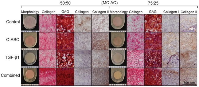
At t = 5 wk, all constructs maintained a flat, disc-shaped morphology (Fig. 1). However, as the ratio of MCs to ACs increased, construct diameter, thickness, and total wet weight decreased (Table 1). Treated constructs from the 50:50 cell ratio group had significantly lower wet weights, diameters, and thicknesses compared to untreated controls. For example, when treated with both C-ABC and TGF-β1, the wet weight, diameter, and thickness of the 50:50 constructs were decreased by 66%, 24%, and 29%, respectively. Similar, decreasing trends were seen in 75:25 constructs, though TGF-β1 alone significantly increased the wet weight 12% compared to controls within this cell ratio. Further, all bioactive treatments increased the thickness of the 75:25 constructs compared to controls. In terms of construct hydration, combination treated constructs of both cell ratio groups had significantly decreased water content compared to controls or either treatment alone, signifying a denser matrix in the combined treated groups. These results are summarized in Table 1.
Fig. 1.
Gross morphology, histology and IHC of constructs at t = 5 wk. Fibrocartilage was treated with either C-ABC alone, TGF-β1 alone, the two agents combined, or left untreated (control). Collagen was stained for using picrosirious red, GAG was stained for using Safranin O/Fast Green, while IHC was used to stain for collagen types I and II. Collagen and GAG staining was stronger in the 50:50 constructs, with the combined treatment producing constructs with the most uniform staining in both cell ratios. IHC staining showed different patterns of collagen types I and II between cell ratios as well as among bioactive treatments within each cell ratio. Scale bar for histology and IHC images is 500 μm and the markings on the morphology images are 1 mm apart.
Table 1.
Construct properties at t = 5 wk.
| Cell Ratio (MC:AC) | Bioactive Treatment | Wet Weight (mg) | Hydration (%) | Diameter (mm) | Thickness (mm) | Cells/Construct (106) |
|---|---|---|---|---|---|---|
|
| ||||||
| 50:50 | None | 30.19 ± 1.49A | 87.25 ± 1.45A | 6.67 ± 0.10A | 0.61 ± 0.08A | 3.05 ± 0.40A |
| C-ABC | 12.38 ± 1.10C | 85.47 ± 0.68B | 5.61 ± 0.11B | 0.33 ± 0.04C | 0.94 ± 0.23B | |
| TGF-β1 | 17.35 ± 1.25B | 84.89 ± 0.66B | 5.72 ± 0.18B | 0.46 ± 0.05B | 3.28 ± 0.30A | |
| Combined | 10.16 ± 0.17D | 82.72 ± 0.64C | 5.08 ± 0.06C | 0.44 ± 0.06B | 0.86 ± 0.33B | |
|
| ||||||
| 75:25 | None | 13.44 ± 0.80b | 88.40 ± 2.10a | 5.20 ± 0.11a | 0.44 ± 0.06c | 1.95 ± 0.18a |
| C-ABC | 8.76 ± 0.16c | 86.80 ± 0.92ab | 4.68 ± 0.09c | 0.47 ± 0.09c | 1.33 ± 0.07b | |
| TGF-β1 | 15.00 ± 0.52a | 86.02 ± 0.82b | 4.99 ± 0.10b | 0.79 ± 0.04a | 2.15 ± 0.41a | |
| Combined | 7.87 ± 0.43c | 82.00 ± 1.25c | 4.24 ± 0.06d | 0.66 ± 0.06b | 1.33 ± 0.11b | |
Values marked with different letters within each category are significantly different (p < 0.05), with A > B > C.
3.2 Histology and IHC
Overall, collagen and GAG staining were stronger in the 50:50 compared to the 75:25 cell ratio constructs. Staining trends within each cell ratio, however, were similar, with the combined bioactive treatment resulting in more uniform, dense collagen and GAG staining compared to all other treatments for both cell ratios (Fig. 1).
To test whether the cell ratio and bioactive treatment factors had an effect on the collagens being produced within a construct, IHC staining specifically for collagen type I and II was performed (Fig. 1). IHC staining found distinct trends to occur among the bioactive treatment levels within each cell ratio. Collagen type I stained positive in the control treatment within the 50:50 cell ratio group, while 50:50 constructs treated with C-ABC alone or in combination with TGF-β1 had much lighter, diffuse collagen type I staining. The 75:25 cell ratio group had positive collagen type I staining in all bioactive treatment levels, with stronger staining appearing in the combination treated constructs. Collagen type II staining was positive in all treatment levels for each cell ratio. Overall, collagen type II staining was slightly more intense in the 50:50 than the 75:25 cell ratio, particularly within the C-ABC treated and combination treated constructs.
3.3 Quantitative Biochemistry
The number of cells per construct was significantly decreased with the addition of C-ABC alone or in combination with TGF-β1, while control and TGF-β1 treated constructs were not significantly different from each other. Overall, the 50:50 cell ratio had higher cellularity within the control and TGF-β1 treatments, at approximately 3 million cells/construct, while the 75:25 control and TGF-β1 treatments had approximately 2 million cells/construct. Following treatment with either C-ABC alone or the combination treatment, cell number decreased by 70% in the 50:50 cell ratio and by 32% in the 75:25 cell ratio (Table 1).
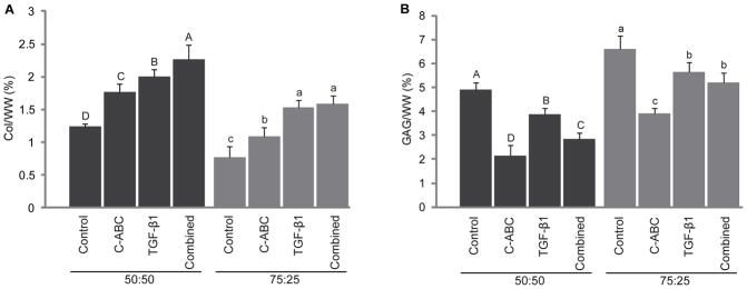
Collagen/WW (Fig. 2A) in the 50:50 cell ratio constructs was significantly affected by the various bioactive treatments employed in this study. C-ABC increased collagen/WW 40% over control values, while a 60% increase over controls was measured in TGF-β1 treated constructs. When the two bioactive agents were combined, collagen content was increased by 80% over control constructs. Furthermore, in the combined treatment, the two factors were found to result in a beneficial or positive significant enhancement of the collagen content in the 50:50 cell ratio compared to treatment with either bioactive agent alone. A similar significant effect was observed for collagen/WW in the 75:25 cell ratio, with values increasing 104% in combination treated constructs over controls. Comparison between the two cell ratios found the 50:50 cell ratio factor to have significantly more collagen/WW compared to the 75:25 cell ratio, while the C-ABC + TGF-β1 treatment factor resulted in significantly higher collagen/WW than any other treatment factor across both cell ratios.
Fig. 2.
Biochemical properties of constructs at t = 5 wk. Fibrocartilage was treated with either C-ABC alone, TGF-β1 alone, the two agents combined, or left untreated (control). Col/WW (A) exhibited significance among bioactive treatments, with constructs from both cell ratios having the greatest collagen content in the combined treatment. C-ABC and TGF-β1 significantly increased the collagen content in the 50:50 constructs in a beneficial or positive manner. Bioactive treatments significantly decreased GAG/WW (B) compared to controls. Bars not connected by the same letter are significantly different (p < 0.05).
GAG/WW (Fig. 2B) was significantly decreased in combination treated 50:50 and 75:25 cell ratios compared to controls. GAG content was significantly greater in the 75:25 cell ratio constructs, ranging from approximately 4 – 6.5%, compared to the 50:50 cell ratio constructs, which ranged from approximately 2 – 5%. Within the bioactive treatment factor, the combined bioactive treatment had significantly less GAG than controls or TGF-β1 treated constructs.
3.4 Biomechanics
Biomechanical testing was run to understand the influence of the cell ratio and bioactive treatment factors on the mechanical properties of the engineered fibrocartilages (Table 2). Similar trends in compressive properties were found among all tested strain levels; therefore, only the 20% strain level results are shown to represent these properties. Stress-relaxation, unconfined compression testing at 20% strain found no significant differences among treatments within the 50:50 cell ratio for the relaxation modulus, with values ranging from 95 ± 16 kPa to 125 ± 40 kPa. The relaxation modulus for the combined treated constructs within the 75:25 cell ratio was significantly lower than controls, at 40 ± 14 kPa compared to 57 ± 8 kPa, respectively. Significant differences were observed among treatments within both cell ratios for the instantaneous modulus and viscosity. At a 20% strain, the instantaneous modulus in TGF-β1 alone treated constructs was 5-fold and 2.4-fold higher in the 50:50 and 75:25 cell ratios compared to respective controls, while similar increases were observed in the viscosity. The combination treatment within either cell ratio, however, was not significantly different than corresponding control values, although they trended higher than their respective controls.
Table 2.
Construct viscoelastic compressive properties at t = 5 wk. Fibrocartilage was treated with either C-ABC alone, TGF-β1 alone, the two agents combined, or left untreated (control). The relaxation modulus showed no significant differences in the 50:50 constructs, but was significantly decreased in the combined treated 75:25 constructs compared to controls. The instantaneous modulus and viscosity was highest in the TGF-β1 alone treatment for both cell ratios.
| Cell Ratio (MC:AC) | Bioactive Treatment | Relaxation Modulus (kPa) | Instantaneous Modulus (kPa) | Viscosity (kPa*s) |
|---|---|---|---|---|
|
| ||||
| 50:50 | None | 52.10 ± 32.8A | 190.18 ± 95.0C | 7742.06 ± 4695.0B |
| C-ABC | 71.33 ± 26.5A | 650.01 ± 189.8B | 14403.08 ± 1682.7B | |
|
| ||||
| 75:25 | TGF-β1 | 86.10 ± 40.0A | 933.70 ± 131.1A | 22395.50 ± 3986.2A |
| Combined | 63.90 ± 10.9A | 397.14 ± 112.7C | 11072.00 ± 3941.8B | |
|
| ||||
| None | 70.40 ± 28.5a | 451.58 ± 122.6bc | 6833.66 ± 2493.9ab | |
| C-ABC | 51.21 ± 19.1ab | 243.32 ± 82.4c | 2681.81 ± 1918.6c | |
|
| ||||
| TGF-β1 | 44.56 ± 10.2ab | 748.53 ± 237.7a | 7747.91 ± 1876.5a | |
| Combined | 30.02 ± 15.6b | 497.48 ± 146.6b | 4230.19 ± 1704.8bc | |
Values marked with different letters within each category are significantly different (p < 0.05), with A > B > C.
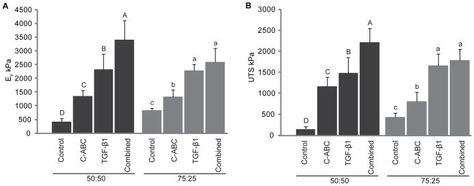
Tensile testing showed the most exciting results of this study (Fig. 3). With significant effects in response to bioactive treatments for both the Young’s modulus (Fig. 3A) and the UTS (Fig. 3B), results mirrored those observed for the collagen/WW data for each cell ratio. Compared to controls, within the 50:50 cell ratio, C-ABC increased the Young’s modulus 226%, while TGF-β1 increased this parameter by an additional 237%. The combined treatment further increased the Young’s modulus by an additional 260%. Overall, the combined treatment synergistically increased the Young’s modulus 724% compared to controls, from 414±121 kPa in the controls to 3410±706 kPa in the combined treated constructs. In a similar fashion, the UTS in combined treated constructs was 12.7-fold higher than control values in the 50:50 cell ratio. Within the 75:25 cell ratio, a 208% increase was observed by the combination treatment over controls, reaching a value of 2295±241 kPa in the combination treated constructs, while the UTS in the combined treated constructs was 4.1-fold higher than that of controls. Overall, the combined treatment was found to additively increase the Young’s modulus and UTS compared to either treatment applied alone, while the 50:50 cell ratio factor had significantly higher tensile properties than the 75:25 cell ratio factor.
Fig. 3.
Tensile properties of constructs at t = 5 wk. Fibrocartilage was treated with either C-ABC alone, TGF-β1 alone, the two agents combined, or left untreated (control). The Young’s modulus (A) and UTS (B) exhibited significance among bioactive treatments, corresponding to trends in the collagen content (Fig. 2, A). Constructs from both cell ratios had the greatest collagen content and tensile properties with the combined treatment. C-ABC and TGF-β1 together synergistically increased the Young’s modulus and UTS of the 50:50 constructs. Bars not connected by the same letter are significantly different (p < 0.05).
3.5 SEM
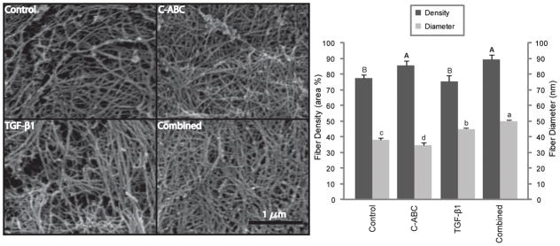
With the 50:50 cell ratio showing the greatest percent increases among bioactive treatments for the collagen/WW and tensile properties, portions of constructs representative of each treatment within this cell ratio were analyzed via SEM to investigate if the different treatments were altering the matrix. Resulting images were quantified for fiber density and diameter (Fig. 4). Overall, fiber density in the SEM images ranged from 75% to 80%, while fiber diameter ranged from 34 to 50 nm. C-ABC treatment was found to significantly increase the density of the fibers 10.2% over controls, while TGF-β1 alone had no effect on the density. For the diameters, TGF-β1 alone was found to significantly increase the fiber diameter 18.4% over control values, although C-ABC alone decreased fiber diameter 9.3%. Interestingly, when constructs were treated with C-ABC and TGF-β1 in combination, both the fiber diameter and the density increased 31.9% and 15.0%, respectively, compared to controls (Fig. 4). Further, these increases were greater than either treatment alone, signifying a synergistic behavior between these two agents in modifying collagen network organization.
Fig. 4.
SEM analysis of 50:50 fibrocartilage constructs at t = 5 wk, with images of control, C-ABC treated, TGF-β1 treated, and C-ABC + TGF-β1 treated constructs. To measure collagen fibril diameter and density, 3 locations on n = 3 fibrocartilage constructs from each bioactive treatment level were quantified. Results found C-ABC in significantly increase collagen density, while C-ABC + TGF-β1 significantly increased collagen fibril diameter over all other treatments. Bars not connected by the same letter are significantly different (p < 0.05). Scale bar is 1 μm.
Discussion
This study focused on generating distinct tissue-engineered fibrocartilages by treating different MC:AC co-culture ratios with the bioactive agents C-ABC and TGF-β1. It was hypothesized that the combined use of C-ABC and TGF-β1 would significantly increase the tensile properties of the co-culture ratio constructs without compromising their compressive properties. Through enhancing their tensile properties, it was further hypothesized that the combined bioactive treatment regimen would promote the development of biomimetic fibrocartilages having co-culture ratio-dependent biochemical and biomechanical properties within the range of native tissue. Through a two-factor, full factorial study design, the hypotheses were confirmed: First, the tensile properties of the engineered tissues for both cell ratio groups were significantly enhanced when C-ABC and TGF-β1 stimuli were combined. The compressive properties of the 50:50 cell ratio were also not compromised, unlike the 75:25 cell ratio, which showed a reduction in the relaxation modulus. Second, by enhancing the tensile properties of the co-cultures, overall construct functional properties fell within the range of native tissue properties for both cell ratio groups by the combined bioactive treatment.
Fibrocartilages generated from different seeding ratios of MCs and ACs were found to have distinct functional properties. This can be correlated with each cell type having different synthetic abilities in vitro. In culture, MCs have been found to produce lower amounts of ECM components compared to ACs. For example, MCs embedded in agarose were found to synthesize 25% less proteoglycans than ACs in the same system. Furthermore, of this 25%, approximately 54% of the GAGs synthesized by the MCs were lost to the surrounding medium [22]. Medium was not assessed in this experiment as some treatments in this study (i.e., all that contained C-ABC) work by causing ECM components to leach out of the constructs. Therefore the media of different treatments are incomparable. In a separate study, the genes encoding for the proteoglycans, collagens, and enzymes important for collagen synthesis and degradation were measured. Results found significantly lower expression levels of α1(I)procollagen, α1(II)procollagen, procollagen-lysine 2-oxoglutarate 5-dioxygenase 1, 2, and 3, and lysyl oxidase genes were measured in freshly isolated MCs than in freshly isolated ACs. Instead, higher expression of the matrix metallopeptidase 13 gene, a member of the matrix metalloproteinase family, was measured in MCs after 18 d of culture in alginate beads [23, 24]. These findings provide much insight as to why MCs alone may not be suitable for producing mechanically sound engineered fibrocartilaginous tissues; without a well-developed matrix, the mechanical integrity of a tissue will be compromised. As shown by this study, incorporating a certain percentage of ACs into co-culture with MCs allows for mechanically robust fibrocartilages having different functional properties to be produced.
The biophysical effect of C-ABC is dependent on its ability to temporarily deplete GAGs, which ultimately allows for the formation of a more functional collagen network with greater tensile strength [18]. Immature cartilage explants were found to experience an imbalance in the normal GAG:collagen ratio when grown in vitro, resulting in a loss of tensile properties [25]. A more physiologic GAG:collagen ratio was reinstated following treatment with C-ABC, recovering the loss of tensile properties [16, 25]. Such studies have led researchers to believe that an imbalance between GAG and collagen occurring in in vitro cartilage growth induces a pre-stress in the tissue, altering the tissue’s mechanical properties. This theory may apply to tissue-engineered fibrocartilages, which tend to have higher than normal GAG levels compared to their native tissue counterparts. This will, in turn, alter the engineered tissue’s GAG:collagen ratio, causing an unnatural pre-stress in the developing matrix of the tissue that may affect its ability to form a mechanically robust matrix. By simultaneously increasing the collagen content while decreasing the GAG content, treatment with C-ABC reinstates a more physiologic GAG:collagen ratio in engineered fibrocartilages. Through significantly decreasing the GAG content and allowing for the development of a more functional collagen network, C-ABC enhanced the tensile properties of the tissue engineered fibrocartilage, as hypothesized.
In addition to generating tissues with a more physiologic GAG:collagen ratio, treatment with C-ABC significantly decreased the cellularity of the engineered fibrocartilage. Previous studies have reported that as cartilage matures in vivo, the cellularity of this tissue decreases via apoptosis by more than half the original number [26–28]. The significant decrease in cellularity of the C-ABC engineered fibrocartilage therefore suggests that C-ABC is expediting the maturation of the tissue in vitro. Further, a study investigating the cell viability of porcine cartilage explants found that loss of GAG via C-ABC treatment did not cause cell death, suggesting the loss of GAG from the engineered fibrocartilage to not be the cause of the decreased cell number [29]. In a separate study on engineered articular cartilage, while C-ABC was found to suppress cell proliferation in the tissue, treatment produced more mature constructs compared to controls [30]. Although the direct cause for decreased cellularity following treatment with C-ABC remains unknown, the resulting maturation of engineered cartilage and fibrocartilage merits future studies to better understand this beneficial phenomenon.
It is important to note that the purpose of this study was to increase the tensile properties without compromising the compressive properties of the tissue-engineered fibrocartilage. In general, compressive properties of the combined treated constructs were not compromised and fell within the range of native tissue for the TMJ disc, intervertebral disc, and knee menisci [31–33]. The only exception was the C-ABC + TGF-β1 treated 75:25 cell ratio constructs, which had a significantly lower relaxation modulus than respective controls. The instantaneous modulus and the viscosity values for this group were not significantly lower than the control values, however. This result appears paradoxical when one considers the accepted structure-function relationship of articular cartilage [31–33]. Past studies on articular cartilage have found a strong correlation between GAG content and compressive properties, as well as between collagen content and tensile properties. In this study, as the percentage of MCs increased, the structure-function behavior deviated from what would be expected based on the articular cartilage paradigm. Along with other recent work, this study challenges the existing structure-function paradigm, showing that it does not necessarily hold for fibrocartilage. Instead, tissues containing low GAG levels, such as fibrocartilages, appear to have a more complex mechanism that has more to do with the interplay between collagen and GAGs, as opposed to each component being independently responsible for a specific mechanical property [32, 34]. Studies must therefore be conducted to explain the intricate interaction between ECM structures and the function of fibrocartilaginous tissues; such data will be useful for optimizing design criteria towards creating tissues able to withstand in vivo loading.
Results from this study shed some light onto the complexity of the tensile behavior of tissue-engineered fibrocartilages. In this study, C-ABC treatment was found to significantly increase the collagen density of the engineered fibrocartilage via SEM analysis. Biochemical analysis, on the other hand, found the collagen/WW to be significantly increased in a positive or beneficial way within the combined treated group. This corroborates the SEM analysis of collagen density, as higher collagen content will lead to a denser collagen network. Together, these results suggest that increased collagen in the engineered constructs is contributing to the increases in tensile properties in the combined treated group, as GAG significantly decreased within this same group. Further, various studies concerning the tensile mechanics of native fibrocartilages have correlated higher tensile properties with greater collagen content and density [6, 12, 35–37]. Together, a strong correlation between tensile properties and collagen content and density can be seen in both engineered and native fibrocartilages.
Another parameter affecting the tensile properties of fibrocartilage is collagen fiber diameter. Native tissue studies have found thicker collagen fibers to correspond to areas of higher tensile strength [38]. Collagen fiber diameter has been found to vary in fibrocartilaginous tissues, ranging between 30 – 70 nm in the TMJ disc and between 50 – 100 nm in the annulus fibrosus and the knee menisci [39–41]. Quantification of fibril diameters within the engineered tissues found them to range between 34 and 50 nm, on par with diameters seen in native TMJ, spine, and knee fibrocartilages. Further, among bioactive treatment levels, the combined bioactive treated constructs had significantly greater fiber diameters than all other treatments. This information provides a mechanistic view for how C-ABC and TGF-β1 alter the developing matrix in engineered fibrocartilage; by increasing collagen fiber diameters, these agents increase the tensile properties of engineered tissue.
The complexity of fibrocartilage is further extended to its anisotropic tensile properties. For instance, the tensile modulus of the annulus fibrosus of the intervertebral disc was found to be 5.6 – 17.4 MPa in the circumferential direction, in contrast to 0.8 MPa in the axial direction [42]. The TMJ disc has also been shown to display anisotropy; both testing direction and location affect the disc’s Young’s modulus, ranging between 3 and 75 MPa [12, 43]. Tensile stiffness values of the menisci are two to three times higher in the circumferential direction compared to values in the radial direction [44]. Although the anisotropic tensile properties of native fibrocartilages are crucial for their function, such properties were not expected, and therefore not assessed for, in this study, as the constructs were not subjected to culture gradients or directional forces. However, it is exciting to note that the tensile properties of the engineered fibrocartilages were synergistically enhanced by treatment with both C-ABC and TGF-β1 to reach levels seen in native tissue. The value of the Young’s modulus for both the 50:50 and 75:25 cell ratios were 3.5 MPa and 2.5 MPa in the combined treated groups, respectively, exceeding the tensile properties measured for the axial direction of intervertebral discs and within the lower range of values seen for the TMJ disc. It is worth repeating that the collagen fiber diameters measured in the engineered tissues were on par with those measured in these two native fibrocartilages. Further work in engineering anisotropy into these tissues, as well as refining the methods identified here to enhance collagen fiber diameters, density and content, will likely have significant effects in further improving the tensile properties to the levels seen in fibrocartilages such as the knee meniscus.
Recent attempts to induce anisotropic mechanical properties in engineered fibrocartilage have used electrospinning to create aligned nanofiber scaffolds. Most studies have created these scaffolds out of polycaprolactone (PCL), a biocompatible polymer that degrades over a course of 1 – 2 years. When electrospun, this material has produced nanofibers ranging from 1 – 4 μm in diameter, having an average tensile modulus of 10 MPa in the pre-dominant direction of fiber alignment [45]. When seeded with cells, aligned nanofibrous scaffolds have been found to direct neotissue formation according to the pre-determined architecture. Further, the tensile modulus has been found to increase by roughly 40 – 50% in seeded vs. unseeded scaffolds [34, 46]. In one example, annulus fibrosus cells were seeded on electrospun, aligned PCL scaffolds. Uniaxial tensile testing revealed no difference in the tensile modulus in the direction parallel or perpendicular to the pre-dominant fiber direction between t = 1 d and t = 4 wk seeded constructs. Testing at an oblique, 30° angle with respect to the pre-dominant fiber direction, however, found the tensile modulus to double, from a value of 3 MPa at t = 1 d to 6 MPa at t = 4 wk post-seeding [47]. While electrospinning shows promise as a tool to induce anisotropy, much work needs to be done to fully understand the effects of this process for tissue engineering applications. Nevertheless, it is exciting to know that anisotropy can be engineered into fibrocartilaginous tissues, as shown with electrospinning studies, suggesting that appropriate methodologies need to be designed to capture anisotropy in the self-assembly process.
While this study has not employed methods to align collagen fibers, treatment with both C-ABC and TGF-β1 nonetheless increased the Young’s modulus of the engineered fibrocartilage 723% and 208% in the 50:50 and 75:25 co-culture ratios, respectively, compared to controls. Modifying the self-assembly process to induce collagen fiber alignment should be explored to further enhance the mechanical properties of scaffold-free engineered fibrocartilage. One approach may be through generating shape-specific constructs; combined with native loading schemes, these constructs may be prompted to capture the complex arrangement of native collagen fibers. Previous work on self-assembled meniscus constructs, for example, has found that geometric constraint by a meniscus-shaped mold guides collagen fibril alignment, resulting in anisotropic tensile properties [8]. Such efforts will lead to the development of a tissue able to adapt to and survive in the native joint environment.
Conclusions
This work speaks to the potential of tissue engineering to create tissues to repair or replace fibrocartilage that is unable to heal on its own accord, an important implication considering the current lack of treatment options for degenerated TMJ discs, knee menisci, and intervertebral discs. Distinct tissue-engineered fibrocartilages with functional properties approaching native tissue were created by treating different MC:AC co-cultures with C-ABC and TGF-β1. SEM analysis showed that these agents together alter the physical structure and organization of the developing collagen network, allowing it to better withstand tensile forces. This alludes to a possible mechanism by which these agents collaboratively augment the collagen network in engineered fibrocartilage. It was found that the engineered fibrocartilages had collagen fibril diameters and tensile properties on par with native tissues. Future work will be focused on subjecting shape-specific, self-assembled constructs to biomechanical stimulation mimicking in vivo loading schemes to generate tissues that capture the anisotropy of native fibrocartilages.
Acknowledgments
The authors would like to acknowledge funding support from NIH 5-T32-G008799 and R01 DE019666.
Footnotes
Publisher's Disclaimer: This is a PDF file of an unedited manuscript that has been accepted for publication. As a service to our customers we are providing this early version of the manuscript. The manuscript will undergo copyediting, typesetting, and review of the resulting proof before it is published in its final citable form. Please note that during the production process errors may be discovered which could affect the content, and all legal disclaimers that apply to the journal pertain.
References
- 1.Tanaka E, van Eijden T. Biomechanical behavior of the temporomandibular joint disc. Crit Rev Oral Biol Med. 2003;14:138–50. doi: 10.1177/154411130301400207. [DOI] [PubMed] [Google Scholar]
- 2.Proctor CS, Schmidt MB, Whipple RR, Kelly MA, Mow VC. Material properties of the normal medial bovine meniscus. J Orthop Res. 1989;7:771–82. doi: 10.1002/jor.1100070602. [DOI] [PubMed] [Google Scholar]
- 3.Ellingson AM, Nuckley DJ. Intervertebral disc viscoelastic parameters and residual mechanics spatially quantified using a hybrid confined/in situ indentation method. J Biomech. 2012;45:491–6. doi: 10.1016/j.jbiomech.2011.11.050. [DOI] [PubMed] [Google Scholar]
- 4.Hu JC, Athanasiou KA. A self-assembling process in articular cartilage tissue engineering. Tissue Eng. 2006;12:969–79. doi: 10.1089/ten.2006.12.969. [DOI] [PubMed] [Google Scholar]
- 5.Acosta FL, Jr, Metz L, Adkisson HD, Liu J, Carruthers-Liebenberg E, Milliman C, et al. Porcine intervertebral disc repair using allogeneic juvenile articular chondrocytes or mesenchymal stem cells. Tissue Eng Part A. 2011;17:3045–55. doi: 10.1089/ten.tea.2011.0229. [DOI] [PMC free article] [PubMed] [Google Scholar]
- 6.Almarza AJ, Bean AC, Baggett LS, Athanasiou KA. Biochemical analysis of the porcine temporomandibular joint disc. Br J Oral Maxillofac Surg. 2006;44:124–8. doi: 10.1016/j.bjoms.2005.05.002. [DOI] [PubMed] [Google Scholar]
- 7.Kalpakci KN, Kim EJ, Athanasiou KA. Assessment of growth factor treatment on fibrochondrocyte and chondrocyte co-cultures for TMJ fibrocartilage engineering. Acta Biomater. 2011;7:1710–8. doi: 10.1016/j.actbio.2010.12.015. [DOI] [PMC free article] [PubMed] [Google Scholar]
- 8.Aufderheide AC, Athanasiou KA. Assessment of a bovine co-culture, scaffold-free method for growing meniscus-shaped constructs. Tissue Eng. 2007;13:2195–205. doi: 10.1089/ten.2006.0291. [DOI] [PubMed] [Google Scholar]
- 9.Ballyns JJ, Wright TM, Bonassar LJ. Effect of media mixing on ECM assembly and mechanical properties of anatomically-shaped tissue engineered meniscus. Biomaterials. 2010;31:6756–63. doi: 10.1016/j.biomaterials.2010.05.039. [DOI] [PMC free article] [PubMed] [Google Scholar]
- 10.Mandal BB, Park SH, Gil ES, Kaplan DL. Multilayered silk scaffolds for meniscus tissue engineering. Biomaterials. 2011;32:639–51. doi: 10.1016/j.biomaterials.2010.08.115. [DOI] [PMC free article] [PubMed] [Google Scholar]
- 11.Wan Y, Feng G, Shen FH, Laurencin CT, Li X. Biphasic scaffold for annulus fibrosus tissue regeneration. Biomaterials. 2008;29:643–52. doi: 10.1016/j.biomaterials.2007.10.031. [DOI] [PubMed] [Google Scholar]
- 12.Allen KD, Athanasiou KA. Viscoelastic characterization of the porcine temporomandibular joint disc under unconfined compression. J Biomech. 2006;39:312–22. doi: 10.1016/j.jbiomech.2004.11.012. [DOI] [PubMed] [Google Scholar]
- 13.Huey DJ, Athanasiou KA. Maturational growth of self-assembled, functional menisci as a result of TGF-beta1 and enzymatic chondroitinase-ABC stimulation. Biomaterials. 2011;32:2052–8. doi: 10.1016/j.biomaterials.2010.11.041. [DOI] [PMC free article] [PubMed] [Google Scholar]
- 14.Natoli RM, Responte DJ, Lu BY, Athanasiou KA. Effects of multiple chondroitinase ABC applications on tissue engineered articular cartilage. J Orthop Res. 2009;27:949–56. doi: 10.1002/jor.20821. [DOI] [PMC free article] [PubMed] [Google Scholar]
- 15.Natoli RM, Revell CM, Athanasiou KA. Chondroitinase ABC treatment results in greater tensile properties of self-assembled tissue-engineered articular cartilage. Tissue Eng Part A. 2009;15:3119–28. doi: 10.1089/ten.tea.2008.0478. [DOI] [PMC free article] [PubMed] [Google Scholar]
- 16.Asanbaeva A, Masuda K, Thonar EJ, Klisch SM, Sah RL. Mechanisms of cartilage growth: modulation of balance between proteoglycan and collagen in vitro using chondroitinase ABC. Arthritis Rheum. 2007;56:188–98. doi: 10.1002/art.22298. [DOI] [PubMed] [Google Scholar]
- 17.Ofek G, Revell CM, Hu JC, Allison DD, Grande-Allen KJ, Athanasiou KA. Matrix development in self-assembly of articular cartilage. PLoS One. 2008;3:e2795. doi: 10.1371/journal.pone.0002795. [DOI] [PMC free article] [PubMed] [Google Scholar]
- 18.Responte DJ, Arzi B, Natoli RM, Hu JC, Athanasiou KA. Mechanisms underlying the synergistic enhancement of self-assembled neocartilage treated with chondroitinase-ABC and TGF-beta1. Biomaterials. 2012;33:3187–94. doi: 10.1016/j.biomaterials.2012.01.028. [DOI] [PMC free article] [PubMed] [Google Scholar]
- 19.Masters KS, Shah DN, Leinwand LA, Anseth KS. Crosslinked hyaluronan scaffolds as a biologically active carrier for valvular interstitial cells. Biomaterials. 2005;26:2517–25. doi: 10.1016/j.biomaterials.2004.07.018. [DOI] [PubMed] [Google Scholar]
- 20.Slinker BK. The statistics of synergism. J Mol Cell Cardiol. 1998;30:723–31. doi: 10.1006/jmcc.1998.0655. [DOI] [PubMed] [Google Scholar]
- 21.Hogg RV, Ledolter J. Applied Statistics for Engineers and Physical Scientists. 2. New York, NY: Macmillan Publishing Company; 1992. [Google Scholar]
- 22.Wilson CG, Nishimuta JF, Levenston ME. Chondrocytes and meniscal fibrochondrocytes differentially process aggrecan during de novo extracellular matrix assembly. Tissue Eng Part A. 2009;15:1513–22. doi: 10.1089/ten.tea.2008.0106. [DOI] [PMC free article] [PubMed] [Google Scholar]
- 23.Vonk LA, Kroeze RJ, Doulabi BZ, Hoogendoorn RJ, Huang C, Helder MN, et al. Caprine articular, meniscus and intervertebral disc cartilage: an integral analysis of collagen network and chondrocytes. Matrix Biol. 2010;29:209–18. doi: 10.1016/j.matbio.2009.12.001. [DOI] [PubMed] [Google Scholar]
- 24.Freije JM, Diez-Itza I, Balbin M, Sanchez LM, Blasco R, Tolivia J, et al. Molecular cloning and expression of collagenase-3, a novel human matrix metalloproteinase produced by breast carcinomas. J Biol Chem. 1994;269:16766–73. [PubMed] [Google Scholar]
- 25.Williamson AK, Masuda K, Thonar EJ, Sah RL. Growth of immature articular cartilage in vitro: correlated variation in tensile biomechanical and collagen network properties. Tissue Eng. 2003;9:625–34. doi: 10.1089/107632703768247322. [DOI] [PubMed] [Google Scholar]
- 26.Williams GM, Klisch SM, Sah RL. Bioengineering cartilage growth, maturation, and form. Pediatr Res. 2008;63:527–34. doi: 10.1203/PDR.0b013e31816b4fe5. [DOI] [PubMed] [Google Scholar]
- 27.Williamson AK, Chen AC, Masuda K, Thonar EJ, Sah RL. Tensile mechanical properties of bovine articular cartilage: variations with growth and relationships to collagen network components. J Orthop Res. 2003;21:872–80. doi: 10.1016/S0736-0266(03)00030-5. [DOI] [PubMed] [Google Scholar]
- 28.Williamson AK, Chen AC, Sah RL. Compressive properties and function-composition relationships of developing bovine articular cartilage. J Orthop Res. 2001;19:1113–21. doi: 10.1016/S0736-0266(01)00052-3. [DOI] [PubMed] [Google Scholar]
- 29.Otsuki S, Brinson DC, Creighton L, Kinoshita M, Sah RL, D’Lima D, et al. The effect of glycosaminoglycan loss on chondrocyte viability: a study on porcine cartilage explants. Arthritis Rheum. 2008;58:1076–85. doi: 10.1002/art.23381. [DOI] [PubMed] [Google Scholar]
- 30.Bian L, Crivello KM, Ng KW, Xu D, Williams DY, Ateshian GA, et al. Influence of temporary chondroitinase ABC-induced glycosaminoglycan suppression on maturation of tissue-engineered cartilage. Tissue Eng Part A. 2009;15:2065–72. doi: 10.1089/ten.tea.2008.0495. [DOI] [PMC free article] [PubMed] [Google Scholar]
- 31.Chia HN, Hull ML. Compressive moduli of the human medial meniscus in the axial and radial directions at equilibrium and at a physiological strain rate. J Orthop Res. 2008;26:951–6. doi: 10.1002/jor.20573. [DOI] [PubMed] [Google Scholar]
- 32.Willard VP, Kalpakci KN, Reimer AJ, Athanasiou KA. The regional contribution of glycosaminoglycans to temporomandibular joint disc compressive properties. J Biomech Eng. 2012;134:011011. doi: 10.1115/1.4005763. [DOI] [PMC free article] [PubMed] [Google Scholar]
- 33.Recuerda M, Cote SP, Villemure I, Preie D. Influence of experimental protocols on the mechanical properties of the intervertebral disc in unconfined compression. J Biomech Eng. 2011;133:071006. doi: 10.1115/1.4004411. [DOI] [PubMed] [Google Scholar]
- 34.Nerurkar NL, Han W, Mauck RL, Elliott DM. Homologous structure-function relationships between native fibrocartilage and tissue engineered from MSC-seeded nanofibrous scaffolds. Biomaterials. 2011;32:461–8. doi: 10.1016/j.biomaterials.2010.09.015. [DOI] [PMC free article] [PubMed] [Google Scholar]
- 35.Ambard D, Cherblanc F. Mechanical behavior of annulus fibrosus: a microstructural model of fibers reorientation. Ann Biomed Eng. 2009;37:2256–65. doi: 10.1007/s10439-009-9761-7. [DOI] [PubMed] [Google Scholar]
- 36.Petersen W, Tillmann B. Collagenous fibril texture of the human knee joint menisci. Anat Embryol (Berl) 1998;197:317–24. doi: 10.1007/s004290050141. [DOI] [PubMed] [Google Scholar]
- 37.Fithian DC, Kelly MA, Mow VC. Material properties and structure-function relationships in the menisci. Clin Orthop Relat Res. 1990:19–31. [PubMed] [Google Scholar]
- 38.Hedlund H, Mengarelli-Widholm S, Reinholt FP, Svensson O. Stereologic studies on collagen in bovine articular cartilage. APMIS. 1993;101:133–40. doi: 10.1111/j.1699-0463.1993.tb00092.x. [DOI] [PubMed] [Google Scholar]
- 39.Berkovitz BK, Robertshaw H. Ultrastructural quantification of collagen in the articular disc of the temporomandibular joint of the rabbit. Arch Oral Biol. 1993;38:91–5. doi: 10.1016/0003-9969(93)90161-e. [DOI] [PubMed] [Google Scholar]
- 40.Iatridis JC, MaClean JJ, Ryan DA. Mechanical damage to the intervertebral disc annulus fibrosus subjected to tensile loading. J Biomech. 2005;38:557–65. doi: 10.1016/j.jbiomech.2004.03.038. [DOI] [PMC free article] [PubMed] [Google Scholar]
- 41.Shibuya S. Meniscus transplantation using a cryopreserved allograft. Histological and ultrastructural study of the transplanted meniscus. J Orthop Sci. 1999;4:135–41. doi: 10.1007/s007760050085. [DOI] [PubMed] [Google Scholar]
- 42.Elliott DM, Setton LA. Anisotropic and inhomogeneous tensile behavior of the human anulus fibrosus: experimental measurement and material model predictions. J Biomech Eng. 2001;123:256–63. doi: 10.1115/1.1374202. [DOI] [PubMed] [Google Scholar]
- 43.Allen KD, Athanasiou KA. A surface-regional and freeze-thaw characterization of the porcine temporomandibular joint disc. Ann Biomed Eng. 2005;33:951–62. doi: 10.1007/s10439-005-3872-6. [DOI] [PubMed] [Google Scholar]
- 44.Tissakht M, Ahmed AM. Tensile stress-strain characteristics of the human meniscal material. J Biomech. 1995;28:411–22. doi: 10.1016/0021-9290(94)00081-e. [DOI] [PubMed] [Google Scholar]
- 45.Koepsell L, Remund T, Bao J, Neufeld D, Fong H, Deng Y. Tissue engineering of annulus fibrosus using electrospun fibrous scaffolds with aligned polycaprolactone fibers. J Biomed Mater Res A. 2011;99:564–75. doi: 10.1002/jbm.a.33216. [DOI] [PubMed] [Google Scholar]
- 46.Baker BM, Nathan AS, Gee AO, Mauck RL. The influence of an aligned nanofibrous topography on human mesenchymal stem cell fibrochondrogenesis. Biomaterials. 2010;31:6190–200. doi: 10.1016/j.biomaterials.2010.04.036. [DOI] [PMC free article] [PubMed] [Google Scholar]
- 47.Nerurkar NL, Elliott DM, Mauck RL. Mechanics of oriented electrospun nanofibrous scaffolds for annulus fibrosus tissue engineering. J Orthop Res. 2007;25:1018–28. doi: 10.1002/jor.20384. [DOI] [PubMed] [Google Scholar]


