Abstract
A shift of approach from ‘clinics trying to fit physiology’ to the one of ‘physiology to clinics’, with interpretation of the clinical phenomena from their physiological bases to the tip of the clinical iceberg, and a management exclusively based on modulation of physiology, is finally surging as the safest and most efficacious philosophy in hemorrhagic shock. ATLS® classification and recommendations on hemorrhagic shock are not helpful because antiphysiological and potentially misleading. Hemorrhagic shock needs to be reclassified in the direction of usefulness and timing of intervention: in particular its assessment and management need to be tailored to physiology.
Keywords: Classification, hemorrhagic shock, management
INTRODUCTION
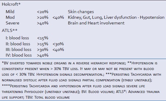
It has always been puzzling trying to understand and accept the rationale and benefits of the ATLS classification[1] especially after having replaced Holcroft more sensible classification,[2] as for the difficulty of practical implementation with reference to timing and optimal management. Both classifications were consequences of experiments done on animals that do not have the same adrenergic receptors distribution and amount on humans, which further varies from individual to individual,[3,4] and a misinterpretation of Shires studies in the 1960s,[5,6] deceptively corroborated by the improvement in renal failure statistics in the Vietnam war with the overload of crystalloids, incidental with a coincidental increase of ARDS.[7,8]
A more useful classification of hemorrhagic shock (HS), individual physiology-tailored and therapeutic/decision-making friendly, which is based on the above two classifications of shock, has been elaborated.
CLASSIFICATION OF HEMORRHAGIC SHOCK
Classifications are meant to summarize the assessment and management of a scenario or of a problem [Table 1]. ATLS ®classification of hemorrhagic shock (HS)[1] is not sensitive and specific enough to help decision-making in reference to the timing of management, being based only on the amount of blood loss that may or may not be rightly estimated, and it is unhelpful and difficult to apply too.[9] The previous physiological classification[2] had advantages overlooked and not re-captured by the ATLS® one, namely the progression of the effects of a hemorrhage on the different organs and systems, a more reliable indicator than the amount of blood itself in guiding timing of intervention. Nevertheless, the physiological classification, despite being more functional and useful does not keep in account the pre-existent different organ physiological reserves or can foresee the level at which hypotension, crucial parameter signaling decompensation, occurs. By recommending the fluid-load of 2 L crystalloids load for adult patients to test the reliability of compensatory mechanisms, as recommended up to recently, classical ATLS® guidelines actually delay the timing of intervention as source control when testing is not required and more crucially end up increasing the ongoing or spontaneously stopped bleeding. The only novelty of the classification is the cutoff at 30% blood loss as level of blood loss always manifesting with hypotension, per se not enough useful information to guide decision making.
Table 1.
Classical clinical classifications of haemorrhagic shock
The new classification [Table 2], which may well be called the "physiological HS classification" or "therapeutical HS classification′, is based on a decision-making that keeps in account hard practice and basic physiological considerations, such as the significance of fluid-blood resistant hypotension and body natural hemostatic mechanisms, the right definition of shock nonetheless the relevance that hemorrhage triggered I-R and SIR have in critical illness scenarios as secondary insult from ischemia.
Table 2.
Therapeutical/physiological classification of HS and first line management of source control
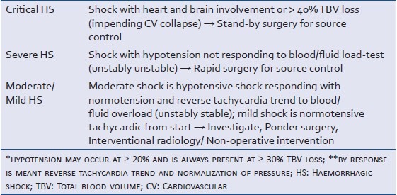
In “critical shock”, in fact there is no much of circulating volume, and brain and heart internal circulations are barely holding as a result of the systemic vasoconstriction from chemoreceptor and central nervous system receptors stimulation, while in “severe shock” there is sufficient blood volume to potentially maintain perfusion despite endogenous compensatory capacity in terms of vasomotion/vasoconstriction has been lost; in moderate shock compensatory capacity instead has not been lost; and mild shock indicates only some blood loss.
The ‘physiological-therapeutical’ classification must be distinguished from the prognostic one, i.e. reversible or irreversible shock and implicitly photographs the levels of shock within a time-frame of reversibility. It must also not be confused with the two hit-model of physiological deterioration either, which describe the time-peaks of clinical downfall.
RATIONALE: TIMING OF INTERVENTION—THE “RESUSCITATION PARADOX”
Timing is everything. The main stem of treatment of shock is removal of the causa prima (source optimization in cardiogenic shock (CS), source control in HS, and source elimination in inflammatory shock (IS). The timing for treatment of HS can be summarized in a “therapeutic paradox”: Intervene soon in a very sick patient to prevent death and accepting the inevitable complications; improve the healthy or moderately sick patient before surgery by optimizing or reinforcing patient physiology with a view to reduce or prevent complications. So, sicker the patient, the earlier more rapid and aggressive the intervention has to be; the less sick the patient more time has to be taken for improvement before intervention. Patient biological and physiological reserves (immunity, nutrition, exercise and age-related cardiovascular reflexes and specific organs homeostatic autoregulatory compensatory mechanisms), pre-existing systemic diseases or derangements (chronic renal failure, hypertension, diabetes, chronic liver disease, and chronic heart disease), and concurrent drug intake (alcohol, antihypertensive, anti-arrhythmic, β-blockers, steroids, vasodilators, inotrops, and insulin) play different significant roles in the overall prognosis of the critical illness by delaying detection, limiting the physiological reserve of the different organs and complicating recovery.
Timing of intervention for source control in HS depends on the clinical severity and degree of compensation that in normal individuals reflects the level of blood loss, and on the response to fluids load. TBV is 70 ml/kg body weight in adults, 80 ml/kg in infant age and 80-90 ml/kg in newborns. HS at the extremes of life is more serious than at the age in between as for the not developed (newborns and infants) or less responsive (elderly) vascular reflexes. Elderly patients as a matter of fact can have shock at seemingly normal blood pressure of 120 mmHg due to atherosclerosis, hypertension, and less functional reflexes maintaining relatively high pressures for perfusion.[10,11]
HS in pregnant women does not manifest with shock signs until 30-35% TBV is lost due to the increase of plasma and cardiac output. The supine position neutralizes the advantages of those preparatory changes to forthcoming intravascular volume-losses as for the uterus compressing on the IVC and impairing VR, phenomenon avoidable by always maintaining a left-oblique positioning when lying down.
Acute blood loss and hypotension with brain and heart disturbances or a blood loss >40% of TBV (critical HS, unstably unstable) require stand-by surgery to stop bleeding; persisting hypotension not responding to blood or fluids load (severe shock) with stable normalization of systolic and reversed HR trend (stably unstable), also requires emergency surgery to stop bleeding.
Heart and brain circulation in “critical shock” are holding because of still functional regional vasomotion, but they have already passed the critical extraction point as by definition ischemic signs are already present.
No response to small volumes of hypertonic/colloid fluid load in “severe shock” signifies a deranged vasomotion due to loss of endogenous compensation as the beginning of a physiological slope with continuing hemorrhage at a rate in which reflex compensatory vasoconstriction cannot maintain pressures.
Anything else other than a fast run to theater and swift anesthesia induction will kill patients in the above two scenarios. To push extra fluids fast or in great quantity will disrupt the balance by counteracting life-saving hypotension, vasoconstriction, vessel retraction, and clot formation, with the end-result of killing the patient or in the less pessimistic scenario causing heart attack or a cerebrovascular accident.[12]
The mechanisms accounting for the worsening of the situation before or in the absence of source control in critical and severe HS are multiple and act in combination evolving in a vicious circle of accelerated exitus.
The chemoreceptor response to low PaO2 at levels of pulse pressure of 70-80 mmHg increases BP by direct stimulation of the vasomotor centers in the reticular activating substance of the medulla oblongata and lower pons, increasing arterioles tone via sympathetic nervous system stimulation. At some stage, without or before source control and in the presence of supplementary fluids and oxygen or hyperoxia, the vasoconstricting reflex gets dampened; eventually, with HbO2reduced to minimal terms from the unarrested bleeding and CO reduced from the decreased venous return, and with dissolved PaO2 that cannot sustain CaO2at a level to maintain sufficient perfusion (DO2), the reflex gets triggered. By the time the chemoreceptor is triggered though, HbO2will have reached minimal levels, and so the DO2, and brain and heart are already suffering of relative hypoxia (critical shock). Coronaries have a very high O2ER at basal conditions (75% vs. 25% of most of the other organs) and are already in pathological supply dependence in critical shock, dangerously near the critical extraction level beyond which anaerobic metabolism and potentially devastating further ischemia ensues.[13] Adjunctive hyperoxia will paradoxically accelerate the physiological slope, particularly if combined with blood or fluids increasing bleeding rate before source control.
As importantly, the ischemic CNS response to pressures <60 mmHg with decreased DO2 delivery, as signaled by an already clinically impalpable level of systolic pressure, would also be counteracted by fluid administration with the paradox of having a situation, otherwise kept compensated by the two reflexes, being instead decompensated by fluids and oxygen administration.
Moreover, the increase of intravascular volumes with fluid transfusion before source control previously advised would further decrease perfusion as it counteracts the three natural physiological mechanisms of hemosthasis, i.e. arterial retraction/spasm, hypotension, and clot, ending up increasing bleeding and decreasing pressures.
Since World War I in fact it has been known that: hypotension, vasoconstriction, vessel retraction, and clot formation prevented continuation of bleeding after wounding; blood or plasma transfusion before surgery was a wasted resource that could cause re-bleeding; and surgery with control of hemorrhage was the most effective resuscitation. The insight of war surgery experience had told us that hypotension, clot formation, and vessels retraction were the reasons for patients’ survival after arterial damage or injury, that an untreated arterial injury or killed rapidly or was savageable if spontaneously stopped, while a venous injury had to rely only on clot formation to stop. This implies, paradoxically, that major venous injury can be more lethal than arterial one in sites such as mediastinum and retroperitoneum where it cannot be compressed.[14–17]
Thus, pushing fluids and hyperoxia and the maintenance-though temporary—of the actual mean arterial pressure (MAP) in critical shock and severe shock before source control, is in principle and de facto deleterious to patient's physiology and outcome. Delay and standard resuscitation with oxygen and fluids will paradoxically result in an earlier ischemia of the two organs than the one that would occur if no transfusion and early surgery were instead implemented.
The fluid-load test is consequently a wasteful and damaging exercise in ‘critical hemorrhagic shock’ as is any delay to fast source control.
In all other cases of hypotensive shock with no heart or brain ischemia the fluid-load test should be carried out as it tells us on the status of compensation present and, importantly, allows distinction between severe and moderate shock, i.e. between a rush to theater or temporizing on further diagnostic or therapeutic strategies. Hypotensive shock without heart or brain involvement, independently whether the loss is 20% (not always accompanied by hypotension) or 30% (always accompanied by hypotension) of TBV, which responds to blood/fluids overload test with normalization of blood pressure and reverse trend in tachycardia (moderate shock), indicates reliably the presence still of a certain physiological reserve in terms of compensatory mechanisms. Such scenarios do not require immediate or rapid surgery but can be investigated before surgery or interventional radiology and considered for conservative management. No investigation should be entertained in the presence of critical or severe shock.
Investigations should be allowed only in not-hypotensive, compensated mild-to-moderate HS, and should be aimed only to identify the origin of the bleeding/s and concomitant pathologies worthy or essential to be picked up before surgery. History, clinical assessment, logistics and equipment dictate the timing of intervention and investigations in compensated shock or consideration for conservative not-operative management. Blood transfusion in these not emergency cases should be given within maximum 4-6 h from insult to prevent I-R complications, till a level judged satisfactory (Hb ≥ 7 g/dL with Hct >21-24%) in healthy patients and ≥9-10 mg/dL with Hct ≥27-30% in cardiac patients, keeping in account SvO2 minimal levels of 70%, and normalized values of BE and LA in the absence of infection. [18,19]
Moderate HS responds well to crystalloids and PRBC i.v.; mild shock can be treated with oral fluids, or i.v. fluids.
For stabilization to be considered established, besides normal BP and reverse or normal pulse, patients must be seen with normal or improved complexion, mental status, urinary output, and comforting indirect signs of perfusion such as PaO2and SaO2.
PITFALLS IN HEMORRHAGIC SHOCK RESUSCITATION
The loading fluid test of 2 L of crystalloids previously recommended by ATLS® was de facto antiphysiological and deleterious especially when indiscriminately implemented and did not bring increase of survival,[20] but an increase in mortality and postoperative complications when compared to no-fluids or less-fluids resuscitation.[21–24] Experimental evidence confirmed the deleterious effects of the crystalloid bolus.[25,26]
At normal heart conditions and healthy valves and myocardium, any increase of venous return will effectively increase MAP which will increase actual bleeding by counteracting hypotension, the physiological vasoconstriction, and the clotting attempts, which are the three natural mechanisms the organism uses to stop hemorrhage. Vasodilatation and decreased viscosity from hemodilution also trigger the same chain of events, leading to increased, continuous or recurrent bleeding. These considerations and observations were tested in animals,[25–36] humans,[20–22,37,38] and computer simulations[39] and confirmed that (i) too much fluid infusion causes hemodilution of platelets and clotting factors, increase of blood pressure, decrease of blood viscosity, vasodilatation, all factors thus leading to a blow out of the hemostatic plug with accentuation of ongoing hemorrhage or/and secondary hemorrhage; (ii) blood loss causes hypothermia, which causes coagulopathy; (iii) in patients with penetrating trunk injury and hypotension and uncontrolled vascular injury, if no fluids in standard fashion are given in prehospital setting before theater, survival is increased, complications decreased and hospital stay shortened compared to standard fluid resuscitation; (iv) surgical hemosthasis is the key therapeutic act for uncontrolled hemorrhage; and (v) limited or moderate resuscitation is superior to aggressive resuscitation in uncontrolled vascular injury. Same hemodynamic and hemostasis derangements occur if hypertonic saline instead of crystalloids is used [Figure 1].[25–27,30,33–34,36]
Figure 1.
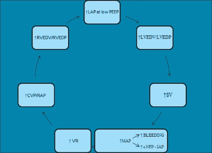
Effects of VR manipulations on haemodynamics in patients with normal cardiac reserve
Moreover, the principle first-crystalloids-then-blood, even worse in a 3:1 ratio, was based on the assumption that noncorpuscolated plasma, the fluid part of plasma, has first to replace the interstitial-intracellular shifts occurring during hemorrhage. Shires studies[5,6] were accurate on the assessment of fluids shifts, not in the way to manage them. The homeostasis of water bidirectionally in the cell-interstitia-blood pathways will be obviously deranged in not-compensated shock whereby definition the grasp on maintaining blood pressure by arteriolar vasoconstriction fails and the ratios in the Starling equilibrium in the capillaries default. It is intuitive that only by restoring vasoconstriction capacity the shifts can re-occur. This assumption was missed when the 3:1 ratio was postulated as the right one, confirming the need to categorize shock for any experiment or study in a more physiological manner. The real message from Shires works is that replenishment to be effective and accurate can only be done after source control where the solution of continuity is repaired and compensatory-physiology restored. Only then fluids infusion will equilibrate the three fluid compartments. It is true that combinations of crystalloids, plasma, and blood in the Vietnam war, increased survival and decreased renal failure at expenses of ARDS (former Da-Nang lung), but that cannot be attributed to the combination fluids blood 3:1 more that it can be attributed to blood only.[7,8] The ratio 3:1 is not only inaccurate in its conception, amount and priority of transfusion but also for its indiscriminate use, i.e. whether shock is compensated and with or without source control. Moreover, patients’ selection was not done in terms of categorizing them with different classes of shock and the decrease of renal failure is likely to have represented a natural selection occurred in survivors with not critical/severe shock cases, eventually some of them evolving in ARDS. Besides the negative effects on bleeding and on viscosity of microcirculation, the classical approach does not make sense intuitively either. If blood loss is the primitive derangement, it is expected blood replacement to be the main and most important corrective action. In other words, blood should be given first and crystalloids should follow once the fluid component of the plasma shifts toward interstitial-intracellular spaces and increases blood hematocrit.[40] The infusion of crystalloids would then replace the lost intravascular component and restore hematocrit. It is simple deduction that unless there is a loss of continuity left unrepaired in the circulation system, which is a closed system, any fluid shift from cells-to-interstitia-to-intravascular space would be reversed in the opposite direction by reproducing the dynamics backwards (Claude Bernard homeostasis concept). In hemorrhage it is blood that is lost and blood needs to be replaced, at least to minimum physiological levels when blood loss trespasses them. An indirect advantage of this shift of policy would be the shortening of time of clot formation compared to the first fluids—then blood current policy that has dominated decades of practice in trauma with deleterious effects.[41]
In hypotensive shock Ringer's lactate should therefore be used only after blood or blood components therapy in an amount tailored to balance osmolality electrolytes and hemathocrit.
Another drawback of an excess of fluid transfusion is RV/LV cardiac failure or CS if the two halves of the heart have pre—existent decreased functional reserve due to valvular or myocardial pathology. Two problems would then be faced—overload and low cardiac output-with treatment of one condition worsening the other one [Figure 2]. Direct inotropic support would then be required in conjunction with blood replenishment and aggressive ICU monitoring of the cardiac output.
Figure 2.
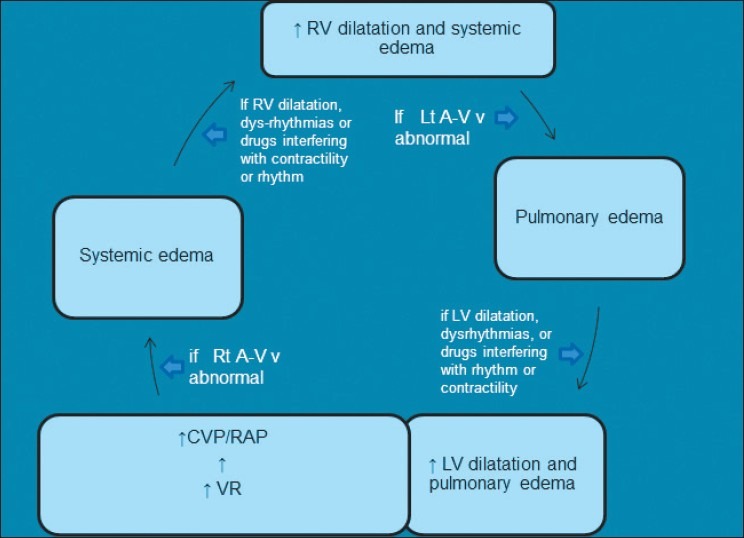
Effects of VR manipulations on haemodynamics in patients with diminished cardiac reserve
Furthermore, excess treatment with fluid or blood overload is deleterious whether is effected before source control or afterwards, as it may cause secondary intra-abdominal compartment syndrome,[42–44] with changes that trigger a second hit SIR or I-R syndromes and ALI helped by SIR and vasodilatation, if resuscitation is done late, or worsened by vasoconstriction with serious effects on abdominal organs perfusion, ventilation, kidney function and venous return, if resuscitation is done early and in excess. Gut edema and increased intraabdominal pressure till abdominal compartment syndrome level will ensue, due the increase of capillary net-filtration-pressure (cNFP) secondary to increased intracapillary pressure result of the increased pressure upstream from increased volume. Starling law ruling the permeable capillaries net filtration pressure states: NFP = [capillary pressure - (interstitial fluid pressure + plasma colloid-osmotic pressure) + interstitial fluid colloid-osmotic pressure].
Inadequate or delayed resuscitation is the other side of inappropriate treatment of HS. Cardiac arrest can occur as a primary hit for insufficient venous return and coronary ischemia due to the dependence of coronary perfusion from blood flow during the diastole phase, which is decreased following the decrease of the stroke volume. The compensatory tachycardia only worsens the situation accelerating heart ischemia. Post-HS SIR or I-R phenomenon is the second serious consequence of inadequate or deficient resuscitation leading to increased morbidity and mortality.[45,46]
STRATEGIES AND TACTICS
Adjuncts in treatment
Compensation of HS occurs by reflex arteriolar vasoconstriction sympathetic-mediated with catecholamines acting on α1 receptors. At some stage hyporeactivity to endogenous catecholamines installs, signaling decompensation, which becomes paralysis when a complete lack of responsiveness occurs even to exogenous vasoconstrictors signaling irreversible shock.
Besides its proven benefits in septic shock (SS), vasopressin alias arginine vasopressin (AVP) alias anti-diuretic hormone (ADH) has shown beneficial effects in CS and in cardiac arrest.[47–51] In HS AVP has been found to reduce the fluid requirement and improve neurological outcome and cardiopulmonary parameters.[52] Vasopressin as bolus (0.4 U once or twice) and/or infusion (0.04-0.1 U/min) reverses intractable or prolonged hypotension in the late phase HS or intraoperative HS with and without cardiac arrest.[53–59] Vasopressin vasoconstrictive effect results from inhibition of KATPchannels and inhibition of nitric oxide-induced accumulation of cGMP. Replacement of depleted stores of vasopressin in the neurohypophysis may also contribute to reversal of shock.[60] ADH may cause problems at higher dosages or when given for several hours.[61]
Noradrenaline (NE) is a potent alpha-adrenergic agonist with minimal β-adrenergic agonist effects. NE increases MAP due to its vasoconstrictive effects with little change in the heart rate and stroke volume, and by doing so increases indirectly the cardiac output as well. Doses of NE going from 0.2 μg/kg/min titrated to response up to 3.3 μg/kg/min are used to maintain CO and BP. Its drawbacks are an increase of workload and oxygen consumption plus coronary vasoconstriction. NE should be used early as neurohormonal augmentation therapy supporting hemodynamic function, rather than as a late rescue therapy to treat shock.[62]
Both NE and ADH combined with hydroxyethyl starch improve cerebral perfusion pressure, oxygenation and metabolism in HS, with AVP being the faster of the two.[63,64] The combination AVP + NE is an effective treatment for uncontrolled HS at the early stage after hemostasis, if blood is unavailable.[65]
NE should be added to AVP if the latter is ineffective, and must be discontinued before ADH.[66]
Likewise in SS, the combination of low doses NE and ADH, or ADH on its own, can be administered from presentation and categorization of critical or severe HS till before source control as a vasoconstriction-maintaining, vasomotor collapse-delaying, drug.
Once arterial pressure is brought to a systolic of at least 90 mmHg and a MAP >65-70 mmHg, and CO still would be low, intravenous dobutamine may take over. In normal hearts there is however scarce need for inotropic support in the postoperative ICU phase.
Dopamine may also be useful in patients with compromised systolic function, low CO, and MAP. At doses of approximately 10-20 μg/kg/min, the prevailing α-adrenergic effect leads to arterial vasoconstriction and elevation in blood pressure. The problem with dopamine however is that before the cardio-vascular system responds to vasoconstricting high dosages it has to pass through low and medium concentrations that may temporarily worsen the situation by increasing the heart rate and regional vasodilatation in a moment where there is not much blood in circulation; furthermore dopamine predisposes to dys-rhythmias.
Titrated hypotensive resuscitation
Too late, too little, too early too much, too late and too much are all harmful resuscitation strategies. To which level then should resuscitation be maintained before imminent surgery or temporarily before surgery or as the only treatment if a conservative nonoperative approach is adopted? This should be a level of blood pressure sufficient enough to maintain perfusion without risking either continuation of bleeding or re-bleeding. These targets are reached with “Titrated Hypotensive Resuscitation” [Table 3].
Table 3.
HS: too much too early, too little and too late. What is the ideal or perfect resuscitation?
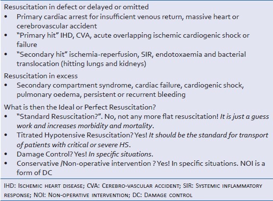
“Hypotensive resuscitation” is an old concept introduced by Cannon around the First World War,[14–17] but implemented only by Crawford in late 1980s for the treatment of ruptured abdominal aortic aneurysm before surgery,[38] recently re-introduced as "permissive or titrated hypotension" by the Israeli Defense Force[67,68] for transported patients with HS where resuscitation monitoring and titration are difficult to achieve and a small volume of infusion is logistically convenient. The systolic pressure was kept less than 90 mmHg to maintain a consciousness level or at the palpable pulse level, i.e. ≥70 and <90 mmHg by titrated prn hourly bolus of 250 ml of RL or HTS; skin complexion and consciousness level direct resuscitation in a conscious patient.
The presence of associated head injury that is not mild (GCS ≤ 12) compounds the clinical picture because of the difficulty or impossibility to use level of consciousness as an indicator of the level of blood loss and compensatory mechanisms efficacy, and the unpredictable loss of the capacity of flow self-regulation following trauma. Patients with HS and HI need therefore a moderate resuscitation in between permissive hypotension and standard resuscitation, i.e. systolic of ≥100 mmHg particularly in view of the fact that the brain loses its circulation self-regulatory capacity at variable levels of HI.[69,70]
Experimental evidence and clinical experience in civilian and military setting have shown benefits and safety of boluses of 250 ml of fluids, whether crystalloids, colloids, or hypertonic saline at different concentrations, as effective and safe initial management of patients with HS[67,68,71–73]HTS should be given not more than 250, maximum 375, ml/h to maximize its merits and diminish its drawbacks on bleeding by interference with coagulation and more importantly following increase of pressure as direct hemodynamic effect.
In a recent study on humans, the no-difference of mortality in patients who received either HTS 7.5%-Dextran 70 at 6% or HTS 7.5% administration boluses of 250 ml compared to NS 0.9% 250 ml as initial fluid treatment before source control,[74] associated to an increase of mortality in the subgroup who did not receive blood transfusion in the first 24 h, appears to contradict some of the conclusions of previous experiences. The inclusion, in the study, of patients in HS with SBP ≤70 mmHg or with SBP between 70 and 90 mmHg + HR ≥110 bpm would fit the profile of critical and severe shock categories if brain or heart were involved and a known loss <40% had occurred, or no consistent response to the fluid bolus was noted; moreover, the observations made in the group with no-blood transfusion indicate patients in whom type and amount of fluids would not have made difference. The study only emphasizes the need of a more accurate and useful categorization of HS.
“Hypotensive resuscitation” and “permissive hypotension on demand” or “titrated hypotension”, more accurately “Titrated Hypotensive Resuscitation” (THR), remains the ideal resuscitation and the way to go as standard initial resuscitation particularly during transport of patients with critical or severe HS independently on the scenario, whether civil or rural or military.
Emergency protocols: The “physiology approach”
In ‘critical shock’ with a known TBV loss of ≥40% or the presence of brain and heart ischemic changes there is no proven effective strategy of resuscitation out-of-hospital that would not contemplate heroics like in situ extreme-outdoor-resuscitation (EOR) by small operative units with or without suspended animation-fastly induced hypothermia techniques. Such techniques with portable femoro-femoral CPBP/ECMO without hypothermia have given dismal results outdoors in CA or CS before refractory CA in normovolemic not-exhanguinating patients even when applied with beating heart, and a survival of 20-30% indoors.[75–76] In trauma scenarios with solution of continuity in the vascular tree and decreased blood volume EOR with the method of suspended animation can only be effective with hypothermia induced by cold saline aortic flush via emergency sternotomy/thoracotomy.[77]
Until methods and techniques of EOR and suspended animation- hypothermia are optimized or perfected, only three options are so far available. ‘One’ is a life-saving run to the nearest medical facility for rapid source control with continuous blood transfusion running in high capacity/flow cannulae, well knowing it is destined to get lost. ‘Two’ is to run at the nearest medical facility with no fluid-treatment whatsoever at all leaving to the patient natural balancing mechanisms to do their best without iatrogenic interference until rapid/swift anesthesia/surgery for source control. "Three" is THR. In the author's view no-resuscitation or THR are the most sensible options to use as initial resuscitation before source control in patients with hypotensive HS both indoor and outdoor while rushing on the way to a medical facility. The no-resuscitation option may indeed be the best option.[21]
THR should also be used in ‘severe’ HS after the initial bolus of the fluid-load test discriminates it from the ‘moderate’ one; if shock turns out to be moderate with normalized systolic and reversed tachycardia trend, no further fluid should be given until source control.
Which fluid to use in “critical HS” in pre-hospital phase and which fluid should be used for load-test in severe HS?
It should be the fluid one would like to use as bridge infusion until source control if blood were unavailable: hyperosmotic-hyperviscous solutions (HHS), HTS or RL combined with alginates, and conjugated albumin solutions appear so far to be excellent choice. There is overwhelming evidence on the importance of maintaining microcirculation function with aim to optimize perfusion in HS by using apt fluid with specific characteristics and composition, particularly viscosity more than colloid-osmotic and oncotic properties, and, in parallel, on the irreplaceable function of blood from microhemodynamics and oxygen-transport end-points.[78–94]
To show predictable benefits of THR on mortality and morbidity,[85,95] specifically in preventing the installing of cryptic shock abutting in a I-R MOD/S clinical picture in the postoperative period, trials[96] should be done on patients in hypotensive shock classifiable as ‘severe’ or ‘critical’.[97]
It is under the same principle of the least interference with physiology during resuscitations that tactics and strategies such as THR, damage control, and damage control resuscitation, have been conceptualized. Likewise general anesthesia should too be titrated to pain/autonomic stimulation response (autonomic controlled-anesthesia) under the comprehensive concept of ‘physiology approach’.
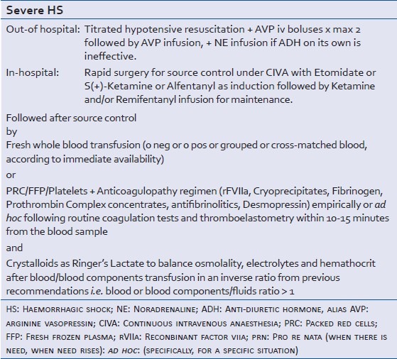
Etomidate, S(+)-ketamine or alfentanyl induction and S(+) ketamine or remifentanyl continuous-intravenous anesthesia (CIVA) titrated to response is the author's suggested method for anesthetizing critical and severe shock patients.[98–100]
Emergency protocols based on a “Physiology approach” comprehensive strategy, i.e. the “Therapeutical Classification of HS”, “THR”, “exogenous vasoconstrictive support”, autonomic-control CIVA, and blood and blood components replenishment,[101–108] are suggested in Tables 4 and 5. In hypotensive shock Ringer's lactate should be used only after blood or blood components therapy in an amount tailored to balance osmolality electrolytes and hematocrit.
Table 4.
Decision making in Critical HS: A comprehensive management
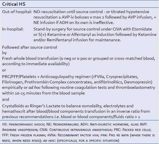
Table 5.
Decision making in Severe HS: A comprehensive management
CONCLUSIONS
The standard ATLS classification of HS is unhelpful, confusing and misleading as it focuses on the amount of blood loss instead of individual physiological response to hemorrhage, which is the core by definition of the derangement we call ‘shock’. A new classification was needed and centered on physiology like the classical classification of Holcroft emphasized.
The era of flat anesthetics and surgery is over and it is time we go toward a tailored-to-individual physiology restoration and reprioritize all critical illness management from the microcirculation stand-point.
Fluids as first modality of treatment, particularly in excess as recommended till recently, are deleterious in hypotensive shock and should be considered second line modality; moreover should be given after blood, and in an amount based on adjusting osmolarity.
Exogenous vasoconstrictors are a valid therapeutic strategy once endogenous vasoconstriction has failed to maintain the grip on the flow-controlling arterioles.
“Save life” is the Commandment in decompensated hypotensive shock not responding to fluid-load test or with a calculated TBV loss > 40% or with brain heart signs of ischemia, accepting inevitable and obligatory I-R and a phase of Cryptic Shock.
“Prevent complications”, namely I-R, is the Commandment in compensated mild or moderate shock.
Playing with the enemy in its own way is the key word of the game. Do not alter the physiological compensatory mechanisms but keep them and reinforce them. Let the body run the orchestra and follow it.
Without the right convenient and physiological classification of HS, we will still continue to generate controversies and contradictory investigator-appeasing results.
Footnotes
Source of Support: Nil.
Conflict of Interest: None declared.
REFERENCES
- 1.Kortbeek JB, Al Turki SA, Ali J, Antoine JA, Bouiloon B, Brasel K, et al. Advanced trauma life support, 8th edition, the evidence for change. J Trauma. 2008;64:1638–50. doi: 10.1097/TA.0b013e3181744b03. [DOI] [PubMed] [Google Scholar]
- 2.Holcroft JW, Shock . Current Surgical Diagnosis and Treatment. In: Dunphy JE, Way DL, editors. 5th ed. Los Altos: Lange Medical Publication; 1981. pp. 172–181. [Google Scholar]
- 3.Ahlquist RP. Present state of alpha and beta-adrenergic drugs.The adrenergic receptor. Am Heart J. 1976;92:661–4. doi: 10.1016/s0002-8703(76)80086-5. [DOI] [PubMed] [Google Scholar]
- 4.Nakai K, Itakura T, Naka Y, Nakakita K, Kamei I, Imai H, et al. The distribution of adrenergic receptors in cerebral blood vessels: an autoradiographic study. Brain Res. 1986;381:148–52. doi: 10.1016/0006-8993(86)90703-1. [DOI] [PubMed] [Google Scholar]
- 5.Shires T, Coln D, Carrico J, Lightfoot S. Fluid therapy in hemorrhagic shock. Arch Surg. 1964;88:688–93. doi: 10.1001/archsurg.1964.01310220178027. [DOI] [PubMed] [Google Scholar]
- 6.Dillon J, Lynch LJ, Jr, Myers R, Butcher HR., Jr The treatment of hemorrhagic shock. Surg Gynecol Obstet. 1966;122:967–78. [PubMed] [Google Scholar]
- 7.Shires GT, Carrico CJ, Baxter CR, Giesecke AH, Jr, Jenkins MT. Principles in treatment of severely injured patients. Adv Surg. 1970;4:255–324. [PubMed] [Google Scholar]
- 8.Cervera AL, Moss G. Progressive hypovolemia leading to shock after continuous hemorrhage and 3: 1 crystalloid replacement. Am J Surg. 1975;129:670–4. doi: 10.1016/0002-9610(75)90343-8. [DOI] [PubMed] [Google Scholar]
- 9.Guly HR, Bouamra O, Little R, Dark P, Coats T, Driscoll P, et al. Testing the validity of the ATLS classification of hypovolaemic shock. Resuscitation. 2010;81:1142–7. doi: 10.1016/j.resuscitation.2010.04.007. [DOI] [PubMed] [Google Scholar]
- 10.Rice IR, Kirkeby OJ. Effect of cerebral ischemia on the cerebrovascular and cardiovascular response to haemorrhage. Acta Neurochir (Wien) 1998;140:699–705. doi: 10.1007/s007010050165. [DOI] [PubMed] [Google Scholar]
- 11.Monge-Garcia MI, Cano AG, Romero MG. Dynamic arterial elastance to predict arterial pressure response to volume loading in preload-dependent patients. Crit Care. 2011;15:R15. doi: 10.1186/cc9420. [DOI] [PMC free article] [PubMed] [Google Scholar]
- 12.Bonanno FG. Pitfalls in surgical resuscitation. Minerva Chir. 2002;57:93–6. [PubMed] [Google Scholar]
- 13.Schumacker PT, Samsel RW. Oxygen delivery and uptake by peripheral tissues: physiology and pathophysiology. Crit Care Clin. 1989;5:255–69. [PubMed] [Google Scholar]
- 14.Cannon WB, Frasen J, Cowel EM. The preventive treatment of wound shock. J Am Med Ass. 1918;70:618–21. [Google Scholar]
- 15.Shaftan GW, Chiu CJ, Dennis C, Harris B. Fundamentals of physiological control of arterial hemorrhage. Surgery. 1965;58:851–6. [PubMed] [Google Scholar]
- 16.Wangensteen SL, Eddy DM, Ludewig RM. Bleeding and blood pressure. Am J Surg. 1969;118:413–4. doi: 10.1016/0002-9610(69)90146-9. [DOI] [PubMed] [Google Scholar]
- 17.Assalia A, Schein M. Resuscitation for haemorrhagic shock. Br J Surg. 1993;80:213. doi: 10.1002/bjs.1800800228. [DOI] [PubMed] [Google Scholar]
- 18.Dutton RP, Carson JL. Indications for early red blood cell transfusion. J Trauma. 2006;60:S35–S40. doi: 10.1097/01.ta.0000199974.45051.19. [DOI] [PubMed] [Google Scholar]
- 19.Paydar S, Fazelzadeh A, Abbasi H, Bolandparvaz S. Base deficit: A better indicator for diagnosis and treatment of shock in trauma patients. J Trauma. 2011;70:1580–1. doi: 10.1097/TA.0b013e318219e07d. [DOI] [PubMed] [Google Scholar]
- 20.Kaweski SM, Sise MJ, Virgilio RW. The effect of pre-hospital fluids on survival in trauma patients. J Trauma. 1990;30:1215–8. doi: 10.1097/00005373-199010000-00005. [DOI] [PubMed] [Google Scholar]
- 21.Bickell WH, Wall MJ, Pepe PE, Martin RR, Ginger VF, Allen MK, et al. Immediate versus delayed fluid resuscitation for hypotensive patients with penetrating torso injuries. N Engl J Med. 1994;331:1105–9. doi: 10.1056/NEJM199410273311701. [DOI] [PubMed] [Google Scholar]
- 22.Heckbert SR, Vedder NB, Hoffman W, Winn RK, Hudson LD, Jurkovich GJ, et al. Outcome after hemorrhagic shock in trauma patients. J Trauma. 1998;45:545–9. doi: 10.1097/00005373-199809000-00022. [DOI] [PubMed] [Google Scholar]
- 23.Cotton BA, Guy JS, Morris JA, Jr, Abumrad NN. The cellular, metabolic, and systemic consequences of aggressive fluid resuscitation strategies. Shock. 2006;26:115–21. doi: 10.1097/01.shk.0000209564.84822.f2. [DOI] [PubMed] [Google Scholar]
- 24.Hußmann B, Lefering R, Taeger G, Waydhas C, Ruchholtz S, Lendemans S. DGU Trauma Registry. Influence of prehospital fluid resuscitation on patients with multiple injuries in hemorrhagic shock in patients from the DGU trauma registry. J Emerg Trauma & Shock. 2011;4:465–71. doi: 10.4103/0974-2700.86630. [DOI] [PMC free article] [PubMed] [Google Scholar]
- 25.Solomonov E, Hirsh M, Yahiya A, Krausz MM. The effect of vigorous fluid resuscitation in uncontrolled hemorrhagic shock after massive splenic injury. Crit Care Med. 2000;28:749–54. doi: 10.1097/00003246-200003000-00024. [DOI] [PubMed] [Google Scholar]
- 26.Krausz MM, Hirsh M. Bolus versus continuous fluid resuscitation and splenectomy for treatment of uncontrolled hemorrhagic shock after massive splenic injury. J Trauma. 2003;55:62–8. doi: 10.1097/01.TA.0000074110.77122.46. [DOI] [PubMed] [Google Scholar]
- 27.Gross D, Landau EH, Assalia A, Krausz MM. Is hypertonic saline safe in “uncontrolled” hemorrhagic shock? J Trauma. 1988;28:751–6. doi: 10.1097/00005373-198806000-00005. [DOI] [PubMed] [Google Scholar]
- 28.Capone AC, Safar P, Stezoski W, Tisherman S, Peitzman AB. Improved outcome with fluid restriction in treatment of uncontrolled hemorrhagic shock. J Am Coll Surg. 1995;180:49–56. [PubMed] [Google Scholar]
- 29.Bickell WH, Bruttig SP, Wade CE. Hemodynamic response to abdominal aortotomy in anesthetized swine. Circ Shock. 1989;28:321–32. [PubMed] [Google Scholar]
- 30.Gross D, Landau EH, Klin B, Krausz MM. Treatment of uncontrolled hemorrhagic shock with hypertonic saline solution. Surg Gyn Obst. 1990;170:106–12. [PubMed] [Google Scholar]
- 31.Bickell WH, Bruttig SP, Millnamow GA, O’Benar J, Wade CE. The detrimental effects of intravenous crystalloids after aortotomy in swine. Surgery. 1991;110:529–36. [PubMed] [Google Scholar]
- 32.Bickell WH, Bruttig SP, Millnamow GA, O’Benar J, Wade CE. Use of hypertonic saline/dextran versus lactated ringer's solution as a resuscitation fluid after uncontrolled aortic hemorrhage in anesthetized swine. Ann Emerg Med. 1992;21:1077–85. doi: 10.1016/s0196-0644(05)80648-1. [DOI] [PubMed] [Google Scholar]
- 33.Krausz MM, Bar-Ziv M, Rabinovici R, Gross D. “Scoop and run” or stabilize hemorrhagic shock with normal or small-volume hypertonic saline? J Trauma. 1992;33:6–10. doi: 10.1097/00005373-199207000-00002. [DOI] [PubMed] [Google Scholar]
- 34.Matsuoka T, Wisner DH. Resuscitation of uncontrolled liver hemorrhage: effects on bleeding, oxygen delivery, and oxygen consumption. J Trauma. 1996;41:439–45. doi: 10.1097/00005373-199609000-00009. [DOI] [PubMed] [Google Scholar]
- 35.Riddez L, Johnson L, Hahn RG. Central and regional hemodynamics during crystalloid fluid therapy after uncontrolled intra-abdominal bleeding. J Trauma. 1998;44:433–9. doi: 10.1097/00005373-199803000-00001. [DOI] [PubMed] [Google Scholar]
- 36.Sukhotnik I, Krausz MM, Brod V, Balan M, Turkieh A, Siplovich L, et al. Divergent effects of oxygen therapy in four models of uncontrolled hemorrhagic shock. Shock. 2002;18:277–84. doi: 10.1097/00024382-200209000-00013. [DOI] [PubMed] [Google Scholar]
- 37.Blair SD, Janvrin SB, McCollum CN, Greenalgh RM. Effect of early blood transfusion on gastrointestinal haemorrhage. Br J Surg. 1986;73:783–5. doi: 10.1002/bjs.1800731007. [DOI] [PubMed] [Google Scholar]
- 38.Crawford ES. Ruptured abdominal aneurysms. J Vasc Surg. 1991;13:348–50. doi: 10.1016/0741-5214(91)90228-m. [DOI] [PubMed] [Google Scholar]
- 39.Hirshberg A, Hoyt DB, Mattox KL. Timing of fluid resuscitation shapes the hemodynamic response to uncontrolled hemorrhage: analysis using dynamic modeling. J Trauma. 2006;60:1221–7. doi: 10.1097/01.ta.0000220392.36865.fa. [DOI] [PubMed] [Google Scholar]
- 40.Takasu A, Minagawa Y, Ando S, Yamamoto Y, Sakamoto T. Improved survival time with combined early blood transfusion and fluid administration in uncontrolled hemorrhagic shock in rats. J Trauma. 2010;68:312–6. doi: 10.1097/TA.0b013e3181c48970. [DOI] [PubMed] [Google Scholar]
- 41.Spoerke NJ, Van PY, Differding JA, Zink KA, Cho SD, Muller PJ, et al. Red blood cells accelerate the onset of clot formation in polytrauma and hemorrhagic shock. J Trauma. 2010;69:1054–61. doi: 10.1097/TA.0b013e3181f9912a. [DOI] [PubMed] [Google Scholar]
- 42.Schein M, Witmann DH, Aprahamian CC, Condon RE. The abdominal compartment syndrome: the physiological and clinical consequences of elevated intra-abdominal pressure. J Am Coll Surg. 1995;180:745–53. [PubMed] [Google Scholar]
- 43.Kirkpatrick AW, Balogh Z, Ball CG, Ahmed N, Chun R, McBeth P, et al. The secondary abdominal compartment syndrome: iatrogenic or unavoidable? J Am Coll Surg. 2006;202:668–79. doi: 10.1016/j.jamcollsurg.2005.11.020. [DOI] [PubMed] [Google Scholar]
- 44.Balogh Z, Moore FA, Moore EE, Biffl WL. Secondary abdominal compartment syndrome: a potential threat for all trauma clinicians. Injury. 2007;38:272–9. doi: 10.1016/j.injury.2006.02.026. [DOI] [PubMed] [Google Scholar]
- 45.Cryer HG, Leong K, McArthur DL, Demetriades D, Bongard FS, Fleming AW, et al. Multiple organ failure: by the time you predict it, it is already there. J Trauma. 1999;46:597–604. doi: 10.1097/00005373-199904000-00007. [DOI] [PubMed] [Google Scholar]
- 46.Douzinas EE, Andrianakis I, Livaditi O, Prigouris P, Paneris P, Villiotou V, et al. The level of hypotension during hemorrhagic shock is a major determinant of the post-resuscitation systemic inflammatory response: an experimental study. BMC Physiol. 2008;18:8–15. doi: 10.1186/1472-6793-8-15. [DOI] [PMC free article] [PubMed] [Google Scholar]
- 47.Wenzel V, Krismer AC, Arntz HR, Sitter H, Stadlbauer KH, Lindner KH. A comparison of vasopressin and epinephrine for out-of-hospital cardiopulmonary resuscitation. N Engl J Med. 2004;350:105–13. doi: 10.1056/NEJMoa025431. [DOI] [PubMed] [Google Scholar]
- 48.Jolly S, Newton G, Horlick E. Effect of vasopressin on hemodynamics in patients with refractory cardiogenic shock complicating acute myocardial infarction. Am J Cardiol. 2005;96:1617–4. doi: 10.1016/j.amjcard.2005.07.076. [DOI] [PubMed] [Google Scholar]
- 49.Treschan TA, Peters J. The vasopressin system: physiology and clinical strategies. Anesthesiology. 2006;105:599–612. doi: 10.1097/00000542-200609000-00026. [DOI] [PubMed] [Google Scholar]
- 50.Krismer AC, Dünser MW, Lindner KH, Stadlbauer KH, Mayr VD, Lienhart HG, et al. Vasopressin during cardiopulmonary resuscitation and different shock states: a review of the literature. Am J Cardiovasc Drugs. 2006;6:51–68. doi: 10.2165/00129784-200606010-00005. [DOI] [PubMed] [Google Scholar]
- 51.Voelckel WG, Convertino VA, Lurie KG, Karlbauer A, Schochl H, Lindner KH, et al. Vasopressin for hemorrhagic shock management: revisiting the potential value in civilian and combat casualty care. J Trauma. 2010;69:S69–S74. doi: 10.1097/TA.0b013e3181e44937. [DOI] [PubMed] [Google Scholar]
- 52.Sanui M, King DR, Feinstein AJ, Varon AJ, Cohn SM, Proctor KG. Effects of arginine vasopressin during resuscitation from hemorrhagic hypotension after traumatic brain injury. Crit Care Med. 2006;34:433–8. doi: 10.1097/01.ccm.0000196206.83534.39. [DOI] [PubMed] [Google Scholar]
- 53.Raedler C, Voelckel WG, Wenzel V, Krismer AC, Schmittinger CA, Herff H, et al. Treatment of uncontrolled hemorrhagic shock after liver trauma: fatal effects of fluid resuscitation versus improved outcome after vasopressin. Anesth Analg. 2004;98:1759–66. doi: 10.1213/01.ANE.0000117150.29361.5A. [DOI] [PubMed] [Google Scholar]
- 54.Haas T, Voelckel WG, Wiedermann F, Wenzel V, Lindner KH. Successful resuscitation of a traumatic cardiac arrest victim in hemorrhagic shock with vasopressin: a case report and brief review of the literature. J Trauma. 2004;57:177–9. doi: 10.1097/01.ta.0000044357.25191.1b. [DOI] [PubMed] [Google Scholar]
- 55.Sharma RM, Setlur R. Vasopressin in hemorrhagic shock. Anesth Analg. 2005;101:833–4. doi: 10.1213/01.ANE.0000175209.61051.7F. [DOI] [PubMed] [Google Scholar]
- 56.Stadlbauer KH, Wenzel V, Krismer AC, Voeckel WG, Lindner KH. Vasopressin during uncontrolled hemorrhagic shock: less bleeding below the diaphragm, more perfusion above. Anest Analg. 2005;101:830–2. doi: 10.1213/01.ANE.0000175217.55775.1C. [DOI] [PubMed] [Google Scholar]
- 57.Tsuneyoshi I, Onomoto M, Yonetani, Kanmura Y. Low dose vasopressin infusion in patients with severe vasodilatory hypotension after prolonged hemorrhage during general anesthesia. J Anest. 2005;19:170–3. doi: 10.1007/s00540-004-0299-4. [DOI] [PubMed] [Google Scholar]
- 58.Roth JV. Bolus vasopressin during hemorrhagic shock? Anesth Analg. 2006;102:1098. doi: 10.1213/01.ANE.0000215135.44887.7E. [DOI] [PubMed] [Google Scholar]
- 59.Raab H, Lindner KH, Wenzel V. Preventing cardiac arrest during hemorrhagic shock with vasopressin. Crit Care Med. 2008;36:S474–S480. doi: 10.1097/ccm.0b013e31818a8d7e. [DOI] [PubMed] [Google Scholar]
- 60.Holmes CL, Landry DW, Granton JT. Science review: vasopressin and the cardiovascular system, Part 2 - clinical physiology. Crit Care. 2004;8:15–23. doi: 10.1186/cc2338. [DOI] [PMC free article] [PubMed] [Google Scholar]
- 61.Dunser MW, Mayr AJ, Tur A, Pajk W, Barbara F, Knotzer H, et al. Ischemic skin lesions as a complication of continuous vasopressin infusion in catecholamine-resistant vasodilatory shock: incidence and risk factors. Crit Care Med. 2003;31:1394–8. doi: 10.1097/01.CCM.0000059722.94182.79. [DOI] [PubMed] [Google Scholar]
- 62.Myburgh J. Norepinephrine: more of a neurohormone than a vasopressor. Crit Care. 2010;14:196. doi: 10.1186/cc9246. [DOI] [PMC free article] [PubMed] [Google Scholar]
- 63.Meybohm P, Cavus E, Bein B, Steinfath M, Weber B, Hamann C, et al. Small volume resuscitation: a randomized controlled trial with either norepinephrine or vasopressin during severe hemorrhage. J Trauma. 2007;62:640–6. doi: 10.1097/01.ta.0000240962.62319.c8. [DOI] [PubMed] [Google Scholar]
- 64.Cavus E, Meybohm P, Doerges V, Hugo HH, Steinfath M, Nordstroem J, et al. Cerebral effects of three resuscitation protocols in uncontrolled haemorrhagic shock: a randomised controlled experimental study. Resuscitation. 2009;80:567–72. doi: 10.1016/j.resuscitation.2009.01.013. [DOI] [PubMed] [Google Scholar]
- 65.Li T, Fang Y, Zhu Y, Fan X, Liao Z, Chen F, et al. A small dose of Arginine Vasopressin in combination with Norepinephrine is a good early treatment for uncontrolled hemorrhagic shock after hemostasis. J Surg Res. 2010 doi: 10.1016/j.jss.2010.02.001. [Epub ahead of print] [DOI] [PubMed] [Google Scholar]
- 66.Bauer SR, Aloi JJ, Ahrens CL, Yeh JY, Culver DA, Reddy AT, et al. Discontinuation of vasopressin before norepinephrine increases the incidence of hypotension in patients recovering from septic shock: a retrospective cohort study. J Crit Care. 2010;25:362. doi: 10.1016/j.jcrc.2009.10.005. [DOI] [PubMed] [Google Scholar]
- 67.Krausz MM. Fluid resuscitation strategies in the Israeli Army. J Trauma. 2003;54:S39–S42. doi: 10.1097/01.TA.0000064506.47688.51. [DOI] [PubMed] [Google Scholar]
- 68.Blumenfeld A, Melamed E, Kalmovich D, et al. Pre-hospital fluid resuscitation in trauma: the IDF-MC panel Summary. J Israeli Mil Med. 2004;1:6–10. [Google Scholar]
- 69.The Brain Trauma foundation, The American Association of Neurological Surgeons and The Joint Section on Neurotrauma and Critical Care. Resuscitation of blood pressure and oxygenation. J Neurotrauma. 2000;17:471–82. doi: 10.1089/neu.2000.17.471. [DOI] [PubMed] [Google Scholar]
- 70.Dar DE, Soustiel JF, Zaaroor M, Broftain EM, Leibowitz A, Shapira Y. Moderate Ringer's lactate solution resuscitation yields best neurological outcome in controlled hemorrhagic shock combined with brain injury in rats. Shock. 2010;34:75–82. doi: 10.1097/SHK.0b013e3181ce2cbc. [DOI] [PubMed] [Google Scholar]
- 71.Holcomb JB. Fluid resuscitation in modern combat casualty care: lessons learnt from Somalia. J Trauma. 2003;54:S46–S51. doi: 10.1097/01.TA.0000051936.91915.A2. [DOI] [PubMed] [Google Scholar]
- 72.Holcroft JW, Vassar MJ, Turner JE, Derlet RW, Kramer GC. 3% NaCl and 7.5% NaCl/dextran 70 in the resuscitation of severely injured patients. Ann Surg. 1987;206:279–88. doi: 10.1097/00000658-198709000-00006. [DOI] [PMC free article] [PubMed] [Google Scholar]
- 73.Wade CE, Kramer GC, Grady JJ, Fabian TC, Younes RN. Efficacy of hypertonic 7.5% saline and 6% dextran 70 in treating trauma: a meta-analysis of controlled clinical trials. Surgery. 1997;122:609–16. doi: 10.1016/s0039-6060(97)90135-5. [DOI] [PubMed] [Google Scholar]
- 74.Bulger EM, May S, Kerby JD, Emerson S, Stiell IG, Schreiber MA, et al. Out-of-hospital Hypertonic Resuscitation After Traumatic Hypovolemic Shock: A Randomized, Placebo Controlled Trial. Ann Surg. 2010 doi: 10.1097/SLA.0b013e3181fcdb22. [DOI] [PMC free article] [PubMed] [Google Scholar]
- 75.Kagawa E, Inoue I, Kawagoe T, Ishihara M, Shimatani Y, Kurisu S, et al. Assessment of outcomes and differences between in- and out-of-hospital cardiac arrest treated with cardiopulmonary resuscitation with extracorporeal life support. Resuscitation. 2010;81:968–73. doi: 10.1016/j.resuscitation.2010.03.037. [DOI] [PubMed] [Google Scholar]
- 76.Le Guen M, Nicolas-Robin A, Carreira S, Raux M, Leprince P, Riou B, et al. Extracorporeal life support following out-of-hospital refractory cardiac arrest. Crit Care. 2011;15:R29. doi: 10.1186/cc9976. [DOI] [PMC free article] [PubMed] [Google Scholar]
- 77.Alam HB, Duggan M, Li Y, Spanioles K, Liu B, Tabbara M, et al. Putting life on hold - for how long.Profound hypothermic cardiopulmonary bypass in a swine model of complex vascular injuries? J Trauma. 2008;64:912–22. doi: 10.1097/TA.0b013e3181659e7f. [DOI] [PubMed] [Google Scholar]
- 78.Cabrales P, Tsai AG, Intaglietta M. Hyperosmotic-hyperoncotic versus hyperosmotic-hyperviscous: small volume resuscitation in hemorrhagic shock. Shock. 2004;22:431–7. doi: 10.1097/01.shk.0000140662.72907.95. [DOI] [PubMed] [Google Scholar]
- 79.Cabrales P, Intaglietta M, Tsai AG. Increase plasma viscosity sustains microcirculation after resuscitation from hemorrhagic shock and continuous bleeding. Shock. 2005;23:549–55. [PubMed] [Google Scholar]
- 80.Wettstein R, Erni D, Intaglietta M, Tsai AG. Rapid restoration of microcirculatory blood flow with hyperviscous and hyperoncotic solutions lowers the transfusion trigger in resuscitation from hemorrhagic shock. Shock. 2006;25:641–6. doi: 10.1097/01.shk.0000209532.15317.15. [DOI] [PubMed] [Google Scholar]
- 81.Cabrales P, Tsai AG, Intaglietta M. Hemorrhagic shock resuscitation with carbon monoxide saturated blood. Resuscitation. 2007;72:306–318. doi: 10.1016/j.resuscitation.2006.06.021. [DOI] [PubMed] [Google Scholar]
- 82.Salazar Vázquez BY, Wettstein R, Cabrales P, et al. Microvascular experimental evidence on the relative significance of restoring oxygen carrying capacity vs blood viscosity in shock resuscitation. Biochem Biophysic Acta. 2008;1784:1421–7. doi: 10.1016/j.bbapap.2008.04.020. [DOI] [PMC free article] [PubMed] [Google Scholar]
- 83.Cheung AT, To PL, Chan DM, et al. Comparison of treatment modalities for hemorrhagic shock. Artif Cells Blood Substit Immobil Biotechnol. 2007;35:173–90. doi: 10.1080/10731190601188257. [DOI] [PubMed] [Google Scholar]
- 84.Tsai AG, Hofmann A, Cabrales P, et al. Perfusion vs. oxygen delivery in transfusion with “fresh” and “old” red blood cells: The experimental evidence. Transfus Apher Sci. 2010;43:69–78. doi: 10.1016/j.transci.2010.05.011. [DOI] [PMC free article] [PubMed] [Google Scholar]
- 85.Kentner R, Safar P, Prueckner S, Behringer W, Wu X, Henchir J, et al. Titrated hypertonic/hyperoncotic solution for hypotensive fluid resuscitation during uncontrolled hemorrhagic shock in rats. Resuscitation. 2005;65:87–95. doi: 10.1016/j.resuscitation.2004.10.012. [DOI] [PubMed] [Google Scholar]
- 86.Hanke AA, Maschler S, Schochl H, Floricke F, Gorlinger K, Zanger K, et al. In vitro impairment of whole blood coagulation and platelets function by Hypertonic Saline Hydroxyethyl Starch. Scand J Trauma Resusc Emerg Med. 2011;19:12. doi: 10.1186/1757-7241-19-12. [DOI] [PMC free article] [PubMed] [Google Scholar]
- 87.Villela NR, Salazar Vázquez BY, Intaglietta M. Microcirculatory effects of intravenous fluids in critical illness: plasma expansion beyond crystalloids and colloids. Curr Opin Anaesthesiol. 2009;22:163–7. doi: 10.1097/ACO.0b013e328328d304. [DOI] [PubMed] [Google Scholar]
- 88.Villela N, Tsai A, Cabrales P, Intaglietta M. Improved Resuscitation From Hemorrhagic Shock With Ringer's Lactate With Increased Viscosity in the Hamster Window Chamber Model. J of Trauma. 2011;71:418–424. doi: 10.1097/TA.0b013e3181fa2630. [DOI] [PubMed] [Google Scholar]
- 89.Matsushita S, Chuang VT, Kanazawa M, Tanase S, Kawai K, Maruyama T et al. Recombinant human serum albumin dimer has high blood circulation activity and low vascular permeability in comparison with native human serum albumin. Pharm Res. 2006;23:882–91. doi: 10.1007/s11095-006-9933-1. [DOI] [PubMed] [Google Scholar]
- 90.Cabrales P, Tsai AG, Intaglietta M. Is resuscitation from hemorrhagic shock limited by blood oxygen-carrying capacity or blood viscosity? Shock. 2007;27:380–9. doi: 10.1097/01.shk.0000239782.71516.ba. [DOI] [PubMed] [Google Scholar]
- 91.Cabrales P, Intaglietta M, Tsai AG. Transfusion restores blood viscosity and reinstates microvascular conditions from hemorrhagic shock independent of oxygen carrying capacity. Resuscitation. 2007;75:124–34. doi: 10.1016/j.resuscitation.2007.03.010. [DOI] [PMC free article] [PubMed] [Google Scholar]
- 92.Martini J, Cabrales P, K A, Acharya SA, Intaglietta M, Tsai AG. Survival time in severe hemorrhagic shock after perioperative hemodilution is longer with PEG-conjugated human serum albumin than with HES 130/0.4: a microvascular perspective. Crit Care. 2008;12:R54. doi: 10.1186/cc6874. [DOI] [PMC free article] [PubMed] [Google Scholar]
- 93.Cabrales P, Tsai AG, Ananda K, Acharya SA, Intaglietta M. Volume resuscitation from hemorrhagic shock with albumin and hexaPEGylated human serum albumin. Resuscitation. 2008;79:139–46. doi: 10.1016/j.resuscitation.2008.04.020. [DOI] [PMC free article] [PubMed] [Google Scholar]
- 94.Exo JL, Shellington DK, Bayir H, Vagni VA, Janesco-Feldman K, Ma L, et al. Resuscitation of traumatic brain injury and hemorrhagic shock with polynitroxylated albumin, hextend, hypertonic saline, and lactated Ringer’s: Effects on acute hemodynamics, survival, and neuronal death in mice. J Neurotrauma. 2009;26:2403–8. doi: 10.1089/neu.2009.0980. [DOI] [PMC free article] [PubMed] [Google Scholar]
- 95.Kowalenko T, Stern SA, Dronen SC, Wang X. Improved outcome with hypotensive resuscitation of uncontrolled hemorrhagic shock in a swine model. J Trauma. 1992;33:349–53. [PubMed] [Google Scholar]
- 96.Dutton RP, Mackenzie CF, Scalea TM. Hypotensive resuscitation during active hemorrhage: impact on in-hospital mortality. J Trauma. 2002;52:1141–6. doi: 10.1097/00005373-200206000-00020. [DOI] [PubMed] [Google Scholar]
- 97.Dronen SC, Stern SA, Wang X, Stanley M. A comparison of the response of near-fatal acute hemorrhage with and without a vascular injury to rapid volume expansion. Am J Emerg Med. 1993;11:331–5. doi: 10.1016/0735-6757(93)90162-5. [DOI] [PubMed] [Google Scholar]
- 98.Kienbaum P, Heuter T, Pavlakovic G, Michel MC, Peters J. S(+)-ketamine increases muscle sympathetic activity and maintains the neural response to hypotensive challenges in humans. Anesthesiology. 2001;94:252–8. doi: 10.1097/00000542-200102000-00014. [DOI] [PubMed] [Google Scholar]
- 99.Bonanno GF. Ketamine in war/tropical surgery (a final tribute to the racemic mixture) Injury. 2002;33:323–7. doi: 10.1016/s0020-1383(01)00209-1. [DOI] [PubMed] [Google Scholar]
- 100.Morris C, Perris A, Klein J, Mahoney P. Anaesthesia in haemodynamically compromised emergency patients does ketamine represent the best choice of induction agent? Anaesthesia. 2009;64:532–9. doi: 10.1111/j.1365-2044.2008.05835.x. [DOI] [PubMed] [Google Scholar]
- 101.Kaur P, Basu S, Kaur G, Kaur R. Transfusion protocol in trauma. J Emerg Trauma Shock. 2011;4:103–8. doi: 10.4103/0974-2700.76844. [DOI] [PMC free article] [PubMed] [Google Scholar]
- 102.Duchesne JC, Barbeau JM, Islam TM, Wahl G, Greiffenstein P, McSwain NE, et al. Damage control resuscitation: from emergency department to the operating room. Am Surg. 2011;77:201–6. doi: 10.1177/000313481107700222. [DOI] [PubMed] [Google Scholar]
- 103.Morrison CA, Carrick MM, Norman MA, Scott BG, Welsh FJ, Tsai P, et al. Hypotensive resuscitation strategy reduces transfusion requirements and severe postoperative coagulopathy in trauma patients with hemorrhagic shock: preliminary results of a randomized controlled trial. J Trauma. 2011;70:652–63. doi: 10.1097/TA.0b013e31820e77ea. [DOI] [PubMed] [Google Scholar]
- 104.Lustenberger T, Talving P, Kobayashi L, Barmparas G, Inaba K, Lam L, et al. Early coagulopathy after isolated severe traumatic brain injury: relationship with hypoperfusion challenged. J Trauma. 2010;69:1410–4. doi: 10.1097/TA.0b013e3181cdae81. [DOI] [PubMed] [Google Scholar]
- 105.Greuters S, van den Berg A, Franschman G, Viersen VA, Beishuizen A, Peederman SM, et al. Acute and delayed mild coagulopathy are related to outcome in patients with isolated traumatic brain injury. Crit Care. 2011:15R2. doi: 10.1186/cc9399. [DOI] [PMC free article] [PubMed] [Google Scholar]
- 106.Kashuk JL, Moore EE, Sawyer M, Le T, Johnson J, Biffl WL, et al. Postinjury coagulopathy management: goal directed resuscitation via POC thrombelastography. Ann Surg. 2010;251:604–14. doi: 10.1097/SLA.0b013e3181d3599c. [DOI] [PubMed] [Google Scholar]
- 107.Schoechl H, Nienaber U, Maegele M, Hochleitner G, Primavesi F, Steitz B, et al. Transfusion in trauma: thromboelastometry-guided coagulation factor concentrate-based therapy versus fresh frozen plasma-based therapy. Crit Care. 2011;15:R83. doi: 10.1186/cc10078. [DOI] [PMC free article] [PubMed] [Google Scholar]
- 108.David JS, Marchal V, Levrat A, Inaba K. Which is the most effective strategy: early detection of coagulopathy with thromboelastometry or use of hemostatic factors or both? Crit Care. 2011;15:433. doi: 10.1186/cc10224. [DOI] [PMC free article] [PubMed] [Google Scholar]


