Abstract
Shiga toxin-producing Escherichia coli (STEC) are zoonotic enteric pathogens associated with human gastroenteritis worldwide. Cattle and small ruminants are important animal reservoirs of STEC. The present study investigated animal reservoirs for STEC in small rural farms in the Culiacan Valley, an important agricultural region located in Northwest Mexico. A total of 240 fecal samples from domestic animals were collected from five sampling sites in the Culiacan Valley and were subjected to an enrichment protocol followed by either direct plating or immunomagnetic separation before plating on selective media. Serotype O157:H7 isolates with the virulence genes stx2, eae, and ehxA were identified in 40% (26/65) of the recovered isolates from cattle, sheep and chicken feces. Pulse-field gel electrophoresis (PFGE) analysis grouped most O157:H7 isolates into two clusters with 98.6% homology. The use of multiple-locus variable-number tandem repeat analysis (MLVA) differentiated isolates that were indistinguishable by PFGE. Analysis of the allelic diversity of MLVA loci suggested that the O157:H7 isolates from this region were highly related. In contrast to O157:H7 isolates, a greater genotypic diversity was observed in the non-O157 isolates, resulting in 23 PFGE types and 14 MLVA types. The relevant non-O157 serotypes O8:H19, O75:H8, O111:H8 and O146:H21 represented 35.4% (23/65) of the recovered isolates. In particular, 18.5% (12/65) of all the isolates were serotype O75:H8, which was the most variable serotype by both PFGE and MLVA. The non-O157 isolates were predominantly recovered from sheep and were identified to harbor either one or two stx genes. Most non-O157 isolates were ehxA-positive (86.5%, 32/37) but only 10.8% (4/37) harbored eae. These findings indicate that zoonotic STEC with genotypes associated with human illness are present in animals on small farms within rural communities in the Culiacan Valley and emphasize the need for the development of control measures to decrease risks associated with zoonotic STEC.
Introduction
Shiga toxin-producing Escherichia coli (STEC) is a group of food- and water-borne pathogens that are known to cause human gastrointestinal illnesses with diverse clinical spectra, ranging from watery and bloody diarrhea to hemorrhagic colitis [1], [2]. In some cases, disease symptoms result in the life-threatening, hemolytic uremic syndrome (HUS), and it is thought that Shiga toxins (Stx1 and Stx2) are the key virulence factors contributing to the development of HUS. Although more than 200 different serotypes of STEC have been isolated, O157:H7 has been the serotype most commonly associated with HUS in North America. Recent epidemiological studies have recognized additional non-O157 serogroups, including O26, O45, O91, O103, O104, O111, O113, O121, and O145, among STEC strains that were linked to severe human disease in the United States, Europe and countries of Latin America [3]–[7].
Epidemiological studies have shown that not all STEC strains producing Stx are clinically relevant. Thus, it has been proposed that accessory STEC genes may also contribute to human disease [4], [8], [9]. For example, a well-characterized adhesin gene is eae that codes for the intimin protein, implicated in attachment to the intestinal epithelial cells prior to lesion formation [10], [11]. Many of the strains implicated in bloody diarrhea and HUS in humans are eae-positive; therefore, eae is recognized as an important risk factor for HUS [1], [12]. Additionally, enterohemolysin, expressed by the ehxA gene, liberates hemoglobin from the red blood cells during infection and has been linked to severe disease symptoms [13]–[15]. Consequently, E. coli strains, recovered from animal reservoirs and harboring stx, eae, and/or ehxA genes, are thought to represent a subpopulation of STEC strains that may pose a higher risk to human health [16], [17].
STEC strains have been isolated from a variety of animals, and cattle are considered to be the major reservoir for STEC strains [1], [18]–[20]. However, recent evidence has indicated that small domestic ruminants, including sheep and goats, are also key reservoirs of STEC [21]–[26]. In particular, sheep and their products have been documented as reservoirs for STECs that belong to a diverse set of non-O157 serogroups (O26, O91, O115, O128, and O130) and that harbor genes encoding key virulence factors that have been implicated in human disease [27]–[33]. STEC strains can also be carried by other domestic and wild animals, such as cats, dogs, rodents, deer, birds, feral pigs, chickens, and insects [18], [34], [35].
A few recent studies have examined the prevalence of O157 and non-O157 STEC strains in products derived from animals or in animal fecal samples at various locations in Mexico [36]–[39]. In particular, a greater prevalence of E. coli O157 was found in swine feces (2.1%) than cattle feces (1.25%), recovered from eight different locations throughout Central Mexico [36]. Furthermore, the presence of O157 as well as non-O157 on beef carcasses was confirmed at slaughter plants in Northeast and Western Mexico [38], [39]. Surveillance studies yielded recovery of non-O157 STEC strains in a large proportion of ready-to-eat meals in Mexico City [40], suggesting that non-O157 could be a potential source of infection in humans. Thus, additional studies about isolation, sources, and prevalence of STEC are needed in other agricultural locations in Mexico.
To determine relevant animal reservoirs for toxigenic E. coli in Mexico, the present study employed a protocol for the selective enrichment of STEC from feces of domestic animals found on small rural farms within the Culiacan Valley. The Culiacan Valley, located in Northwest Mexico, has large and fertile agricultural fields and a successful packaging industry for horticultural commodities (tomatoes, cucumbers, and bell peppers) [41]. The small rural farms, sampled in the present study, were located in rural communities where the primary purpose of raising livestock is for local consumption [42]. The recovered STEC isolates were further examined for O- and H-antigens and several important virulence factors. Furthermore, the genetic relatedness of the isolates was analyzed by the genotyping methods pulse-field gel electrophoresis (PFGE) and multiple-locus variable-number tandem repeat analysis (MLVA) to obtain a better understanding of the geographical distribution and relevant sources of zoonotic STEC in the Culiacan Valley.
Results
Isolation of STEC from Feces of Farm Animals in the Culiacan Valley
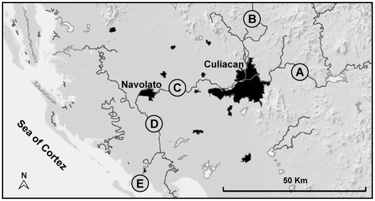
The aim of the present study was to identify both O157 and non-O157 STEC recovered from feces of domestic farm animals in the Culiacan Valley, a region in Northwest Mexico. Fecal samples were recovered from small farms at five sampling sites in the Culiacan Valley, each located in close proximity to rivers used for irrigation (Figure 1). During the first six months of the sampling (July to December 2008), enrichment broths were prepared from a total of 120 fecal samples from all sampling sites and were plated directly onto the indicator media (see Materials and Methods). Presumptive STEC colonies were recovered from 5% (6/120) of the fecal samples after plating on both indicator media (Table 1). Sampling sites C, D, and E had 8.3% (either 1/12 or 2/24) positive samples when compared to 4.2% (1/24) for sampling site B. Presumptive STEC colonies were predominantly detected in 11.1% (4/36) of the sheep fecal samples and, secondly, in 3.3% (2/60) of the cattle fecal samples (Table 1). No positive samples were obtained from sampling site A after plating directly from the enrichment broths onto the two indicator media.
Figure 1. Sampling sites in the Culiacan Valley in Northwest Mexico.
Map of the five sampling sites, Jotagua (A), Agua Caliente (B), Cofradia de Navolato (C), Iraguato (D), and El Castillo (E) that were selected to be 14–38 km apart and to represent the study area in the Culiacan Valley, Sinaloa, Mexico. Dark areas in the map indicate urban zones with more than 20,000 inhabitants. Scale bar corresponds to 50 km.
Table 1. Proportion of fecal samples positive for presumptive STEC.
| Method1 | Source | Number of positive samples/Number of fecal samples tested per sampling site2 | |||||
| Site A | Site B | Site C | Site D | Site E | Total | ||
| Direct plating | Cattle | 0/12 | 1/12 | 0/12 | 1/12 | 0/12 | 2/60 |
| Chicken | 0/12 | 0/12 | NA3 | NA | NA | 0/24 | |
| Sheep | 0/12 | NA | 2/12 | NA | 2/12 | 4/36 | |
| Total | 0/36 | 1/24 | 2/24 | 1/12 | 2/24 | 6/120 | |
| IMS | Cattle | 0/12 | 0/12 | 4/12 | 3/12 | 2/12 | 9/60 |
| Chicken | 0/12 | 1/12 | NA | NA | NA | 1/24 | |
| Sheep | 2/12 | NA | 5/12 | NA | 2/12 | 9/36 | |
| Total | 2/36 | 1/24 | 9/24 | 3/12 | 4/24 | 19/120 | |
Enrichment broths were subjected to direct plating from July to December 2008 or to immunomagnetic separation (IMS) from January to June 2009.
Sampling sites correspond to regions in the Culiacan Valley, Sinaloa, Mexico, as shown in Figure 1.
NA, None available.
In the subsequent sampling period (January to June 2009), the isolation method was modified for the recovery of STEC. A second set of 120 fecal samples were collected from farm animals at the same five sampling sites (Figure 1), and enrichment broths from the fecal samples were subjected to an immunomagnetic separation (IMS) method, followed by plating onto the indicator media. By using the IMS method, presumptive E. coli colonies were recovered from 15.8% (19/120) of the fecal samples tested (Table 1). Although all sampling sites had at least one enrichment broth that yielded presumptive colonies, 37.5% (9/24) of the positive samples were collected at farms in sampling site C, followed by sampling site D with 25.0% (3/12) of the positive samples. A lower number of positive samples were obtained from the other sampling sites (sites A, B, and E) (Table 1). Presumptive STEC colonies were mostly identified in 25.0% (9/36) of the enrichment broths from sheep feces, followed by 15.0% (9/60) from cattle feces and 4.2% (1/24) from chicken feces. From 25 enrichment broths that were positive by using both direct plating and IMS methods (Table 1), a total of 65 presumptive STEC isolates were isolated, and 83.1% (54/65) of these isolates were recovered from samples processed by the IMS method (Tables 2 and 3).
Table 2. List of E. coli O157 isolates used in this study.
| Isolate | Fecal Sample | Serotype1 | Sampling date2 | Source | Sampling site3 | Genes identified | |||||
| stx1 | stx2 | eae | ehxA | ||||||||
| RM8744 | Dc10 | O157:H7 | 18-Nov-08 | Cattle | D | – | + | + | + | ||
| RM8753 | Cs11 | O157:H7 | 02-Dec-08 | Sheep | C | – | + | + | + | ||
| RM8754 | Dc10 | O157:H7 | 18-Nov-08 | Cattle | D | – | + | + | + | ||
| RM8759 | Cs14 | O157:H7 | 20-Jan-09 | Sheep | C | – | + | + | + | ||
| RM8767 | Cc14 | O157:H7 | 20-Jan-09 | Cattle | C | – | + | + | + | ||
| RM8768 | Cc14 | O157:H7 | 20-Jan-09 | Cattle | C | – | + | + | + | ||
| RM8769 | Cc14 | O157:H7 | 20-Jan-09 | Cattle | C | – | + | + | + | ||
| RM8771 | Cc14 | O157:H7 | 20-Jan-09 | Cattle | C | – | + | + | + | ||
| RM8781 | Cs17 | O157:H7 | 25-Feb-09 | Sheep | C | – | + | + | + | ||
| RM8920 | Cs17 | O157:H7 | 25-Feb-09 | Sheep | C | – | + | + | + | ||
| RM8921 | Cs17 | O157:H7 | 25-Feb-09 | Sheep | C | – | + | + | + | ||
| RM8922 | Cc18 | O157:H7 | 10-Mar-09 | Cattle | C | – | + | + | + | ||
| RM8927 | Ec18 | O157:NT | 10-Mar-09 | Cattle | E | – | – | + | – | ||
| RM8928 | Ec18 | O157:H4 | 10-Mar-09 | Cattle | E | – | – | + | – | ||
| RM9450 | Cs18 | O157:H7 | 10-Mar-09 | Sheep | C | – | + | + | + | ||
| RM9451 | Cs18 | O157:H7 | 10-Mar-09 | Sheep | C | – | + | + | + | ||
| RM9452 | Cs23 | O157:H7 | 26-May-09 | Sheep | C | – | + | + | + | ||
| RM9453 | Cs23 | O157:H7 | 26-May-09 | Sheep | C | – | + | + | + | ||
| RM9454 | Cc23 | O157:H7 | 26-May-09 | Cattle | C | – | + | + | + | ||
| RM9455 | Cc23 | O157:H7 | 26-May-09 | Cattle | C | – | + | + | + | ||
| RM9456 | Cc20 | O157:H7 | 07-Apr-09 | Cattle | C | – | + | + | + | ||
| RM9457 | Cc20 | O157:H7 | 07-Apr-09 | Cattle | C | – | + | + | + | ||
| RM9458 | Bh19 | O157:H7 | 24-Mar-09 | Chicken | B | – | + | + | + | ||
| RM9459 | Bh19 | O157:H7 | 24-Mar-09 | Chicken | B | – | + | + | + | ||
| RM9460 | Cc20 | O157:H7 | 07-Apr-09 | Cattle | C | – | + | + | + | ||
| RM9461 | Cc20 | O157:H7 | 07-Apr-09 | Cattle | C | – | + | + | + | ||
| RM9462 | Dc24 | O157:H7 | 03-Jun-09 | Cattle | D | – | + | + | + | ||
| RM9463 | Cs18 | O157:H7 | 10-Mar-09 | Sheep | C | – | + | + | + | ||
NT, H-antigen non-typeable; ONT, O-antigen non-typeable.
Enrichment broths subjected to direct plating from July to December 2008 or to immunomagnetic separation from January to June 2009.
Sampling sites correspond to regions in the Culiacan Valley, Sinaloa, Mexico, as show in Figure 1.
Table 3. List of E. coli non-O157 isolates used in this study.
| Isolate | FecalSample | Serotype1 | Sampling date2 | Source | Sampling site3 | Genes identified | |||||
| stx1 | stx2 | eae | ehxA | ||||||||
| RM8745 | Es11 | O73:H4 | 02-Dec-08 | Sheep | E | + | + | – | + | ||
| RM8746 | Es11 | O73:H4 | 02-Dec-08 | Sheep | E | + | + | – | + | ||
| RM8747 | Bc2 | O15:NT | 22-Jul-08 | Cattle | B | – | + | + | + | ||
| RM8748 | Bc2 | O73:NT | 22-Jul-08 | Cattle | B | – | + | + | + | ||
| RM8749 | Es3 | O20:H4 | 12-Aug-08 | Sheep | E | – | + | – | – | ||
| RM8750 | Es3 | O20:H4 | 12-Aug-08 | Sheep | E | – | + | – | – | ||
| RM8751 | Es3 | O20:H4 | 12-Aug-08 | Sheep | E | – | + | – | – | ||
| RM8752 | Cs7 | O75:H8 | 07-Oct-08 | Sheep | C | + | + | – | + | ||
| RM8755 | Cs14 | O111:H8 | 20-Jan-09 | Sheep | C | + | – | + | + | ||
| RM8756 | As14 | O146:H21 | 20-Jan-09 | Sheep | A | + | + | – | + | ||
| RM8757 | As14 | O146:H21 | 20-Jan-09 | Sheep | A | + | + | – | + | ||
| RM8758 | As14 | O146:H21 | 20-Jan-09 | Sheep | A | + | + | – | + | ||
| RM8760 | Cs14 | O75:H8 | 20-Jan-09 | Sheep | C | + | + | – | + | ||
| RM8761 | Es14 | O146:H21 | 20-Jan-09 | Sheep | E | + | + | – | + | ||
| RM8762 | Es14 | O146:H8 | 20-Jan-09 | Sheep | E | + | + | – | + | ||
| RM8763 | Cs15 | O75:H8 | 03-Feb-09 | Sheep | C | + | + | – | + | ||
| RM8764 | Cs15 | O75:H8 | 03-Feb-09 | Sheep | C | + | + | – | + | ||
| RM8765 | Cs15 | O75:H8 | 03-Feb-09 | Sheep | C | + | + | – | + | ||
| RM8766 | Cs15 | O8:NT | 03-Feb-09 | Sheep | C | – | + | – | + | ||
| RM8770 | As13 | ONT:NT | 08-Jan-09 | Sheep | A | + | + | – | + | ||
| RM8772 | Ec16 | O8:H19 | 17-Feb-09 | Cattle | E | + | + | – | + | ||
| RM8773 | Ec16 | O8:H19 | 17-Feb-09 | Cattle | E | + | + | – | + | ||
| RM8774 | Ec16 | O8:H19 | 17-Feb-09 | Cattle | E | + | + | – | + | ||
| RM8775 | Ec16 | O8:H19 | 17-Feb-09 | Cattle | E | + | + | – | + | ||
| RM8776 | Dc15 | O8:H19 | 03-Feb-09 | Cattle | D | + | + | – | + | ||
| RM8778 | Cs17 | O75:H8 | 25-Feb-09 | Sheep | C | + | + | – | + | ||
| RM8779 | Cs17 | O75:H8 | 25-Feb-09 | Sheep | C | + | + | – | + | ||
| RM8780 | Cs17 | O75:H8 | 25-Feb-09 | Sheep | C | + | + | – | + | ||
| RM8916 | Cs14 | O111:H8 | 20-Jan-09 | Sheep | C | + | – | + | + | ||
| RM8917 | Dc14 | O168:NT | 20-Jan-09 | Cattle | D | – | + | – | – | ||
| RM8923 | Cc18 | O75:H8 | 10-Mar-09 | Cattle | C | + | + | – | + | ||
| RM8924 | Es18 | ONT:H4 | 10-Mar-09 | Sheep | E | + | + | – | + | ||
| RM8925 | Es18 | ONT:H4 | 10-Mar-09 | Sheep | E | + | + | – | + | ||
| RM8926 | Es18 | ONT:H4 | 10-Mar-09 | Sheep | E | + | + | – | + | ||
| RM8929 | Cs18 | O75:H8 | 10-Mar-09 | Sheep | C | + | + | – | + | ||
| RM8930 | Cs18 | O75:H8 | 10-Mar-09 | Sheep | C | + | + | – | + | ||
| RM13865 | Cc20 | O75:H8 | 07-Apr-09 | Cattle | C | + | + | – | – | ||
NT, H-antigen non-typeable; ONT, O-antigen non-typeable.
Enrichment broths subjected to direct plating from July to December 2008 or to immunomagnetic separation from January to June 2009.
Sampling sites correspond to regions in the Culiacan Valley, Sinaloa, Mexico, as show in Figure 1.
Identification of Serotypes and Virulence Factors
The presumptive STEC isolates were examined by PCR for the presence of O-antigen and H-antigen genes that have been proposed to be associated with human disease. In addition, isolates were examined by PCR for the presence of the virulence genes stx1, stx2, eae, and ehxA (Tables 2 and 3). The results from these analyses showed that 43.1% (28/65) of all isolates were positive for the O157 O-antigen serogroup, and 40.0% (26/65) of all isolates were specifically serotype O157:H7 from 13 positive fecal samples that yielded at least one O157 isolate (Table 2). Interestingly, O157:H7 was a predominant serotype in the Culiacan Valley, being isolated from 3 of 5 sampling sites. E. coli O157:H7 isolates were recovered from chicken, cattle and sheep feces and were found to harbor the same virulence genes stx2, eae and ehxA (Table 2). Also identified were two eae-positive O157 isolates (O157:H4 and O157:NT) without stx genes, recovered from the same cattle fecal sample from sampling site E.
Several important non-O157 serotypes, previously associated with a significant number of human infections [4], [19], were also isolated from 17 positive fecal samples that yielded at least one non-O157 isolate (Table 3). Two O111:H8 isolates were recovered from sheep feces at sampling site C and were positive by PCR for stx1, eae and ehxA genes (Table 3). Serotypes O8:NT and O8:H19, implicated in human illness but less frequently than serotype O111 [4], [19], were recovered from sheep feces at site C and from cattle feces at sites D and E, respectively. Furthermore, serotypes O146:H21 and O146:H8 together were identified in 7.7% (5/65) of the recovered isolates in the Culiacan Valley. All isolates with the O146 serogroup were isolated from sheep feces at sites A and E, corresponding to the two sampling locations that were furthest apart geographically (Table 3 and Figure 1). The most predominant non-O157 serotype, identified in 18.5% (12/65) of the recovered isolates, was O75:H8. Most of the O75:H8 isolates were recovered from sheep feces from site C and were positive by PCR for stx1, stx2, and ehxA, the same profile as O8 and O146 isolates (Table 3). Other identified serotypes, not associated previously with human illness [19], were O15:NT, O20:H4, O73:H4, O73:NT, O168:NT (Table 3).
Genotyping O157 STEC Isolates by PFGE and MLVA
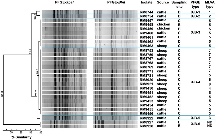
A PFGE analysis was conducted to identify the genetic relatedness in the recovered O157 isolates from the Culiacan Valley. The results indicated six unique PFGE types (Figure 2). Five of the six PFGE types were found in isolates recovered exclusively from one sampling site. The PFGE types X/B-1 and X/B-2 had a 96.3% similarity and were found in cattle isolates from sampling site D, and two closely-related types (X/B-3 and X/B-4) were identified in 82.1% (23/28) of the O157 isolates with >98% homology (Figure 2). In particular, the PFGE type X/B-4 was observed in 57.1% (16/28) of the O157 isolates from cattle and sheep feces from sampling site C alone. In contrast, isolates with the PFGE type X/B-3 were recovered from the three types of animal feces at sampling sites B, C, and D. The stx-negative O157 isolates RM8927 and RM8928 (PFGE type X/B-6) were recovered only from site E and showed the greatest divergence with only 61.8% similarity when compared to all other O157 isolates (Figure 2).
Figure 2. Analysis of the genetic relatedness among the O157 STEC isolates by PFGE and MLVA methods.
Dendograms of the combined PFGE types with XbaI and BnlI restriction enzymes were constructed using Bionumerics software v6.1 with the Dice coefficient and the UPGMA method. MLVA types were designated to each unique profile after analysis of variable tandem repeats in 11 genomic loci using Bionumerics software. ND, Not determined.
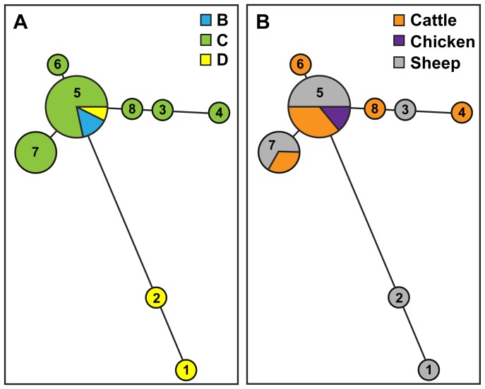
As an alternate method for further differentiating the O157:H7 STEC isolates, MLVA was performed to identify polymorphisms in 11 variable number tandem repeat (VNTR) loci. The results indicated that the 11-loci MLVA method was able to discriminate among O157:H7 isolates that were indistinguishable by PFGE analysis (Figure 2). Among the 26 E. coli O157:H7 isolates that were examined, eight unique profiles were identified by MLVA as compared to only five PFGE types. Furthermore, MLVA type 5 (MLVA profile 35-10-12-15-7-6-13-5-8-2-4; Table S1) was the most common, representing 53.8% (14/26) of the O157:H7 isolates, and MLVA type 7 (MLVA profile 34-10-12-14-7-6-13-5-8-2-4; Table S1) was identified in 23% (6/26) of the O157:H7 isolates (Figure 2). To examine the relatedness among the identified MLVA types, a minimum spanning tree was constructed (Figure 3). The analysis revealed that five MLVA types were unique to isolates recovered from sampling site C (Figure 3A) but from various sources (Figure 3B). Only isolates with MLVA type 5 were identified at multiple sampling sites (Figure 3A). Furthermore, the distantly-related MLVA types 1 and 2 were identified in sheep isolates from site D.
Figure 3. Phylogenetic relationships of MLVA types for the 26 O157:H7 STEC isolates.
A minimum-spanning tree of MLVA types was generated with Bionumerics software v6.1 and the Manhattan distance algorithm. Each circle in the tree represents a different MLVA type, and the number in the circle indicates the MLVA type number. The circle size corresponds to the number of isolates with an identical MLVA type. Colors represent the sampling sites (A) or the sampling source (B) for each MLVA type.
The MLVA assay also revealed that the genetic diversity varied considerably at some VNTR loci in the O157:H7 isolates. For example, the number of tandem repeats at Vhec1 locus varied from 21 to 45 for isolates RM8744 and RM9450, respectively (Table S1). By contrast, the number of tandem repeats at Vhec5 locus was 7 for all isolates examined (Table S1). To further quantify the variation in the number of repeats at each VNTR locus among the O157 isolates, Nei’s diversity index was calculated (Table 4). As indicated by the diversity index, the degree of polymorphism ranged from 0 to 0.61 (Table 4). Vhec1 was the most variable locus with a total of 7 alleles. Vhec4 locus had intermediate diversity with 3 alleles, whereas no diversity was observed for Vhec5 with only one allele. The remaining eight VNTR loci that were included in this MLVA method were of low diversity for this set of O157:H7 isolates with diversity index values ranging from 0.07 to 0.21 (Table 4).
Table 4. Characterization of 11 variable-number tandem repeat loci in the O157:H7 STEC isolates used in this study.
| Variable Number Tandem Repeat loci | |||||||||||
| Vhec 1 | Vhec 2 | Vhec 3 | Vhec 4 | Vhec 5 | Vhec 6 | Vhec 7 | O157-17 | O157-19 | O157-25 | O157-35 | |
| Number of alleles | 7 | 2 | 4 | 3 | 1 | 2 | 2 | 2 | 2 | 4 | 3 |
| Tandem repeats range | 21–45 | 9–10 | 8–12 | 14–17 | 7 | 6–9 | 7–13 | 3–5 | 7–8 | 1–5 | 4–8 |
| Null alleles | No | No | No | No | No | No | No | No | No | No | No |
| Nei’s diversity index1 | 0.61 | 0.14 | 0.21 | 0.41 | 0 | 0.14 | 0.14 | 0.14 | 0.07 | 0.21 | 0.15 |
Nei’s diversity index for each locus was calculated as 1-∑(allele frequency)2 [67].
Genotyping Non-O157 STEC Isolates by PFGE and MLVA
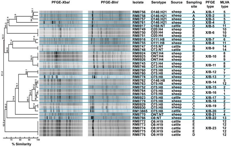
The PFGE analysis of 37 non-O157 STEC isolates revealed 23 distinct PFGE types with 61.4% similarity (Figure 4). Twenty-one of the PFGE types corresponded to non-O157 isolates that were recovered exclusively from only one of the sampling sites and from either cattle or sheep feces. Only PFGE types X/B-14 and X/B-23 were identified among non-O157 isolates from two different sampling sites (Figure 4). Some PFGE types were associated with a particular serotype. In particular, PFGE types X/B-6, X/B-11 and X/B-23 were only identified in isolates of serotypes O20:H4, O73:H4 and O8:H19, respectively, and predominantly from sampling site E. To further examine the genetic relatedness of the isolates, MLVA was performed by examining the polymorphisms in 10 genomic repeat-containing loci (Figure 4), targeting different loci than those used for the O157:H7 serotype (Table S2). The 10-loci MLVA identified 14 different MLVA types among the 37 non-O157 isolates analyzed and differentiated non-O157 isolates that could not be discriminated by PFGE (type X/B-14) (Figure 4). By contrast, this MLVA method was less discriminating than PFGE for distinguishing serotype O146:H21 isolates since the different PFGE types found in O146:H21 isolates were all MLVA type 5. MLVA type 10 was only identified in serotype O20:H4 isolates from sheep, and no other MLVA type was observed to be serotype specific (Figure 4). Multiple PFGE and MLVA types were identified in O75:H8 isolates, indicating that isolates with this serotype were the most diverse.
Figure 4. Analysis of the genetic relatedness among the non-O157 STEC isolates by PFGE and MLVA methods.
Dendograms of the combined PFGE types with XbaI and BnlI restriction enzymes were constructed using Bionumerics software v6.1 with the Dice coefficient and the UPGMA method. MLVA types were designated to each unique profile after analysis of tandem repeats in 10 genomic loci using Bionumerics software.
MLVA type 12 (MLVA profile 8-3-NA-8-3-10-1-15-NA-21; Table S2) was identified in 16.2% (6/37) of the non-O157 isolates. The other common MLVA types, MLVA type 2 (MLVA profile 6-3-NA-7-3-7-1-2-NA-15) and MLVA type 5 (MLVA profile 6-3-NA-8-3-4-1-2-NA-15), were each detected in 13.5% (5/37) of the non-O157isolates (Table S2). The number of alleles detected per locus ranged from 1 (locus CVN015) to 9 (locus CVN014) (Table 5). In the set of non-O157 isolates analyzed in this study, five loci (CVN002, CVN003, CVN016, CVN017 and CCR001) had null alleles (Tables 5 and S2). Interestingly, locus CVN003 could only be amplified in serotypes that were positive for the flagellin H4 antigen (O20:H4, O73:H4 and ONT:H4) (Table S2), suggesting either sequence polymorphisms at priming sites in serotypes that had null alleles or the absence of this locus. Further analysis of the allelic polymorphisms by calculating Nei’s diversity index revealed that three loci (CVN004, CVN014 and CVN016) were among the most diverse (Table 5). In particular, locus CVN014 was the most polymorphic locus, as has been observed in other studies [43]–[45]. The diversity index calculation also indicated that two loci had intermediate diversity (CVN001 and CCR001), whereas five loci resulted in low or no diversity (CVN002, CVN003, CVN007, CVN015 and CVN017) (Table 5).
Table 5. Characterization of 10 genomic-repeat containing loci in the non-O157 STEC isolates used in this study.
| Genomic repeat-containing loci | ||||||||||
| CVN001 | CVN002 | CVN003 | CVN004 | CVN007 | CVN014 | CVN015 | CVN016 | CVN017 | CCR001 | |
| Number of alleles | 3 | 3 | 3 | 6 | 2 | 9 | 1 | 6 | 2 | 3 |
| Tandem repeats range | 5–8 | 1–3 | 2–5 | 4–13 | 3–4 | 4–15 | 1 | 1–15 | 9 | 15–21 |
| Null alleles | No | Yes | Yes | No | No | No | No | Yes | Yes | Yes |
| Nei’s diversity index1 | 0.43 | 0.20 | 0.36 | 0.74 | 0.10 | 0.83 | 0 | 0.70 | 0.10 | 0.51 |
Nei’s diversity index for each locus was calculated as 1−∑(allele frequency)2 [67].
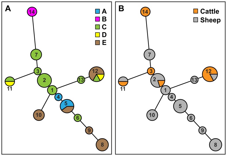
A minimum spanning tree of MLVA types indicated that MLVA type 14 was more distantly-related when compared to the other MLVA types (Figure 5). Furthermore, MLVA type 14 was only identified in isolates recovered from sampling site B (Figure 5A) and only from cattle feces (Figure 5B). The minimum spanning tree also revealed that sampling site C was a source of MLVA types 1, 2, 3, and 7 that were closely related (Figure 5A). Interestingly, sheep were a primary source of non-O157 isolates with diverse MLVA types (Figure 5B).
Figure 5. Phylogenetic relationships of MLVA types for the 37 non-O157 STEC isolates.
A minimum-spanning tree was generated with Bionumerics software v61 and the Manhattan distance algorithm. Each circle in the tree represents a different MLVA type, and the number in the circle indicates the MLVA type number. The circle size corresponds to the number of isolates with an identical MLVA type. Colors represent the sampling sites (A) or the sampling source (B) for each MLVA type.
Discussion
The present study investigated the prevalence of zoonotic STEC in small farms within rural communities, located in the agricultural Culiacan Valley along the coastal northwestern part of Mexico. The Culiacan Valley is considered to be one of the most important agricultural regions in Mexico [41]. The farms, examined in this study, were located in rural communities where livestock was often raised without an efficient management of animal wastes [42]. Thus, a lack of good agricultural practices could potentially contribute to the dispersal and transmission of pathogens throughout the environment. An understanding of the primary animal reservoirs in rural communities within an agricultural region would be imperative for the development of control measures of any risk factors that could lead to human infections with zoonotic STEC.
Only a limited number of studies have previously documented the prevalence of STEC in animal reservoirs in various locations in Mexico. The contamination of animal carcasses with O157:H7 and non-O157 STEC was detected in slaughter plants sampled in northeast and western Mexico [38], [39]. The recovered STEC isolates from these slaughter plants were found positive for the virulence factors intimin and hemolysin. A separate study confirmed the presence of E. coli O157:H7 in animal feces collected from dairy and beef cattle farms and from swine-farrowing facilities throughout Central Mexico [36]. Whether the recovered O157 isolates from the farms in Central Mexico harbor genes associated with severe human illness is still to be determined. Thus, additional studies are needed to identify the prevalence and phylogenetic relatedness of O157 and non-O157 STEC in other relevant agricultural regions of Mexico.
Given that cattle and other ruminants are considered relevant reservoirs of zoonotic STEC [1], [18], [19], [21], the present study is the first report that has examined the prevalence of O157 and non-O157 STEC from feces of domestic animals that were raised on the sampled rural farms in the agricultural Culiacan Valley. The results from this study indicated that O157 STEC were detected in 5.4% (13/240) and non-O157 STEC were detected in 7.1% (17/240) of the animal fecal samples. The observed STEC prevalence in farm animals in Mexico’s Culiacan Valley appears to be lower when compared to some countries. Moreover, a published report that comprehensively examined the worldwide prevalence of O157 and non-O157 in livestock feces reported significant wide ranges [46]. In particular, the prevalence of O157 ranged from 0.2% to 27.8% and of non-O157 ranged from 2.1% to 70.1% [46]. These observations have also indicated that multiple factors, including sampling locations, hosts, periods of study and methods, may contribute to the dynamic nature observed among studies of pathogen prevalence in animals [18], [35], [46], [47].
Due to the complexity of detecting and enumerating toxigenic E. coli in naturally-contaminated samples [18], [48], an enrichment method and plating on selective media was employed to increase the efficiency for isolating both O157 and non-O157 STEC. The use of the IMS method facilitated significantly higher recovery of isolates when compared to direct plating. The number of fecal samples that were positive for presumptive STEC using the IMS method was 2.1-times higher than those subjected to direct plating on selective media. These findings are in agreement with other studies that have reported an increased effectiveness in STEC detection in animal feces by using the IMS method [23], [49]–[51]. Furthermore, the increased recovery of STEC isolates that was observed during the second sampling period can be attributed to performing the enrichment step without antibiotics, a procedure proposed to increase the number of STEC cells before exposure to the selective conditions in the isolation media [52].
The O157:H7 serotype was identified in 40% (26/65) of the E. coli isolates that were recovered from the domestic animals sampled in this study. Most O157 isolates were recovered from cattle feces and sheep feces. Interestingly, two O157:H7 isolates were recovered from chicken feces, considered to be a rare animal source for this serotype [18]. Genotyping studies revealed that most O157:H7 isolates grouped into two PFGE 2-enzyme profiles with 98.6% homology, thus, the use of MLVA as a genotyping tool facilitated further discrimination of isolates that were indistinguishable by PFGE. Further analysis of the allelic polymorphisms of the 11 VNTR loci of the O157 MLVA system indicated that most alleles had low or intermediate diversity in the O157:H7 isolates from the Culiacan Valley. Vhec1 was the only VNTR locus that was identified to have high allelic diversity. This finding is in agreement with previous reports that documented a particularly high polymorphism at Vhec1 [53]–[55]. The limited allelic diversity in conjunction with the observation that all O157:H7 isolates had the same virulence profile (positive for stx2, eae, ehxA) suggests that the O157:H7 isolates from this region in Mexico are highly related. It also appears that a small number of genetic types may predominate and persist in different animal reservoirs on the rural farms. In particular, the O157:H7 sheep isolate RM8753 was recovered by direct plating, and six months later, genotypically-identical isolates by MLVA and PFGE were recovered by the IMS method from the same sampling site (site C), but from both sheep and cattle. This finding may suggest persistence of the O157:H7 isolates in the Culiacan Valley and a potential transmission between species.
The non-O157 serotypes O8:H19, O75:H8, O111:H8 and O146:H21 were identified in 35.4% (23/65) of the STEC isolates that were recovered in the Culiacan Valley. These relevant serotypes have been previously implicated with disease outbreaks or with severe symptoms in humans [3], [4], [19]. The non-O157 isolates were predominantly recovered from sheep and were identified to harbor either one or both stx genes. With the exception of five isolates, all other non-O157 isolates (86.5%, 32/37) were identified to be positive for ehxA. However, only 10.8% (4/37) of the non-O157 isolates harbored eae gene. Similar to what was observed in the analysis of O157:H7 isolates, a diverse set of non-O157 isolates was identified at sampling site C, located in a valley between two large urban cities (Culiacan and Navolato) and proximal to the mountain ranges along the east.
Among the non-O157 serotypes that were identified in this study, serotype O75:H8 was the most variable serotype by both PFGE and MLVA. The O75:H8 isolates, representing 18.5% (12/65) of the isolates recovered from the Culiacan Valley, were a common sheep serotype. Another sheep serotype was O146:H21. The predominant isolation of O75:H8 and O146:H21 isolates from sheep was in agreement with previous reports that documented the same animal source for these non-O157 serotypes in several countries such as Australia, Brazil, New Zealand, and Spain [16], [29], [30], [56], [57]. Moreover, to our knowledge, the present study is the first report of sheep feces as an animal source of STEC O111:H8, a serotype previously identified to be recovered exclusively from cattle [4], [56], [58], [59].
Genotypic analysis by PFGE and MLVA revealed that cattle isolates with serotype O8:H19 from the Culiacan Valley were closely related. By contrast, a high genotypic diversity was identified in O8:H19 isolates from beef products in Argentina [44]. In the present study, the low genomic diversity that was observed in the O8:H19 isolates may indicate that a prevalent genotype may be circulating in this geographical region in Mexico. Additional surveys would be needed to determine whether other STEC serotypes may also represent stable and predominant genotypes in this agricultural region in Mexico. Future work will investigate other sources of O157 and non-O157 STEC by examining wildlife and environmental samples (plants, soil, sediment, and irrigation water) to obtain a better understanding of pathogen diversity, persistence, and movement. Further assessment of additional molecular risk factors associated with STEC pathogenesis would also provide relevant information regarding the virulence potential of toxigenic E. coli recovered from the Culiacan Valley in Mexico.
Materials and Methods
Ethics Statement
No specific permits were obtained for the described field studies. Access to privately-owned land was provided after verbal agreement with land owners. The field studies did not involve endangered or protected species. No animals were harmed in the acquisition of fecal samples.
Sample Collection
From July 2008 to June 2009, a total of 240 fecal samples were collected from different sampling sites in the Culiacan Valley in Northwest Mexico. The five sampling sites, Jotagua (A), Agua Caliente (B), Cofradia de Navolato (C), Iraguato (D), and El Castillo (E), were selected to be 14–38 km apart and to represent the study area in the Culiacan Valley (Figure 1). Sites A and B were located in the mountain hillsides with a semi-humid climate. Sites C and D were in close proximity to the urban areas of Navolato and Culiacan municipalities, and site E was located nearest to the coast (Figure 1). Sites C, D, and E had a semi-arid climate. Each sampling site was visited every two weeks and consisted of small farms of about 20 to 50 farm animals that were located in rural communities nearby rivers used for irrigation, where livestock raising is a common activity among local families. A fecal sample was collected from each species of animal present at each sampling site. Approximately, 100 g of fecal samples from asymptomatic cattle and sheep and 30 g of fecal samples from chicken were collected from different areas within each sampling site by using a sterile spatula and gloves, and placed into labeled sterile plastic bags, as in previous studies [42]. All collected fecal samples were transported immediately under refrigeration to the Food and Environmental Microbiology Laboratory (CIAD, Culiacan, Mexico) and then processed within the next 6 h.
Isolation Method for STEC from Animal Fecal Samples
Fecal samples were collected during two sequential periods and were analyzed by separate isolation methods. From July to December 2008, STEC were isolated from 120 fecal samples by employing a direct plating method. A total of 25 g of animal feces were removed with sterile spatula and placing it in a sterile plastic bag. The sample was homogenized manually for 5 to 10 min, followed by addition of 225 ml of tryptic soy broth (TSB) (BD Bioxon, Nuevo Leon, Mexico), supplemented with 1.5 g/l bile salts (Sigma-Aldrich Chemie GmbH, Munich, Germany), 1.5 g/l dipotassium phosphate (Sigma-Aldrich), 0.0125 mg/l cefixime (Sigma-Aldrich), 10 mg/l cefsulodin sodium salt hydrate (Sigma-Aldrich), and 8 mg/l vancomycin hydrochloride (Sigma-Aldrich). The enrichment culture was incubated at 37°C for 18–24 h with constant shaking. A 50 µl aliquot of the enrichment cultures were spread on sorbitol MacConkey agar (CT-SMAC) (Difco, Detroit, MI, USA), containing 0.5 mg/l cefixime (Sigma-Aldrich) and 2.5 mg/l potassium tellurite hydrate (Sigma-Aldrich) [34] as well as on CHROMagar™ O157 (CHROMagar, Paris, France). Agar plates were then incubated for 18–24 h at 37°C. Presumptive colonies were selected based on purple colony color on CHROMagar or either a colorless or light gray color on CT-SMAC.
A total of 120 fecal samples were collected from January to June 2009 and were processed by including an IMS method, modified from a method described previously for isolation of O157 STEC from environmental samples [34]. Briefly, a total of 10 g of animal feces were homogenized as described above and were then added to 90 ml of TSB without antibiotics, as in previous studies [34]. The enrichment broths were incubated for 2 h at 25°C, then at 42°C for 8 h with constant shaking, and were held at 4°C until the following morning without shaking. A 500 µl sample of the enrichment culture and 500 µl of 1× phosphate buffer solution (PBS) were incubated with 10 µl of Dynabeads® anti-E. coli O157 magnetic beads (Life Technologies, Grand Island, NY, USA) for 30 min with constant mixing. The magnetic beads were then washed twice with PBS containing 0.05% Tween-20 by using a Dynal® BeadRetriever Tube Rack (Life Technologies) and finally resuspended in 100 µl of PBS. A 50 µl sample was plated on CHROMagar O157 and on Rainbow® Agar O157 (Biolog, Hayward, CA, USA). Presumptive colonies were selected based on purple color CHROMagar O157 and on the several colors for typical colonies on Rainbow Agar O157, as in previous studies [60].
Identification of Serotypes and Virulence Gene Profiles
To determine whether the isolates, grown on selective indicator media, were authentic E. coli O157, a colony immunoblot assay was performed. Presumptive colonies and positive control O157 STEC strain RM1484 were patched on plates of Difco™ Luria Bertani (LB) agar (Fisher Scientific, Pittsburg, PA, USA) by using a numbered grid and grown at 37°C for 18 h and were then blotted onto Whatman Protran® BA85 nitrocellulose membrane filter discs (0.45 µm pore size, VWR International, Radnor, PA, USA). The nitrocellulose membranes were washed, blocked, incubated with anti-O157 IgG monoclonal antibody 13B3, followed by incubation with alkaline phosphatase-conjugated goat anti-mouse IgG antibody, following previously described procedures [34]. Positive signals on the membranes were detected after incubation with SIGMAFAST™ BCIP®/NBT colorimetric substrates (Sigma-Aldrich) [34].
Positive colonies with the anti-O157 antibody were then examined by real-time polymerase chain reaction (RT-PCR) after amplification of a 497-bp region of the rfbE gene, encoding the O157 O-antigen transporter, as described elsewhere [34]. As template for the RT-PCR reaction, bacteria were transferred from the patch plate directly into the PCR tube by using a sterile toothpick. The RT-PCR reaction was performed in a Stratagene MX3000P Real-Time PCR machine (Agilent Technologies, Santa Clara CA, USA) with the following conditions: 95°C for 5 min, and 60 cycles of 95°C for 15 sec and 60°C for 45 sec. Samples were considered positive for rfbE gene when the cycle threshold value of the RT-PCR assay was below 20, as in previous studies [34].
Presumptive colonies on selective media with atypical colony colors for O157 STEC were analyzed further by PCR to detect the wzx and wzy genes in the O-antigen gene cluster of 10 non-O157 serogroups (O26, O45, O91, O103, O104, O111, O113, O121, O128, and O145) using PCR primers published previously [61]. All isolates were screened for the presence of the flagellin fliC H7 gene, and the virulence genes encoding Shiga toxins (stx1, stx2), intimin (eae), and enterohemolysin (exhA) [61]. Detection of fliC genes, targeting additional H-types (H2, H8, H1, H19, H21, and H28), was performed as described previously [62]. Detection of flagellin H4 antigen was performed with primers 5′-GATAACCAGACGATCAGCATTGG-3′ (forward) and 5′-CTTCCGCTGCACCAACAGT-3′ (reverse). PCR reagents were supplied by Promega Corporation (Madison, WI, USA), and PCR primers were purchased from Eurofins MWG Operon (Huntsville, AL, USA). As template for the PCR reaction, bacterial cultures of the isolates were grown aerobically in TSB broth (Difco) for 24 h with constant shaking (200 rpm) at 37°C, and 100 µl of the bacterial cultures were collected by centrifugation at 2000×g for 5 min. Cell pellets were resuspended in 100 µl of HyPure™ molecular biology-grade water (HyClone Laboratories, Inc., Logan, UT, USA) and incubated at 95°C for 20 min, as in previous studies [63]. The lysates were centrifuged at 2000×g for 5 min, and the supernatants were collected and frozen until further use. PCR amplifications were performed in a 25 µl reaction mixture, each containing 5 µl of the bacterial crude lysate, 0.5µM of each primer and 12.5 µl of 2× GoTaq® Green Master Mix (Promega Corporation). The reaction mixtures were placed in a Dyad Peltier Thermal Cycler (Bio-Rad Laboratories, Hercules, CA, USA) with the following settings: 5 min at 94°C, followed by 25 cycles of 45 sec at 94°C, 1 min 60°C, 1 min at 72°C, and a final extension time of 7 min at 72°C. Amplified products were analyzed in 2% agarose gels containing 0.04 µl/ml GelRed Nucleic Acid Stain (Phenix Research, Candler, NC, USA). When the PCR assays for O- antigen were inconclusive, bacterial isolates were sent to the E. coli Reference Center at Pennsylvania State University, University Park, PA, USA.
PFGE Molecular Subtyping
STEC isolates were typed with the standardized rapid PFGE protocol used by PulseNet laboratories, according to previous reports [64]. Isolates were grown for 14–18 h on LB agar and were resuspended in a cell suspension buffer (100 mM Tris, 100 mM EDTA, pH 8.0) to an OD600 0.8–1.0, and an agarose plug was prepared by mixing 400 µl of the cell suspension with 400 µl of 1% SeaKem® Gold agarose (Lonza, Rockland, ME, USA) and 20 µl of proteinase K (Roche Diagnostics, Indianapolis, IN, USA) [64]. Agarose-embedded cells were lysed, and intact genomic DNA was digested with either XbaI or BlnI restriction enzymes (Roche Diagnostics) for 4 h at 37°C. The fragments were separated with a CHEF Mapper™ Pulsed-Field Electrophoresis System (BioRad, Hercules, CA, USA) for 20.5 h with switch times at 6 V/cm ranging from 2.16 to 54.17 s. The PulseNet universal standard strain, Salmonella enterica Braenderup H9812, was used as a molecular reference marker [64]. Gels were stained with ethidium bromide and were visualized under UV trans-illumination (Alpha Innotech Corp., San Leandro, CA, USA). PFGE profiles of the isolates with both digests were compared by performing band matching, and dendograms were constructed using Bionumerics software version 6.1 (Applied Maths, Austin, TX, USA) with the Dice coefficient and the unweighted pair-group method with arithmetic mean (UPGMA).
MLVA Molecular Subtyping
MLVA typing of O157 STEC isolates was performed by using capillary electrophoresis after amplification of 11 VNTR loci in three multiplex PCR reactions [34], [53], [65]. Multiplex PCR forward primers were 5′-fluorescently labeled with 6FAM, NED, and VIC dyes [34], and all primers were purchased from Applied Biosystems-Life Technologies (Carlsbad, CA, USA). PCR reaction 1 amplified VNTR loci Vhec1, Vhec3, Vhec4, and Vhec5, and reaction 2 amplified VNTR loci Vhec1, Vhec2, Vhec6, and Vhec7, as in previous studies [34], [65]. VNTR loci O157–17, O157–19, O157–25, and O157–37 were amplified in reaction 3 [53]. PCR reaction mix and thermal cycling parameters were performed as described in a previous study [34]. For performing the MLVA typing in non-O157 STEC, ten genomic-repeat containing loci were amplified in four multiplex PCR reactions with fluorescent dye-conjugated primers [45], [66]. VNTR loci CVN003 and CVN014 were amplified in reaction 1, and loci CVN001, CVN004, CVN007 and CVN015 were amplified in reaction 2, as described previously [45]. PCR reaction 3 only amplified CVN002 locus [45], and loci CVN016, CVN017 and CCR001 were amplified in reaction 4 [66]. PCR reaction mix and thermal cycling parameters were performed as described in a previous study [45], [66].
After the PCR amplifications, the multiplex reactions for O157 STEC were pooled and diluted 1∶50 into distilled water. For non-O157 STEC, the multiplex PCR reactions were pooled as follows: 10 µl of reaction 1, 1.75 µl of reaction 2 and 5 µl of reaction 3. One µl of the pooled PCR reactions was added to 12 µl HiDi formamide (Applied Biosystems-Life Technologies) and 0.08 µl of ROX-labeled MapMarker 1000 size standard (Bioventures, Inc., Murfreesboro, TN). For reaction 4, 1 µl of a 1∶50 dilution was added to 10 µL HiDi formamide (Applied Biosystems-Life Technologies) and 0.04 µL of ROX-labeled MapMarker 1000 size standard (Bioventures, Inc.). The mixtures were size-fractionated with the Applied Biosystems 3130xl Genetic Analyzer, and the fragments sizes were determined with the GeneMapper® software v3.7 (Applied Biosystems-Life Technologies) and were then converted to number of tandem repeats [34], [53]. Each new multiple of tandem repeat was assigned to a distinct allele number, and the allele numbers per locus were determined using BioNumerics software v6.1 to designate MLVA types to each unique MLVA profile. For data analysis, an arbitrary number (−2) was assigned to null alleles, where no amplification product was detected [53]. Cluster analysis and minimal spanning trees of MLVA types were constructed using BioNumerics software v6.1 and the Manhattan distance algorithm to determine the genetic relationships among the isolates. The Nei’s marker diversity index, ranging from 0 (no diversity) to 1 (complete diversity), was calculated for each locus by using the equation 1−∑(allele frequency)2 [67], as in previous studies [55], [68]. Null alleles were included in the diversity index calculation.
Supporting Information
MLVA profiles for each unique MLVA type identified in the STEC O157:H7 isolates used in this study.
(DOCX)
MLVA profiles for each unique MLVA type identified in the STEC non-O157 isolates used in this study.
(DOCX)
Acknowledgments
The authors would like to thank Michelle S. Swimley, Kimberly Nguyen, Diana Carychao, Samarpita Fontanoz, and Ronak N. Patel (USDA-ARS-WRRC in Albany, CA) as well as Célida Martínez, and Gabriela Gaxiola (CIAD in Culiacan, Sinaloa, Mexico) for excellent technical assistance. Dr. Jennifer L. Kyle is greatly acknowledged for critical reading of the manuscript.
Funding Statement
This work was supported by the U.S. Department of Agriculture, Agricultural Research Service CRIS project number 5325-42000-047 and by a Postgraduate Studies Scholarship from The National Council of Science and Technology in Mexico (CONACyT grant #234885) to BAAL. The funders had no role in study design, data collection and analysis, decision to publish, or preparation of the manuscript.
References
- 1. Gyles CL (2007) Shiga toxin-producing Escherichia coli: an overview. J Anim Sci 85: E45–62. [DOI] [PubMed] [Google Scholar]
- 2.Karmali MA (2009) Host and pathogen determinants of verocytotoxin-producing Escherichia coli-associated hemolytic uremic syndrome. Kidney Int Suppl: S4–7. [DOI] [PubMed]
- 3. Brooks JT, Sowers EG, Wells JG, Greene KD, Griffin PM, et al. (2005) Non-O157 Shiga toxin-producing Escherichia coli infections in the United States, 1983–2002. J Infect Dis 192: 1422–1429. [DOI] [PubMed] [Google Scholar]
- 4. Bettelheim KA (2007) The non-O157 Shiga-toxigenic (verocytotoxigenic) Escherichia coli; under-rated pathogens. Crit Rev Microbiol 33: 67–87. [DOI] [PubMed] [Google Scholar]
- 5. Caprioli A, Morabito S, Brugère H, Oswald E (2005) Enterohaemorrhagic Escherichia coli: emerging issues on virulence and modes of transmission. Vet Res 36: 289–311. [DOI] [PubMed] [Google Scholar]
- 6. Mathusa EC, Chen Y, Enache E, Hontz L (2010) Non-O157 Shiga toxin-producing Escherichia coli in foods. J Food Prot 73: 1721–1736. [DOI] [PubMed] [Google Scholar]
- 7. Beutin L, Martin A (2012) Outbreak of Shiga toxin-producing Escherichia coli (STEC) O104:H4 infection in Germany causes a paradigm shift with regard to human pathogenicity of STEC strains. J Food Prot 75: 408–418. [DOI] [PubMed] [Google Scholar]
- 8. Bolton DJ (2011) Verocytotoxigenic (Shiga toxin-producing) Escherichia coli: virulence factors and pathogenicity in the farm to fork paradigm. Foodborne Pathog Dis 8: 357–365. [DOI] [PubMed] [Google Scholar]
- 9. Coombes BK, Gilmour MW, Goodman CD (2011) The evolution of virulence in non-O157 Shiga toxin-producing Escherichia coli . Front Microbiol 2: 90. [DOI] [PMC free article] [PubMed] [Google Scholar]
- 10. Jerse AE, Yu J, Tall BD, Kaper JB (1990) A genetic locus of enteropathogenic Escherichia coli necessary for the production of attaching and effacing lesions on tissue culture cells. Proc Natl Acad Sci U S A 87: 7839–7843. [DOI] [PMC free article] [PubMed] [Google Scholar]
- 11. Frankel G, Phillips AD, Rosenshine I, Dougan G, Kaper JB, et al. (1998) Enteropathogenic and enterohaemorrhagic Escherichia coli: more subversive elements. Mol Microbiol 30: 911–921. [DOI] [PubMed] [Google Scholar]
- 12. Ethelberg S, Olsen KE, Scheutz F, Jensen C, Schiellerup P, et al. (2004) Virulence factors for hemolytic uremic syndrome, Denmark. Emerg Infect Dis 10: 842–847. [DOI] [PMC free article] [PubMed] [Google Scholar]
- 13. Cookson AL, Bennett J, Thomson-Carter F, Attwood GT (2007) Molecular subtyping and genetic analysis of the enterohemolysin gene (ehxA) from Shiga toxin-producing Escherichia coli and atypical enteropathogenic E. coli . Appl Environ Microbiol 73: 6360–6369. [DOI] [PMC free article] [PubMed] [Google Scholar]
- 14. Boerlin P, McEwen SA, Boerlin-Petzold F, Wilson JB, Johnson RP, et al. (1999) Associations between virulence factors of Shiga toxin-producing Escherichia coli and disease in humans. J Clin Microbiol 37: 497–503. [DOI] [PMC free article] [PubMed] [Google Scholar]
- 15. Schmidt H, Karch H (1996) Enterohemolytic phenotypes and genotypes of Shiga toxin-producing Escherichia coli O111 strains from patients with diarrhea and hemolytic-uremic syndrome. J Clin Microbiol 34: 2364–2367. [DOI] [PMC free article] [PubMed] [Google Scholar]
- 16. Djordjevic SP, Ramachandran V, Bettelheim KA, Vanselow BA, Holst P, et al. (2004) Serotypes and virulence gene profiles of Shiga toxin-producing Escherichia coli strains isolated from feces of pasture-fed and lot-fed sheep. Appl Environ Microbiol 70: 3910–3917. [DOI] [PMC free article] [PubMed] [Google Scholar]
- 17. Hornitzky MA, Vanselow BA, Walker K, Bettelheim KA, Corney B, et al. (2002) Virulence properties and serotypes of Shiga toxin-producing Escherichia coli from healthy Australian cattle. Appl Environ Microbiol 68: 6439–6445. [DOI] [PMC free article] [PubMed] [Google Scholar]
- 18. Ferens WA, Hovde CJ (2011) Escherichia coli O157:H7: animal reservoir and sources of human infection. Foodborne Pathog Dis 8: 465–487. [DOI] [PMC free article] [PubMed] [Google Scholar]
- 19. Hussein HS (2007) Prevalence and pathogenicity of Shiga toxin-producing Escherichia coli in beef cattle and their products. J Anim Sci 85: E63–72. [DOI] [PubMed] [Google Scholar]
- 20. Karmali MA, Gannon V, Sargeant JM (2010) Verocytotoxin-producing Escherichia coli (VTEC). Vet Microbiol 140: 360–370. [DOI] [PubMed] [Google Scholar]
- 21. La Ragione RM, Best A, Woodward MJ, Wales AD (2009) Escherichia coli O157:H7 colonization in small domestic ruminants. FEMS Microbiol Rev 33: 394–410. [DOI] [PubMed] [Google Scholar]
- 22. Chapman PA, Siddons CA, Gerdan Malo AT, Harkin MA (1997) A 1-year study of Escherichia coli O157 in cattle, sheep, pigs and poultry. Epidemiol Infect 119: 245–250. [DOI] [PMC free article] [PubMed] [Google Scholar]
- 23. Heuvelink AE, van den Biggelaar FL, de Boer E, Herbes RG, Melchers WJ, et al. (1998) Isolation and characterization of verocytotoxin-producing Escherichia coli O157 strains from Dutch cattle and sheep. J Clin Microbiol 36: 878–882. [DOI] [PMC free article] [PubMed] [Google Scholar]
- 24. Meng J, Zhao S, Doyle MP (1998) Virulence genes of Shiga toxin-producing Escherichia coli isolated from food, animals and humans. Int J Food Microbiol 45: 229–235. [DOI] [PubMed] [Google Scholar]
- 25. Fegan N, Desmarchelier P (1999) Shiga toxin-producing Escherichia coli in sheep and pre-slaughter lambs in eastern Australia. Lett Appl Microbiol 28: 335–339. [DOI] [PubMed] [Google Scholar]
- 26. Ogden ID, MacRae M, Strachan NJ (2005) Concentration and prevalence of Escherichia coli O157 in sheep faeces at pasture in Scotland. J Appl Microbiol 98: 646–651. [DOI] [PubMed] [Google Scholar]
- 27. Djordjevic SP, Hornitzky MA, Bailey G, Gill P, Vanselow B, et al. (2001) Virulence properties and serotypes of Shiga toxin-producing Escherichia coli from healthy Australian slaughter-age sheep. J Clin Microbiol 39: 2017–2021. [DOI] [PMC free article] [PubMed] [Google Scholar]
- 28. Cookson AL, Hayes CM, Pearson GR, Roe JM, Wales AD, et al. (2002) Isolation from a sheep of an attaching and effacing Escherichia coli O115:H− with a novel combination of virulence factors. J Med Microbiol 51: 1041–1049. [DOI] [PubMed] [Google Scholar]
- 29. Cookson AL, Taylor SC, Bennett J, Thomson-Carter F, Attwood GT (2006) Serotypes and analysis of distribution of Shiga toxin-producing Escherichia coli from cattle and sheep in the lower North Island, New Zealand. N Z Vet J 54: 78–84. [DOI] [PubMed] [Google Scholar]
- 30. Blanco M, Blanco JE, Mora A, Rey J, Alonso JM, et al. (2003) Serotypes, virulence genes, and intimin types of Shiga toxin (verotoxin)-producing Escherichia coli isolates from healthy sheep in Spain. J Clin Microbiol 41: 1351–1356. [DOI] [PMC free article] [PubMed] [Google Scholar]
- 31. Aktan I, Sprigings KA, La Ragione RM, Faulkner LM, Paiba GA, et al. (2004) Characterisation of attaching-effacing Escherichia coli isolated from animals at slaughter in England and Wales. Vet Microbiol 102: 43–53. [DOI] [PubMed] [Google Scholar]
- 32. Kalchayanand N, Arthur TM, Bosilevac JM, Brichta-Harhay DM, Guerini MN, et al. (2007) Microbiological characterization of lamb carcasses at commercial processing plants in the United States. J Food Prot 70: 1811–1819. [DOI] [PubMed] [Google Scholar]
- 33. Rey J, Sánchez S, Blanco JE, Hermoso de Mendoza J, Hermoso de Mendoza M, et al. (2006) Prevalence, serotypes and virulence genes of Shiga toxin-producing Escherichia coli isolated from ovine and caprine milk and other dairy products in Spain. Int J Food Microbiol 107: 212–217. [DOI] [PubMed] [Google Scholar]
- 34. Cooley M, Carychao D, Crawford-Miksza L, Jay MT, Myers C, et al. (2007) Incidence and tracking of Escherichia coli O157:H7 in a major produce production region in California. PLoS One 2: e1159. [DOI] [PMC free article] [PubMed] [Google Scholar]
- 35.Mandrell RE (2011) Tracing pathogens in fruit and vegetable production chains. In: Brul S, Fratamico PM, McMeekin T, editors. Tracing Pathogens in the Food Chain. Philadelphia, PA: Woodhead Publishing. 548–595.
- 36. Callaway TR, Anderson RC, Tellez G, Rosario C, Nava GM, et al. (2004) Prevalence of Escherichia coli O157 in cattle and swine in central Mexico. J Food Prot 67: 2274–2276. [DOI] [PubMed] [Google Scholar]
- 37. Sosa Hernández LA, Del Río García JC, Córdova Ponce R, Cuenca Verde NM, Valdivia Lara EG, et al. (2008) Isolation in Mexico of Shiga (Stx) toxin-producing Escherichia coli and Salmonella enterica from turtles (Trachemis scripta) maintained in captivity. RECVET-Revista Electrónica de Clínica Veterinaria III: 1–13. [Google Scholar]
- 38. Varela-Hernández JJ, Cabrera-Diaz E, Cardona-López MA, Ibarra-Velázquez LM, Rangel-Villalobos H, et al. (2007) Isolation and characterization of Shiga toxin-producing Escherichia coli O157:H7 and non-O157 from beef carcasses at a slaughter plant in Mexico. Int J Food Microbiol 113: 237–241. [DOI] [PubMed] [Google Scholar]
- 39. Gallegos M, Morales A, Álvarez G, Vásquez J, Morales L, et al. (2009) Characterization of Escherichia coli O157:H7 isolates obtained from bovine and porcine carcasses. Revista Científica-FCV-LUZ XIX: 139–146. [Google Scholar]
- 40. López-Saucedo C, Cerna JF, Estrada-García T (2010) Non-O157 Shiga toxin-producing Escherichia coli is the most prevalent diarrheagenic Escherichia coli pathotype in street-vended taco dressings in Mexico City. Clin Infect Dis 50: 450–451. [DOI] [PubMed] [Google Scholar]
- 41.Siller-Cepeda JH, Chaidez-Quiroz C, Castro-del Campo N (2009) Produce contamination issues in Mexico and Central America. In: Sapers GM, Solomon EB, Matthews KR, editors. The Produce Contamination Problem: Causes and Solutions. Burlington, MA: Elsevier. 309–326.
- 42. Jiménez M, Martínez-Urtaza J, Chaidez C (2011) Geographical and temporal dissemination of salmonellae isolated from domestic animal hosts in the Culiacan Valley, Mexico. Microb Ecol 61: 811–820. [DOI] [PubMed] [Google Scholar]
- 43. Bustamante AV, Sanso AM, Lucchesi PM, Parma AE (2010) Genetic diversity of O157:H7 and non-O157 verocytotoxigenic Escherichia coli from Argentina inferred from multiple-locus variable-number tandem repeat analysis (MLVA). Int J Med Microbiol 300: 212–217. [DOI] [PubMed] [Google Scholar]
- 44. Franci T, Sanso AM, Bustamante AV, Lucchesi PM, Parma AE (2011) Genetic characterization of non-O157 verocytotoxigenic Escherichia coli isolated from raw beef products using multiple-locus variable-number tandem repeat analysis. Foodborne Pathog Dis 8: 1019–1023. [DOI] [PubMed] [Google Scholar]
- 45. Lindstedt BA, Brandal LT, Aas L, Vardund T, Kapperud G (2007) Study of polymorphic variable-number of tandem repeats loci in the ECOR collection and in a set of pathogenic Escherichia coli and Shigella isolates for use in a genotyping assay. J Microbiol Methods 69: 197–205. [DOI] [PubMed] [Google Scholar]
- 46. Hussein HS, Bollinger LM (2005) Prevalence of Shiga toxin-producing Escherichia coli in beef cattle. J Food Prot 68: 2224–2241. [DOI] [PubMed] [Google Scholar]
- 47. Doane CA, Pangloli P, Richards HA, Mount JR, Golden DA, et al. (2007) Occurrence of Escherichia coli O157:H7 in diverse farm environments. J Food Prot 70: 6–10. [DOI] [PubMed] [Google Scholar]
- 48. Muniesa M, Jofre J, García-Aljaro C, Blanch AR (2006) Occurrence of Escherichia coli O157:H7 and other enterohemorrhagic Escherichia coli in the environment. Environ Sci Technol 40: 7141–7149. [DOI] [PubMed] [Google Scholar]
- 49. Wright DJ, Chapman PA, Siddons CA (1994) Immunomagnetic separation as a sensitive method for isolating Escherichia coli O157 from food samples. Epidemiol Infect 113: 31–39. [DOI] [PMC free article] [PubMed] [Google Scholar]
- 50. Chapman PA, Wright DJ, Siddons CA (1994) A comparison of immunomagnetic separation and direct culture for the isolation of verocytotoxin-producing Escherichia coli O157 from bovine faeces. J Med Microbiol 40: 424–427. [DOI] [PubMed] [Google Scholar]
- 51. Verstraete K, De Zutter L, Messens W, Herman L, Heyndrickx M, et al. (2010) Effect of the enrichment time and immunomagnetic separation on the detection of Shiga toxin-producing Escherichia coli O26, O103, O111, O145 and sorbitol positive O157 from artificially inoculated cattle faeces. Vet Microbiol 145: 106–112. [DOI] [PubMed] [Google Scholar]
- 52. Hussein HS, Bollinger LM (2008) Influence of selective media on successful detection of Shiga toxin-producing Escherichia coli in food, fecal, and environmental samples. Foodborne Pathog Dis 5: 227–244. [DOI] [PubMed] [Google Scholar]
- 53. Hyytiä-Trees E, Smole SC, Fields PA, Swaminathan B, Ribot EM (2006) Second generation subtyping: A proposed PulseNet protocol for multiple-locus variable-number tandem repeat analysis of Shiga toxin-producing Escherichia coli O157 (STEC O157). Foodborne Pathogens and Disease 3: 118–131. [DOI] [PubMed] [Google Scholar]
- 54. Keys C, Kemper S, Keim P (2005) Highly diverse variable number tandem repeat loci in the E. coli O157:H7 and O55:H7 genomes for high-resolution molecular typing. J Appl Microbiol 98: 928–940. [DOI] [PubMed] [Google Scholar]
- 55. Noller AC, McEllistrem MC, Pacheco AG, Boxrud DJ, Harrison LH (2003) Multilocus variable-number tandem repeat analysis distinguishes outbreak and sporadic Escherichia coli O157:H7 isolates. J Clin Microbiol 41: 5389–5397. [DOI] [PMC free article] [PubMed] [Google Scholar]
- 56. Brett KN, Ramachandran V, Hornitzky MA, Bettelheim KA, Walker MJ, et al. (2003) stx1c is the most common Shiga toxin 1 subtype among Shiga toxin-producing Escherichia coli isolates from sheep but not among isolates from cattle. J Clin Microbiol 41: 926–936. [DOI] [PMC free article] [PubMed] [Google Scholar]
- 57. Vettorato MP, Leomil L, Guth BE, Irino K, Pestana de Castro AF (2003) Properties of Shiga toxin-producing Escherichia coli (STEC) isolates from sheep in the State of São Paulo, Brazil. Vet Microbiol 95: 103–109. [DOI] [PubMed] [Google Scholar]
- 58. Hornitzky MA, Bettelheim KA, Djordjevic SP (2001) The detection of Shiga toxin-producing Escherichia coli in diagnostic bovine faecal samples using vancomycin-cefixime-cefsulodin blood agar and PCR. FEMS Microbiol Lett 198: 17–22. [DOI] [PubMed] [Google Scholar]
- 59. Tristão LC, Gonzalez AG, Coutinho CA, Cerqueira AM, Gomes MJ, et al. (2007) Virulence markers and genetic relationships of Shiga toxin-producing Escherichia coli strains from serogroup O111 isolated from cattle. Vet Microbiol 119: 358–365. [DOI] [PubMed] [Google Scholar]
- 60. Fratamico PM, Bagi LK, Cray WC Jr, Narang N, Yan X, et al. (2011) Detection by multiplex real-time polymerase chain reaction assays and isolation of Shiga toxin-producing Escherichia coli serogroups O26, O45, O103, O111, O121, and O145 in ground beef. Foodborne Pathog Dis 8: 601–607. [DOI] [PubMed] [Google Scholar]
- 61. Quiñones B, Swimley MS, Narm K-E, Patel RN, Cooley MB, et al. (2012) O-antigen and virulence profiling of Shiga toxin-producing Escherichia coli by a rapid and cost-effective DNA microarray colorimetric method. Front Cell Inf Microbio 2: 61 doi: 10.3389/fcimb.2012.00061. [DOI] [PMC free article] [PubMed] [Google Scholar]
- 62. Bugarel M, Beutin L, Martin A, Gill A, Fach P (2010) Micro-array for the identification of Shiga toxin-producing Escherichia coli (STEC) seropathotypes associated with Hemorrhagic Colitis and Hemolytic Uremic Syndrome in humans. Int J Food Microbiol 142: 318–329. [DOI] [PubMed] [Google Scholar]
- 63. Quiñones B, Swimley MS, Taylor AW, Dawson ED (2011) Identification of Escherichia coli O157 by using a novel colorimetric detection method with DNA microarrays. Foodborne Pathog Dis 8: 705–711. [DOI] [PMC free article] [PubMed] [Google Scholar]
- 64. Ribot EM, Fair MA, Gautom R, Cameron DN, Hunter SB, et al. (2006) Standardization of pulsed-field gel electrophoresis protocols for the subtyping of Escherichia coli O157:H7, Salmonella, and Shigella for PulseNet. Foodborne Pathog Dis 3: 59–67. [DOI] [PubMed] [Google Scholar]
- 65. Lindstedt BA, Vardund T, Kapperud G (2004) Multiple-Locus Variable-Number Tandem-Repeats Analysis of Escherichia coli O157 using PCR multiplexing and multi-colored capillary electrophoresis. J Microbiol Methods 58: 213–222. [DOI] [PubMed] [Google Scholar]
- 66. Løbersli I, Haugum K, Lindstedt BA (2012) Rapid and high resolution genotyping of all Escherichia coli serotypes using 10 genomic repeat-containing loci. J Microbiol Methods 88: 134–139. [DOI] [PubMed] [Google Scholar]
- 67. Nei M (1973) Analysis of gene diversity in subdivided populations. Proc Natl Acad Sci U S A 70: 3321–3323. [DOI] [PMC free article] [PubMed] [Google Scholar]
- 68. Lindstedt BA, Heir E, Gjernes E, Vardund T, Kapperud G (2003) DNA fingerprinting of Shiga-toxin producing Escherichia coli O157 based on Multiple-Locus Variable-Number Tandem-Repeats Analysis (MLVA). Ann Clin Microbiol Antimicrob 2: 12. [DOI] [PMC free article] [PubMed] [Google Scholar]
Associated Data
This section collects any data citations, data availability statements, or supplementary materials included in this article.
Supplementary Materials
MLVA profiles for each unique MLVA type identified in the STEC O157:H7 isolates used in this study.
(DOCX)
MLVA profiles for each unique MLVA type identified in the STEC non-O157 isolates used in this study.
(DOCX)


