

Abstract
Objectives
The aim was to investigate the effect of changes in horizontal X-ray beam angulation in intraoral radiography on the detection accuracy of furcation defects in the mandibular first molar, and to examine the anatomical relationship between the roots and furcation area as a possible cause of changes in detectability.
Methods
Simulated furcation defects with various depths were created in five mandibular first molars. Intraoral radiographs were taken at various horizontal angulations of the projection beams. The diagnostic accuracies were determined based on receiver operating characteristic analysis. The geometric relationship that might influence the accuracy was investigated through use of a compact cone beam CT in 59 first molar areas.
Results
Although the horizontal angulations showing the highest accuracies were shifted mesially, no differences were found between the angles of −10° and 20°. The relationship between the roots and the furcation area was relevant to the range of angulations showing high detectabilities.
Conclusions
The angulations traditionally used for detecting proximal caries are also suitable for detecting furcation defects.
Keywords: radiography, dental; furcation defects; molar; receiver operating characteristic curve; cone beam computed tomography
Introduction
As furcation involvement in molars is one of the major problems in periodontal treatment1 and is directly related to the survival of the teeth,2 it should be detected accurately at an early stage. Usually, it is assessed by using a probing procedure, but the anatomical complexity in this region often reduces its accuracy.3, 4 Although radiographic diagnosis is likely to underestimate the furcation bone loss in comparison with the probing procedure,4, 5 intraoral radiography plays an adjunctive role in the diagnosis.4, 6 It is recommended that intraoral radiographs should be taken at different X-ray beam angles to reduce the risk of missing furcation involvement.6 However, there are no recommendations for suitable angulations to detect furcation involvement. Moreover, the influence of change in angulation has not been investigated in the detection of bifurcation involvement of the mandibular molars.
Even for furcation disease, the horizontal angle of the X-ray beam is recommended to be tangential to the interproximal surface of the contact point.6 Several studies confirm that this traditional horizontal angulation shows the best detectability in the evaluation of proximal caries. However, there have been no studies verifying that the best angulation for proximal caries is also suitable for detecting bifurcation involvement in the mandibular first molar.
Diagnostic accuracies are generally determined with receiver operating characteristic (ROC) analysis, which requires a gold standard, but only a few studies have addressed the furcation involvement in intraoral radiography.7, 8 This may be partially attributed to the difficulty of verifying the actual size of bony defects. A recently available compact cone beam CT (CBCT) apparatus enables measurement of the actual size9 and can provide a sufficent gold standard.
On the other hand, the configuration of the furcation area10–12 shows wide variations, and the geometrical relationship between the mesial and distal roots differs among subjects.13, 14 This relationship may influence the detectability of a furcation defect. CBCT can also clearly depict this relationship.9
The purposes of the present study were to find the effective horizontal X-ray beam angulation of intraoral radiography for the detection of artificially created furcation defects of the mandibular first molar and to examine the geometric relationship between the roots and furcation area based on an investigation with compact CBCT.
Materials and methods
Furcation disease model
Three adult dried mandibles (age and gender unknown) with bilateral first molars were used. No periodontal bone loss was found around the first molars. These molars were extracted carefully to preserve the periodontal bony structures. Five of them were successfully extracted without any fractures and were used in the following experiments. They could be easily reinserted into the extraction socket. One molar was excluded because of irrecoverable damage.
The present study was carried out according to the method described by Gürgan et al,8 which is the only previous study based on artificially created mandibular furcation defects. Simulated furcation defects were created step-by-step from the buccal side in the interradicular bone of the first molars with a 1 mm round burr, to mimic the clinical progression of plaque-induced periodontal bone defect12, 15 (Figure 1). Initially, the buccal bony structure, including the cortical plate, was shaved vertically until the buccolingual width of the remaining plate reached approximately 2 mm (Figure 1). However, it was stopped at the highest level where the furcation entrance could be seen from the horizontal plane regardless of the plate thickness. The interradicular bone was then cut horizontally from the buccal furcation entrance to the lingual cortical plate with a vertical depth of 2 mm (Figure 1). Whenever new cutting with an approximately 1 mm reduction of bone was performed, intraoral radiographs were taken in various directions with a special device, described below. The number of radiographs obtained differed among the five molar areas used for this experiment because of the difference in the buccolingual width of the five areas. To measure the actual size and shape of the bone defects, three-dimensional images were obtained simultaneously using a compact CBCT unit (3DX multi-image micro CT, Morita, Kyoto Japan) with exposure conditions of 80 kV and 1.2 mA. The imaging area had a cylindrical volume, with radius 20 mm and height 30 mm. The spatial resolution was set to 0.125 mm. A dried mandible was set on a specially fabricated table and the first molar was positioned at the centre of the imaging area.
Figure 1.

Preparation of furcation defects and a typical image obtained by compact cone beam CT
Acquisition of intraoral radiographs
Radiographs were taken using an MTX-90 dental radiograph machine (Asahi Roentgen Ind. Co., Ltd., Kyoto, Japan) at 60 kV and 5.25 mAs, and with a 1.5 mm Al filter. The films used were Insight IP-22 (Eastman Kodak Co., Rochester, NY). All radiographs were processed immediately after exposure with an automatic processor (Excel, DENT-X Corporation, Elmsford, NY). All the processing chemicals and machines were properly managed by radiological technicians.
A device was fabricated to standardize the projection angle, and consisted of a wooden base, two rings for fixing the cone and a rotation table (Figure 2). The projection angle was determined using the rotation table with reference to a protractor. The mandible, a soft tissue equivalent material and a film-fixing device made of acrylic resin were mounted on the rotation table by adhesive tape. The mandibular occlusal plane was adjusted horizontally. A film was placed perpendicular to the occlusal plane and parallel to the line connecting the interproximal contact points of the first molar. The axis of horizontal rotation was set at the furcation entrance of the first molar. The distance between the focus and film was set at 37.5 cm. An angle of 0° was defined as when the centre of the X-ray beam was directed at a right angle to the film through the rotation axis. The projection angle was varied horizontally from −30° to +30° using the rotation table. A positive angle was defined as a projection from the mesially shifted position. Radiographs were taken every 5°, and 13 radiographs were obtained for each step of bone reduction (Figure 3).
Figure 2.

Device for standardizing the projection angle
Figure 3.

Examples of radiographs obtained at different angulations. A furcation defect with Grade 1 depth between the roots of the first molar is more clearly observed on the radiograph obtained at 10° angulation (b) than on that at 0° angulation (a). Note the difference in the findings regarding the proximal surface between the first and second molars
Evaluation of radiographs
Simulated defects were classified into four grades according to the degree of bone loss determined by CT examinations (Figure 1) with reference to the classification suggested by Hamp et al16 and Tarnow and Fletcher.12 This classification was carried out by two examiners (TH and MN) using i-View software (Morita, Kyoto, Japan). Almost all defects could be classified into the same grade between the two examiners. When their opinions were different, the final decisions were reached by consensus after discussion. These classifications were used as the gold standard for ROC analyses.
The definitions are as follows:
Grade 0: Absorption is seen only in the buccal cortical bone and is not observed in the interradicular bone.
Grade 1: Interradicular bone absorption is less than one-third of the buccolingual width of the tooth crown.
Grade 2: Absorption is greater than one-third but less than two-thirds of the buccolingual width of the tooth crown.
Grade 3: Absorption is greater than two-thirds of the buccolingual width of the tooth crown.
With the CT verifications of 5 molar areas, 19, 14, 16 and 17 defects were classified as Grades 0, 1, 2 and 3, respectively. Consequently, a total of 247 radiographs were obtained at 13 different angulations of 19 defects for Grade 0. A total of 182, 208 and 221 radiographs were obtained for Grades 1, 2 and 3, respectively.
All radiographs were randomly set in paper mounting frames (Hanshin, Osaka, Japan). Images were evaluated on a light box without magnification. Five observers (three periodontists and two radiologists) interpreted the radiographs independently under dark and quiet conditions. Each observer's interpretation was divided into three sessions to provide rest and refreshment for the observers. During the 3 sessions, a total of 858 radiographs were assessed. Short breaks were taken every 100 interpretations, and long rests were added between sessions of approximatery 300 interpretations.
For the ROC analysis, each observer was asked to define the probability of the presence or absence of a furcation defect according to a five-point rating scale: 1, definitely absent; 2, probably absent; 3, unsure; 4, probably present; 5, definitely present.
Before viewing sessions, instructions were given to each observer using radiographs that were not used for actual assessements.
Anatomical measurements on CT images
To investigate anatomical characteristics of furcation areas in the mandibular first molar, 59 areas of the first molar with 2 roots in 30 adult dried mandibles (race, gender and age unknown) were investigated. The exposure conditions of CT examination were the same as for the furcation disease model examinations mentioned above. The angles of the tangential lines were measured on CT images to clarify the influence of root morphology on the detection of furcation defects (Figure 4). Two representative slices of 1 mm thickness were selected from the slice data parallel to the occlusal plane. One was the slice in which the crown showed the maximum area, and the other was the adjacent slice just beneath the furcation roof. Thereafter, the outlines were traced (Figure 4). The line with 0° projection angulation was determined as the line perpendicular to the line connecting the mesial and distal contact points. These slices were superimposed and the angles of the two tangential lines (Lines A and B) relative to the projection angulation of 0° were measured. These two lines passed through the inner surfaces of the distal and mesial roots. The projection directions between the angles of the two lines might be adequate for depicting furcation defects.
Figure 4.

Anatomical measurements of the horizontal angles that are thought to be suitable for detecting furcation defects. Black lines show the traced outline of the roots just below the furcation. Grey lines show the traced outline of the teeth crowns at the level of mesiodistal contact points of the first molar. Lines A and B show the mesial and distal limits, respectively
The traces and measurements were operated by TH and the reproducibility of these measurements (SE) was ensured using Dahlberg's formula:
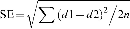 |
where d1 and d2 are measured values and n is the sample size.
The mean of the measurement error for the five randomly selected areas was 0.174°, which was sufficiently small, and the measurement was verified to have high reproducibility.
Statistical analysis
ROC curve analysis was used to investigate the rating data. SPSS II software (SPSS Inc., Chicago, IL) was used to perform the calculation for each observer, each angle and each grade of bone loss. The area under the ROC curve (Az) was calculated as an index of accuracy. The Az values for each angle in each grade were statistically compared using Welch's test.
Results
Effects of angulation
The diagnostic accuracy, as expressed by Az values, was lower in Grade 1 than in Grades 2 and 3 at all angulations (Figure 5). In Grade 2 and Grade 3, the accuracies of each angulation were almost the same. The accuracies were decreased dramatically in Grades 2 and 3 at angulations of −20° or less, and at −30° they were almost the same as that in Grade 1.
Figure 5.

Diagnostic accuracy (Az value) in relation to the horizontal projection angle. The highest Az values are observed at angulations of 15°, 10° and 20° for Grades 1 (∗), 2 (∗∗) and 3 (∗∗∗), respectively. Grey vertical dotted lines are the means of Lines A and B, which indicate the mesial and distal limits, respectively, of adequate angulation
In Grade 1, the Az values at −30°, 30° (P < 0.01), −25° and −20° (P < 0.05) were significantly lower than the highest value (0.770) at 15°. At 0°, the Az value was 0.659.
In Grades 2 and 3, high accuracies were found over a range of angulations between −15° and 25°. The highest values were 0.976 and 0.988 at angulations of 10° and 20° in Grades 2 and 3, respectively. The Az values at −30°, −25°, −20° (P < 0.01), −15° and 30° (P < 0.05) in Grade 2, and at −30°, −25° (P < 0.01), −20°, −15°, 0°, and 5° (P < 0.05) in Grade 3 were significantly lower than the highest values noted above. At 0° angulation, the Az values in Grades 2 and 3 were 0.939 and 0.952, respectively.
The highest Az values were shifted towards the mesial direction in all grades (15°, 10° and 20° for Grades 1, 2 and 3, respectively), as were the ranges of angulations that showed high accuracy (Figure 5).
Anatomical measurements
The average mesial limit (Line A) was 25.8° (SD 5.5°) relative to the 0° angulation that was perpendicular to the line connecting the mesial and distal contact points. The mean distal limit (Line B) was −9.7° (SD 5.7°). The range is added in Figure 5. The accuracy (Az value) was fairly constant within this range and decreased in either direction outside of this range.
Discussion
Because it can be difficult to measure the depth of artificially created defects with a caliper due to a thin interradicular trabecula, a new compact CBCT apparatus was used, the accuracy of which had already been verified.9 With this CT, it was possible to acquire a sufficient number of images to perform a ROC study and the defects could be accurately classified into one of four grades.
Although this study used the classification suggested by Hamp et al16 because of practical use in our clinics, Gürgan et al8 also reported that the horizontal depth of the defect had the greatest effect on observer performance when the beam angle was limited to the interdental area between the first and second molars and perpendicular to the film. In their study, defects with a horizontal depth from one-third to one-half of the width of the interradicular area were compared with those with depths less than one-third of the width. The angle used in their study corresponded to the 0° angulation in the present study and a Lesion 1 defect in their study was almost the same as a Grade 1 defect in the present study. The results of the present study support their finding because Grade 1 defects (less than one-third depth) showed lower detectability than Grade 2 and 3 defects. However, the lack of a difference between Grade 2 and 3 defects indicated that the depth did not affect the detectability of deeper defects. The Az value (0.676) in their study was similar to that of a Grade 1 defect (0.659) in the present study. As for Lesion 2 defects, the Az value (0.862) was somewhat lower than that for Grade 2 defects in the present study (0.949). This discrepancy may be due to the difference in the horizontal depth of the defects. Their Lesion 2 defects ranged from one-third to one-half of the buccolingual width of the interradicular area, whereas the present study included defects beyond the midpoint. The variance in Az values was found to be large in the Grade 1 defects, whereas it was relatively small in Grade 2 and Grade 3. This might be due to the difference in interpretation ability among observers. In Grade 3, the Az value of 30° angulation was larger than that of 25° angulation. The cause was not clear, but there was no statistical difference between the Az values of each angulation.
Based on the Az value (0.659), the Grade 1 furcation involvement is somewhat difficult to detect by intraoral radiograph, and it may also be difficult by probing procedure because the bone loss is not accompanied with severe gingival recession or soft tissue attachment loss. For such defects, a compact CBCT could be effective. Actually, the evaluations of furcation defect grade on dried mandibles were almost consistent between the two observers. Although the compact CBCT is a promising tool for detecting furcation defects, we should pay attention to the radiation dose to the patients.
The present study verified that the X-ray beam projection angle affects the accuracy of the diagnosis of a furcation defect. It is evident that changes in horizontal angulation cause geometric distortion in intraoral radiography.17 Moreover, morphological factors, such as the shape of the furcation entrance, the width of the periodontal ligament space and root configurations, are considered to be related to image distortion, and they may alter the detectability of furcation involvement.
Although the angle with the highest accuracy was shifted mesially, the best projection angle for detecting proximal caries was included within the range showing high accuracy for the detection of furcation defects. As in the detection of caries in the proximal surface, the greatest accuracy is seen with 0° angulation in which the beam projects tangentially to the surface, and decreases symmetrically on both sides of 0° angulation.18 In the present study, higher detectability was observed in mesial angulations and the highest values were obtained with angulations of 10–20°. This may be due to the anatomical relationship of the mesial and distal roots with the interradicular bone between them. To investigate this morphological relationship, reconstructed CT images were used. The angle that was thought to be appropriate for depicting the furcation area was 0° angulation, which was shifted mesially to a slight degree, similar to the Az values (diagnostic accuracy), for the created defects.
In conclusion, the detectability of furcation defects was related to the morphological relationship between the roots and interradicular area, and the traditionally used angulation for proximal caries detection appeared to be suitable also for furcation disease detection in the first molar area.
References
- 1.Hirschfeld L, Wasserman B. A long-term survey of tooth loss in 600 treated periodontal patients. J Periodontol 1978;49:225–237 [DOI] [PubMed] [Google Scholar]
- 2.McGuire MK, Nunn ME. Prognosis versus actual outcome. III. The effectiveness of clinical parameters in accurately predicting tooth survival. J Periodontol 1996;67:666–674 [DOI] [PubMed] [Google Scholar]
- 3.Zappa U, Grosso L, Simona C, Graf H, Case D. Clinical furcation diagnoses and interradicular bone defects. J Periodontol 1993;64:219–227 [DOI] [PubMed] [Google Scholar]
- 4.Muller H-P , Eger T. Furcation diagnosis. J Clin Periodontol 1999;26:485–498 [DOI] [PubMed] [Google Scholar]
- 5.Suomi JD, Plumbo J, Barbano JP. A comparative study of radiographs and pocket measurements in periodontal disease evaluation. J Periodontol 1968;39:311–315 [DOI] [PubMed] [Google Scholar]
- 6.Perschbacher S. Periodontal diseases. In: White SC, Pharoah MJ (eds). Oral radiology, principles and interpretation, 6th edn. St. Louis, MO: Mosby, 2009, pp 282–294 [Google Scholar]
- 7.Rees TD, Biggs NL, Collings CK. Radiographic interpretation of periodontal osseous lesions. Oral Surg Oral Med Oral Pathol 1971;32:141–153 [DOI] [PubMed] [Google Scholar]
- 8.Gürgan C, Gröndahl K, Wennström JL. Radiographic detectability of bone loss in the bifurcation of mandibular molars: an experimental study. Dentomaxillofac Radiol 1994;23:143–148 [DOI] [PubMed] [Google Scholar]
- 9.Naitoh M, Katsumata A, Mitsuya S, Kamemoto H, Ariji E. Measurement of mandibles with microfocus x-ray computerized tomography and compact computerized tomography for dental use. Int J Oral Maxillofac Implants 2004;19:239–246 [PubMed] [Google Scholar]
- 10.Bower RC. Furcation morphology relative to periodontal treatment. Furcation root surface anatomy. J Periodontol 1979;50:366–374 [DOI] [PubMed] [Google Scholar]
- 11.Carnevale G, Pontoriero R, Lindhe J. Treatment of furcation-involved teeth. In: Lindhe J, Lang NP (eds). Clinical periodontology and implant dentistry, 5th edn. Copenhagen: Munksgaard, 2008, pp 823–847 [Google Scholar]
- 12.Tarnow D, Fletcher PJ. Classification of the vertical component of furcation involvement. J Periodontol 1984;55:283–284 [DOI] [PubMed] [Google Scholar]
- 13.Fuhrmann RA, Bucker A, Diedrich PR. Furcation involvement: comparison of dental radiographs and HR-CT-slices in human specimens. J Periodontal Res 1997;32:409–418 [DOI] [PubMed] [Google Scholar]
- 14.Hou GL, Chen YM, Tsai CC, Weisgold AS. A new classification of molar furcation involvement based on the root trunk and horizontal and vertical bone loss. Int J Periodontics Restorative Dent 1998;18:257–265 [PubMed] [Google Scholar]
- 15.Waerhaug J. The infrabony pocket and its relationship to trauma from occlusion and subgingival plaque. J Periodontol 1979;50:355–365 [DOI] [PubMed] [Google Scholar]
- 16.Hamp SE, Nyman S, Lindhe J. Periodontal treatment of multirooted teeth. Results after 5 years. J Clin Periodontol 1975;2:126–135 [DOI] [PubMed] [Google Scholar]
- 17.Jenkins SM, Dummer PM, Addy M. An in vitro study of the influence of X-ray beam angulation on the radiographic images of the amelocemental junction and simulated alveolar crest. J Oral Rehabil 1992;19:629–637 [DOI] [PubMed] [Google Scholar]
- 18.Ariji Y, Shimizu Y, Okano T, Matsui O, Naitoh M, Yuasa H, et al. Influence of X-ray beam angulation in the detection of proximal caries: interobserver agreement in the CCD system. Oral Radiol 1999;15:27–35 [Google Scholar]