Abstract
Objective
The purpose of this study was to assess the in vitro diagnostic ability of visual inspection, film, charge-coupled device (CCD) sensor, photostimulable phosphor (PSP) sensor and cone beam CT in the detection of proximal caries in posterior teeth compared with the histological gold standard.
Methods
Visual inspection, film, CCD, PSP and cone beam CT images were used to detect proximal caries in the mesial and distal surfaces of 138 teeth (276 surfaces). Visual inspection and evaluation of all intraoral digital and conventional radiographs and cone beam CT images were performed twice by three oral radiologists. Weighted kappa coefficients were calculated to assess intra- and interobserver agreement for each image set, and scores were compared with the histological gold standard using receiver operating characteristic (ROC) analysis to evaluate diagnostic ability.
Results
Intraobserver kappa coefficients calculated for each observer for each method of detecting caries ranged from 0.739 to 0.928. Strong interobserver agreement ranging from 0.631 to 0.811 was found for all detection methods. The highest Az values for all three observers were obtained with the cone beam CT images; however, differences between detection methods were not statistically significant (P > 0.05).
Conclusion
Visual inspection, film, CCD, PSP plates and cone beam CT performed similarly in the detection of proximal caries.
Keywords: detection, cone beam computed tomography, digital radiography, film, proximal caries
Introduction
Dental caries are among the most common problems encountered in clinical dentistry, with a very high incidence of caries observed in the population.1 Early and accurate diagnosis of caries is essential for clinicians, who require exact knowledge of the depth of caries in order to determine the appropriate type of restoration and treatment planning.2,3
Among the various types of methods used today in the diagnosis of caries, probing, visual examination, intraoral film and digital sensors are the ones most commonly used in routine clinical practice. Several studies have shown that between 25% and 42% of caries lesions remain undetected by clinical examination performed without radiographic examination.4–6 Interproximal caries lesions develop between the contacting proximal surfaces of two adjacent teeth. They first appear clinically as opaque regions caused by the loss of enamel transparency at the outermost enamel between the contact point and the top of the free gingival margin.7–9 As the caries lesion progresses through the enamel, it takes on a triangular configuration, with the top of the triangle located at the enamel–dentine border. When it reaches the enamel–dentine border, it expands laterally and towards the inner dentine, forming another triangle in the dentine, with the base of the triangle located in the enamel–dentine border and the top of the triangle pointing towards the pulpal space.9
Owing to the difficulties in detecting proximal caries lesions, clinical examination should be supplemented with radiographic examination. Radiographic diagnosis is based upon the amount of demineralization needed to create a change in radiographic density, with a minimum of 40% mineral loss needed for radiographic visibility.1 Owing to the large size of the proximal surfaces of posterior teeth and the subtle mineral loss initially presented by lesions on these surfaces, proximal caries on posterior teeth are usually very difficult to visualize on radiographs. Conventional intraoral film, solid state detectors and photostimulable phosphor (PSP) plates are the image receptors most commonly used to diagnose proximal caries.10 Solid state detectors consist of either a charge-coupled device (CCD) that uses a thin wafer of silicon as the basis for image recording or a light-sensitive complementary metal oxide semiconductor (CMOS) chip and a scintillator layer that converts X-rays to light.10–12 A PSP plate consists of a polyester base coated with crystalline halide composed of europium-activated barium fluorohalide compounds.13,14 All these systems are able to provide two-dimensional information about dental tissue and disease.
Cone beam CT dedicated to dentomaxillofacial imaging was introduced in response to the high demand for a technique that could provide three-dimensional data at a lower cost and with lower absorbed doses than the conventional CT used in medical radiology. Dental cone beam CT has since gained broad acceptance in dentistry, with the most promising results obtained in relation to caries diagnosis and endodontic applications.15 The use of cone beam CT in clinical practice offers a number of potential advantages over conventional tomography, including easier image acquisition, higher image accuracy, reduced artefacts, lower effective radiation doses, faster scan times and greater cost-effectiveness.16,17 Rather than the fan-shaped beam emitted by conventional CT technology, cone beam CT units, as the name implies, emit a cone-shaped X-ray beam that covers the entire region of interest, allowing images to be acquired in only one pass or less around the patient's head.18
In view of the importance of radiological diagnosis of proximal caries and the potential difference in diagnostic performance of different caries detection methods, the aim of the present study was to assess the in vitro diagnostic ability of visual inspection, film, CCD sensors, PSP sensors and cone beam CT in the detection of proximal caries in posterior teeth in comparison with the histological gold standard.
Materials and methods
Our study comprised 230 human premolar and molar teeth with and without caries that had been extracted for periodontal or orthodontic reasons. Teeth were cleaned of calculus and debris, disinfected in 2% sodium hypochlorite solution for 20 min and stored in distilled water. Teeth were embedded in acrylic blocks in groups of five, with the proximal surfaces in contact; all blocks and teeth were numbered. The mesial and distal aspects of the 3 teeth located in the centre of each block were assessed for caries, for a total of 276 surfaces of 138 teeth embedded in 46 blocks. Detection of proximal caries was performed using visual inspection and film, CCD, PSP and cone beam CT images. Visual inspection was conducted under daylight using a mirror and probe, and surfaces were scored according to the following scale: 0, no caries lesion; 1, opacity or cavitation in enamel; 2, cavitation in dentine; 3, cavitation in dentine extending to pulp.
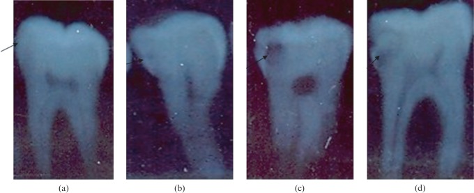
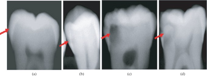
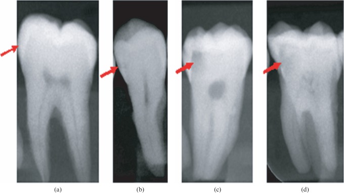
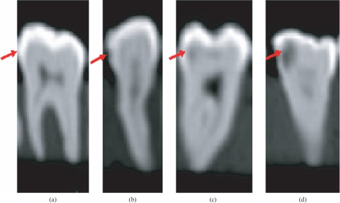
All blocks were imaged using three different intraoral radiography techniques and cone beam CT. Intraoral digital and conventional images were exposed with a Trophy Trex X-ray unit (Croissy, Beaubourg, France) operated at 65 kVp and 8 mA with a standardized paralleling technique and a focus–receptor distance of 20 cm. A 1 cm thick acrylic block was placed behind the teeth in each block in order to simulate soft tissue. Film radiographs were obtained using Kodak E speed (size 2) film (Kodak, Rochester, NY) with an image exposure time of 0.40 s (Figure 1). Films were processed immediately after exposure using a Velopex, Extra-X automatic processing machine (Medivance Instruments Limited, London, UK) and fresh chemicals according to the manufacturer's specifications. Direct digital images were obtained using a Progeny Vision DX (size 1) direct digital intraoral CCD sensor (Progeny Dental, Buffalo Grove, IL) with an image exposure time of 0.2 s. The Progeny CCD offers 1.25 million pixels, a pixel size of 22 μm × 22 μm and a theoretical resolution of 23 lp mm−1) (Figure 2). Semi-direct digital images were recorded using a Digora Optime (Soredex, Tuusula, Finland) PSP digital intraoral system, which includes a feature that automatically erases residual image signals. Image recording was set at a 40 μm (super) pixel size, 14-bit greyscale, 12.5 lp mm−1 spatial resolution and an image exposure time of 0.20 s. A size 2 imaging plate was used, and the exposed phosphor plates were scanned immediately after exposure (Figure 3). Cone beam CT images were obtained using an ILUMA ultra cone beam CT scanner (Imtec Imaging, Ardmore, OK) with a 24.4 × 19.5 cm amorphous silicon flat-panel image detector and a cylindrical volume of reconstruction up to 21.1 × 14.2 cm. Images were obtained at 120 kVp, 3.8 mA and a voxel size of 0.3 mm, with an exposure time of 40 s. Volumetric data were reconstructed to provide serial coronal and sagittal sections (Figure 4).
Figure 1.
Film radiographs obtained using Kodak E speed: (a) score 0, (b) score 1, (c) score 2, (d) score 3 (indicated by arrows)
Figure 2.
Images obtained by charge-coupled device sensor: (a) score 0, (b) score 1, (c) score 2, (d) score 3 (indicated by arrows)
Figure 3.
Images obtained by photostimulable phosphor plate sensor: (a) score 0, (b) score 1, (c) score 2, (d) score 3 (indicated by arrows)
Figure 4.
Images obtained by cone beam CT: (a) score 0, (b) score 1, (c) score 2, (d) score 3 (indicated by arrows)
Visibility of the pulpal root canal, dentine and enamel was used as an indication of optimal image quality for all images. All images were viewed at random in a dimly lit room. Images were evaluated separately by three oral radiologists experienced in image interpretation. Intraobserver agreement was assessed by having each observer view all images twice, with a 2 week interval between viewing to eliminate memory bias. Conventional film radiographs were evaluated using a light box and magnifier (× 2). Digital images were viewed on a 22 inch LG Flatron monitor (LG, Seoul, Korea) set at a screen resolution of 1440 × 900 pixels and 32 bit colour depth using each system's own software. The presence or absence of proximal caries was assessed according to radiographic criteria using the following scale: 0, no caries detected in the proximal surface; 1, proximal radiolucency in enamel (enamel caries); 2, proximal radiolucency in dentine (dentine caries); 3, proximal radiolucency extending to pulp (deep dentine caries).
Histological validation of caries status was performed by serially sectioning each tooth mesiodistally in parallel to the long axis of the crown using an Accutom-50 (Struers, Ballerup, Denmark). The average thickness of the serial sections was 400 μm. The section with the deepest carious lesion was assessed using a high-resolution scanner (Epson Perfection V750-M Pro Scanner Epson, Nagano, Japan) at 600 dpi resolution and × 15 magnification. Histological status was determined by consensus of one of the researchers and a histology specialist. Teeth were recorded as either sound or as having a caries lesion, which was defined as a demineralized white or yellowish-brown discoloured area in the enamel or dentine. Histological sections were assessed using the following scale: 0, no caries lesion in the proximal surface; 1, proximal caries in enamel; 2, proximal caries extending to the enamel–dentine junction or in the outer half of the dentine; 3, proximal caries in the inner half of the dentine. Histological examination of 276 proximal tooth surfaces revealed no caries in 142 surfaces (51.5%) and caries in 134 surfaces (48.5%). When analysed by caries level, 40 surfaces (14.5%) were found to have enamel caries, 45 surfaces (16%) had dentine caries confined to the outer half of the dentine and 49 surfaces (18%) had deep dentine caries extending to the inner half of the dentine.
Weighted kappa coefficients were calculated to assess intra- and interobserver agreement for each image set, and scores were compared with the histological gold standard using receiver operating characteristic (ROC) analysis to evaluate diagnostic ability. The areas under the ROC curves (Az values) for each image type, observer and reading were calculated using the MedCalc statistical software. Az values were compared using t-tests, with a significance level of 0.05, and Bonferroni correction was applied. Kappa values were calculated to assess intra- and interobserver agreement according to the following criteria: < 0.10, no agreement; 0.10–0.40, poor agreement; 0.41–0.60, significant agreement; 0.61–0.80, strong agreement; 0.81–1.00, excellent agreement. McNemar's test was applied to compare sensitivity and specificity values of radiographic methods obtained from each level of caries.
Results
Intraobserver kappa coefficients calculated for each observer by caries detection method ranged between 0.739 and 0.928 (Table 1). Considering the very high intraobserver kappa coefficients suggestive of strong and excellent intraobserver agreement, interobserver kappa coefficient and Az value calculations were based on the first readings only. Interobserver kappa coefficients by caries detection method ranged from 0.631 to 0.811, with strong interobserver agreement found for all detection methods (Table 2).
Table 1. Intraobserver kappa coefficients calculated for each observer by caries detection method.
| Observer 1 |
Observer 2 |
Observer 3 |
|
| Weighted kappa (SD) | Weighted kappa (SD) | Weighted kappa (SD) | |
| Visual inspection | 0.739 (0.052) | 0.840 (0.031) | 0.914 (0.029) |
| Intraoral film | 0.827 (0.035) | 0.868 (0.025) | 0.893 (0.027) |
| Intraoral CCD sensor | 0.810 (0.047) | 0.856 (0.031) | 0.794 (0.047) |
| Intraoral PSP sensor | 0.819 (0.035) | 0.878 (0.026) | 0.827 (0.046) |
| Cone beam CT | 0.846 (0.035) | 0.759 (0.039) | 0.928 (0.022) |
CCD, charge-coupled device; PSP, photostimulable phosphor; SD, standard deviation
Table 2. Interobserver kappa coefficients by caries detection method.
| Observers 1 and 2 |
Observers 1 and 3 |
Observers 2 and 3 |
|
| Weighted kappa (SD) | Weighted kappa (SD) | Weighted kappa (SD) | |
| Visual inspection | 0.631 (0.062) | 0.657 (0.064) | 0.665 (0.057) |
| Intraoral film | 0.699 (0.049) | 0.750 (0.044) | 0.759 (0.042) |
| Intraoral CCD sensor | 0.746 (0.045) | 0.769 (0.048) | 0.777 (0.041) |
| Intraoral PSP sensor | 0.774 (0.039) | 0.790 (0.040) | 0.811 (0.036) |
| Cone beam CT | 0.695 (0.051) | 0.802 (0.047) | 0.802 (0.039) |
CCD, charge-coupled device; PSP, photostimulable phosphor; SD, standard deviation
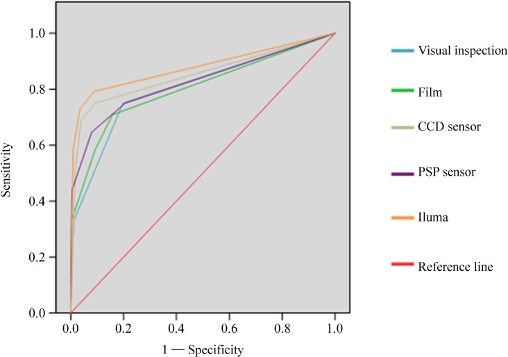
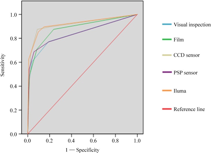
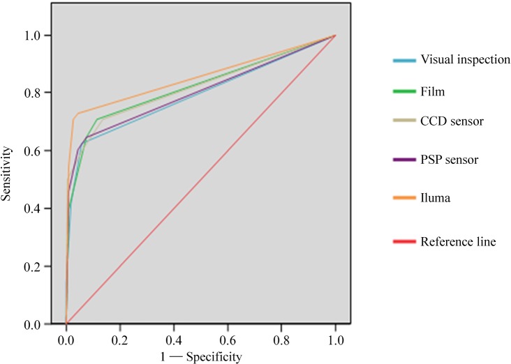
The areas under the ROC curves (Az values), their standard errors and significance levels for the first readings of each observer are given in Table 3. The highest Az values for all three observers were obtained with the Iluma cone beam CT images. The lowest Az values for Observers 2 and 3 were obtained with visual inspection, whereas the lowest Az value for Observer 1 was obtained with intraoral film; however, there were no statistically significant differences (P > 0.05) between any of the Az values obtained with the different methods of caries detection. Figures 5, 6 and 7 show the ROC curves for the first reading for each method for Observers 1, 2 and 3, respectively.
Table 3. Az values, standard errors and significance levels for each observer's first reading.
| Observer 1 |
Observer 2 |
Observer 3 |
||||
| Az (SE) | P-value | Az (SE) | P-value | Az (SE) | P-value | |
| Visual inspection | 0.805 (0.040) | <0.001 | 0.838 (0.039) | <0.001 | 0.792 (0.044) | <0.001 |
| Intraoral film | 0.803 (0.042) | <0.001 | 0.882 (0.032) | <0.001 | 0.819 (0.041) | <0.001 |
| Intraoral CCD sensor | 0.850 (0.039) | <0.001 | 0.914 (0.029) | <0.001 | 0.820 (0.042) | <0.001 |
| Intraoral PSP sensor | 0.825 (0.041) | <0.001 | 0.844 (0.039) | <0.001 | 0.801 (0.044) | <0.001 |
| Cone beam CT | 0.877 (0.036) | <0.001 | 0.920 (0.028) | <0.001 | 0.853 (0.040) | <0.001 |
CCD, charge-coupled device; PSP, photostimulable phosphor; SE, standard error
Figure 5.
Receiver operating characteristic curves for Observer 1 for the first reading for each caries detection method. CCD, charge-coupled device; PSP, photostimulable phosphor
Figure 6.
Receiver operating characteristic curves for Observer 2 for the first reading for each caries detection method. CCD, charge-coupled device; PSP, photostimulable phosphor
Figure 7.
Receiver operating characteristic curves for Observer 3 for the first reading for each caries detection method. CCD, charge-coupled device; PSP, photostimulable phosphor
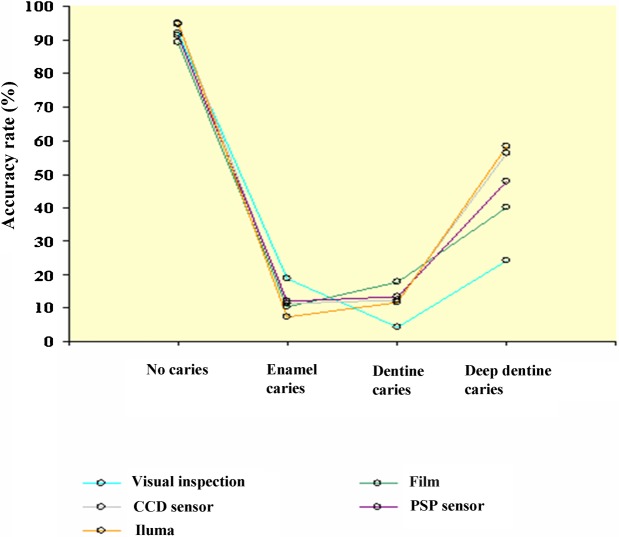
Table 4 shows the average percentage of correct diagnoses for each observer for each caries detection method and each level of caries. Teeth free from caries, followed by teeth with caries in the inner half of the dentine, yielded the best diagnoses rates for all observers and all caries detection methods, whereas the worst diagnoses were obtained from teeth with enamel caries, followed by teeth with caries in the outer half of the dentine. Figure 8 shows the accuracy rates of the different detection methods in diagnosing different caries levels. Table 5 shows the true positive ratio and false positive ratio of the first readings of three observers obtained using different methods. When sensitivity and specificity values of radiographic methods obtained from each level of caries were assessed, no statistical significance (P > 0.05) was found.
Table 4. Average percentage of true diagnoses of three observers using different detection methods.
| Visual inspection | Film | CCD | PSP | Iluma | |
| No caries (%) | 92 | 89.2 | 94.6 | 91.3 | 94.8 |
| Enamel caries (%) | 18.7 | 10.6 | 11.3 | 12.2 | 7.3 |
| Dentine caries (%) | 4.4 | 17.8 | 12.6 | 13.3 | 11.8 |
| Deep dentine caries (%) | 24.3 | 40.2 | 56.2 | 47.9 | 58.3 |
CCD, charge-coupled device; PSP, photostimulable phosphor
Figure 8.
Accuracy rates of caries diagnosis obtained by different methods. CCD, charge-coupled device; PSP, photostimulable phosphor
Table 5. True positive ratio (TPR) and false positive ratio (FPR): percentage of the first readings of three observers obtained using different methods.
|
Observer 1 |
Observer 2 |
Observer 3 |
||||
| TPR (%) | FPR (%) | TPR (%) | FPR (%) | TPR (%) | FPR (%) | |
| Visual inspection | 14.6 | 9.9 | 43.8 | 10.6 | 14.6 | 3.5 |
| Film | 33.3 | 9.2 | 47.9 | 17.6 | 39.6 | 5.6 |
| CCD | 52.1 | 3.5 | 66.7 | 7.7 | 50 | 4.9 |
| PSP | 43.8 | 12.7 | 54.2 | 11.3 | 45.8 | 2.1 |
| Cone beam CT | 58.3 | 2.8 | 62.5 | 11.3 | 54.2 | 1.4 |
CCD, charge-coupled device; PSP, photostimulable phosphor
Discussion
The present study found no differences in the diagnostic ability of visual inspection, film, CCD, PSP and cone beam CT images in the detection of proximal caries. Although observers obtained the highest Az values from cone beam CT images, the differences between modalities were not statistically significant (P > 0.05). It should be kept in mind that the cone beam CT images used in the present study were obtained at the lowest available resolution voxel size (0.3 mm) that is routinely used in clinical practice. Moreover, because of the beam hardening that occurs with the presence of metal such as amalgam, this study was limited to non-restored teeth.
The very high intraobserver kappa coefficients found in the present study suggest excellent intraobserver agreement and strong interobserver agreement among experienced observers for all detection methods. Intraobserver kappa coefficients ranged from 0.739 to 0.928, and interobserver kappa coefficients ranged from 0.631 to 0.811. The level of intra- and interobserver agreement reported by previous studies examining the use of different intraoral radiography techniques in the detection of caries has varied.19–23 Excellent interobserver agreement (kappa coefficient = 0.89) has been reported using bitewing film radiographs in the detection of proximal caries,19 and a kappa coefficient of 0.79 was reported for intraobserver agreement using bitewing film in the in vitro detection of proximal caries.20 In a study in which experienced observers assessed proximal caries depth using direct digital radiography and film, the average intraobserver kappa coefficient was 0.77 for digital and 0.78 for film, whereas the interobserver agreement was 0.44 for digital and 0.42 for film, suggesting no difference between the two modalities.21 Intraobserver coefficients of 0.74 for film and 0.67 for digital radiography and interobserver coefficients of 0.60 for film and 0.54 for digital radiography have been reported.22 Similar interobserver agreement kappa coefficients ranging between 0.57 and 0.60 have been reported, suggesting significant agreement in the detection of proximal caries using bitewing film.23 The differences in intra- and interobserver agreement kappa values among the different studies may be related to observer experience, radiographic quality, viewing conditions, study design and study material, all of which are important factors in determining observer agreement.
The use of visual examination and probing is a practical method for the clinical diagnosis of proximal caries. Although an earlier study found the use of visual inspection insufficient in the detection of superficial proximal caries,24 the present study found no statistically significant differences (P > 0.05) between visual inspection and other caries detection modalities.
When film was used in the detection of caries in posterior teeth, 50% of proximal caries lesions were diagnosed correctly and 93% of caries-free surfaces were correctly identified.25 Similarly, in our study, 89.2% of caries-free surfaces were identified correctly. When looked at by the level of caries, correct diagnoses were found for 10.6% of enamel caries, 17.8% of dentine caries and 40.2% of deep dentine caries.
Several studies have compared the diagnostic ability of intraoral digital sensors with film in the detection of caries lesions.26 In line with our results, most previous studies comparing film and CCD sensors in the detection of proximal caries have revealed similar results for both systems,4,19,27–30 although some studies have found film to be more accurate than direct digital radiography.31,32 Our study found that CCD sensors (56.2%) had a higher rate than film (40.2%) and PSP plates (47.9%) but a lower rate than cone beam CT (58.3%) in correctly diagnosing deep dentine caries; however, the differences between methods were statistically insignificant. Four different intraoral storage phosphor plate systems (DenOptix, Denstply/Gendex, Chicago, IL; Cd-dent, DigiDent, Nesher, Israel; Digora blue plate and Digora white plate, Soredex, Helsinki, Finland) exposed with two different plate exposure times (10% and 25% of the film exposure time) were compared with traditional film images (Ektaspeed Plus, Eastman Kodak, Rochester, NY) in the detection of proximal caries.33 A total of 365 surfaces were imaged. Using the longer exposure time, no significant differences in diagnostic accuracy were found among the DenOptix, Digora blue, Digora white and Ektaspeed Plus, all of which were significantly more accurate than the Cd-dent. Moreover, the accuracy of the DenOptix and Digora blue systems in diagnosing proximal caries improved significantly with the longer exposure time compared with the shorter exposure time.33 It was found that, in comparison with film (Ektaspeed Plus) images, PSP (Digora) images using a caries-specific enhancement feature significantly improved accuracy and reduced observer variability in assessing the depth of caries in the outer half of the enamel.34 In the present study, only one of three observers had better Az values with PSP (Digora) than with film images, although the difference was not statistically significant.
Several other studies have been conducted in order to evaluate and compare different intraoral radiographic modalities in the detection of proximal caries. 56 surfaces were radiographed using two different E-speed dental films (Ektaspeed Plus, Eastman Kodak, and Dentus M2 Comfort, Agfa-Gevaert, Mortsel, Belgium), two CCD systems (Sidexis, Sirona, Bensheim, Germany, and Visualix Gendex, Milan, Italy) and two PSP systems (Digora, Soredex, and DenOptix, Denstply/Gendex).35 No significant differences were found in diagnostic accuracy between the two dental films and the Sidexis and Digora systems. Lesion depth was found to significantly affect observer performance and was, in general, underestimated. The authors concluded that, rather than imaging modality, the main factor affecting diagnostic accuracy was observer ability.35 Extracted human teeth were radiographed using two CCD systems (Dixi, Planmeca, Helsinki, Finland, and Sidexis, Sirona) and two PSP systems (Digora, Soredex, and DenOptix, Gendex), and the Dixi and Digora systems were found to be more accurate than the Sidexis and DenOptix systems in measuring the depth of proximal caries lesions.36 In the present study, no differences were found between the Digora Optime and the other systems tested. The accuracy of older and newer versions of a CMOS system (Schick CDR/Schick CDR Wireless, Schick Technologies, Long Island City, NY) and a PSP system (Digora FMX/Digora Optime, Soredex) were compared in the detection of proximal carious lesions.37 The new systems (Digora Optime and Schick CDR Wireless) were found to have significantly higher sensitivities than their predecessors.37 No significant differences in specificity were found among the Digora FMX, Schick CDR and Schick CDR Wireless systems, all of which had a significantly higher specificity than the Digora Optime (P < 0.02). Fewer false positive diagnoses occurred with the Digora FMX than with the Digora Optime, which also had a lower positive predictive value than that of both CCD systems assessed (P < 0.02). However, in terms of overall accuracy, the differences between the older and newer versions of the PSP and CCD systems were not statistically significant.37
The diagnostic accuracy of conventional tomograms has also been found to be similar to that of conventional bitewing and digital intraoral radiographs in the detection of proximal caries.38 Although no difference was found between local CT (LCT) (Sirona Dental Systems, Bensheim, Germany) and conventional radiography in the detection of proximal caries, LCT was found to be more accurate in assessing lesion depth.39
A number of studies have compared the diagnostic ability of different dental cone beam CT systems and intraoral modalities in the detection of proximal caries.40–42 The accuracy of proximal caries depth measurement using limited cone beam CT (3DX Accuitomo, J Morita MFG, Kyoto, Japan) was compared with PSP digital (Digora, Soredex) and F-speed film radiography (Eastman Kodak).40 Significant differences (P < 0.01) were found between histological and film measurements and between histological and PSP measurements, but no significant differences (P > 0.05) were found between histological and limited cone beam CT measurements. Interobserver agreement was 0.998 for the limited cone beam CT. The mean difference in measurements between observers using cone beam CT was approximately 0.02 mm, which was regarded as clinically insignificant.40 The authors concluded that cone beam CT was a promising tool for the detection and monitoring of proximal caries lesions that provided highly accurate measurements of caries depth.40 In contrast to Akdeniz et al's40 study, the present study found no differences between cone beam CT and intraoral modalities. Differences in findings between the two studies may be due to differences in devices, settings and materials used.
The diagnostic accuracy of two cone beam CT systems, the NewTom 3G (Quantitative Radiology, Verona, Italy) and the 3DX Accuitomo (Morita), were compared with two intraoral receptors, one digital (Digora FMX, Soredex) and one film (Insight, Kodak).41 Images were obtained with the NewTom 3G using three different fields of view (FOVs), 12 inches (0.36 mm pixel size), 9 inches (0.25 mm pixel size) and 6 inches (0.16 mm pixel size), and with the 3DX Accuitomo using a 4 cm FOV (0.125 mm pixel size). For proximal surfaces, the 3DX Accuitomo had significantly higher sensitivity (P < 0.02) than the NewTom 12 inch and 9 inch images, and the film and Digora FMX images had significantly higher specificity (P < 0.04) than the NewTom 9 inch and 6 inch images, whereas there were no significant differences among the film, Digora FMX and Accuitomo images for any of the variables tested (P > 0.2). For occlusal surfaces, the Accuitomo showed higher sensitivity than all the other systems tested, whereas specificity and overall true score did not differ (P > 0.06) among the modalities.41 The authors concluded that the diagnostic accuracy of the NewTom 3G cone beam CT was lower than both intraoral modalities and the 3DX Accuitomo cone beam in the detection of caries lesions. Although the Accuitomo cone beam CT had a higher sensitivity than the intraoral systems in the detection of dentine lesions, the overall true score was not higher.41 Although the cone beam CT Iluma system used in the present study does not offer different FOVs, it does offer different voxel sizes for image capture. We used a voxel size of 0.3 mm, which is the standard for routine clinical use.
The diagnostic ability of a limited cone beam CT imaging system (3DX Accuitomo, Morita) in detecting incipient proximal caries was assessed.42 No difference was found in the accuracy of the cone beam CT system and traditional film (Insight, Eastman Kodak). Cone beam CT images of 100 surfaces from 50 extracted premolars detected a total of 71 proximal carious lesions, 47 of which were enamel caries and 24 of which were dentine caries. Analysis of variance showed no statistically significant difference between the 3DX Accuitomo and film images. Mean Az values were 0.63 ± 0.02 for 3D Accuitomo and 0.63 ± 0.03 for film.42 Compared with our study, the Az value of the cone beam CT system in this study was lower, which can be explained by the fact that most of the caries lesions in this study were confined to the enamel.
In the present study, none of the modalities tested showed both high sensitivity and high specificity, and none of the modalities significantly outperformed the others in the detection of proximal caries. Considering these results, digital intraoral systems can be recommended over film because of their lower levels of radiation exposure. The best diagnostic outcomes are likely to be achieved through the combined use of different caries detection modalities. Further research is also needed to assess the performance of newly developed cone beam CT systems in proximal caries detection and other sensitive diagnostic tasks.
In conclusion, visual inspection, film, CCD, PSP and cone-beam CT chosen for this study performed similarly in the detection of proximal caries in vitro.
Acknowledgements
The authors would like to thank Tomoloji Dentomaxillofacial Radiology centre (Ankara, Turkey) for providing the iluma unit.
References
- 1.Pereira AC, Verdonschot EH, Huysmans MC. Caries detection methods: can they aid decision making for invasive sealant treatment? Caries Res 2001;35:83–89 [DOI] [PubMed] [Google Scholar]
- 2.Attrill DC, Ashley PF. Occlusal caries detection in primary teeth: a comparison of DIAGNOdent with conventional methods. Br Dent J 2001;190:440–443 [DOI] [PubMed] [Google Scholar]
- 3.Ohki M, Okano T, Nakamura T. Factors determining the diagnostic accuracy of digitized conventional intra-oral radiographs. Dentomaxillofac Radiol 1994;23:77–82 [DOI] [PubMed] [Google Scholar]
- 4.Haak R, Wicht MJ, Noack MJ. Conventional, digital and contrast-enhanced bite-wing radiographs in the decision to restore proximal carious lesions. Caries Res 2001;35:193–199 [DOI] [PubMed] [Google Scholar]
- 5.Moystad A, Svanaes DB, Risnes S, Larheim TA, Gröndahl HG. Detection of proximal caries with a storage phosphor system. A comparison of enhanced digital images with dental x-ray film. Dentomaxillofac Radiol 1996;25:202–206 [DOI] [PubMed] [Google Scholar]
- 6.Tam LE, McComb D. Diagnosis of occlusal caries: Part II. Recent diagnostic technologies. J Can Dent Assoc 2001;67:459–463 [PubMed] [Google Scholar]
- 7.Kidd EA, Pitts NB. A reappraisal of the value of the bite-wing radiograph in the diagnosis of posterior proximal caries. Br Dent J 1990;169:195–200 [DOI] [PubMed] [Google Scholar]
- 8.Svenson B, Gröndahl HG, Petersson A, Olving A. Accuracy of radiographic caries diagnosis at different kilovoltages and two film speeds. Swed Dent J 1985;9:37–43 [PubMed] [Google Scholar]
- 9.Hansen BF. Clinical and roentgenologic caries detection. A comparison. Dentomaxillofac Radiol 1980;9:34–36 [DOI] [PubMed] [Google Scholar]
- 10.Ludlow JB, Mol A. Digital imaging. White SC, Pharoah MJ.Oral radiology. Principles and interpretation (6th edn). St Louis: Mosby, 2009, pp 78–99 [Google Scholar]
- 11.Kitagawa H, Scheetz JP, Farman AG. Comparison of complementary metal oxide semiconductor and charge-coupled device intra-oral X-ray detectors using subjective image quality. Dentomaxillofac Radiol 2003;32:408–411 [DOI] [PubMed] [Google Scholar]
- 12.Farman AG, Farman TT. A comparison of 18 different x-ray detectors currently used in dentistry. Oral Surg Oral Med Oral Pathol Oral Radiol Endod 2005;99:485–489 [DOI] [PubMed] [Google Scholar]
- 13.Hildebolt CF, Couture RA, Whiting BR. Dental photostimulable phosphor radiography. Dent Clin North Am 2000;44:273–297 [PubMed] [Google Scholar]
- 14.Borg E, Attaelmanan A, Gröndahl HG. Image plate systems differ in physical performance. Oral Surg Oral Med Oral Pathol Oral Radiol Endod 2000;89:118–124 [DOI] [PubMed] [Google Scholar]
- 15.Tyndall DA, Rathore S. Cone-beam CT diagnostic applications: caries, periodontal bone assessment, and endodontic applications. Dent Clin North Am 2008;52:825–841 [DOI] [PubMed] [Google Scholar]
- 16.Scarfe WC, Farman AG, Sukovic P. Clinical applications of cone-beam computed tomography in dental practice. J Can Dent Assoc 2006;72:75–80 [PubMed] [Google Scholar]
- 17.Scarfe WC, Farman AG. What is cone-beam CT and how does it work? Dent Clin North Am 2008;52:707–730 [DOI] [PubMed] [Google Scholar]
- 18.White SC. Cone-beam imaging in dentistry. Health Physics 2008;95:628–637 [DOI] [PubMed] [Google Scholar]
- 19.Holt RD, Azevedo MR. Fibre-optic transillumination and radiographs in diagnosis of proximal caries in primary teeth. Community Dent Health 1989;6:239–247 [PubMed] [Google Scholar]
- 20.Peers A, Hill FJ, Mitropoulos CM, Holloway PJ. Validity and reproducibility of clinical examination, fibre-optic transillumination, and bite-wing radiology for the diagnosis of small proximal carious lesions: an in vitro study. Caries Res 1993;27:307–311 [DOI] [PubMed] [Google Scholar]
- 21.Naitoh M, Yuasa H, Toyama M, Shiojima M, Nakamura M, Ushida M, et al. Observer agreement in the detection of proximal caries with direct digital intra-oral radiography. Oral Surg Oral Med Oral Pathol Oral Radiol Endod 1998;85:107–112 [DOI] [PubMed] [Google Scholar]
- 22.Abreu Júnior M, Tyndall DA, Platin E, Ludlow JB, Phillips C. Two-and three-dimensional imaging modalities for the detection of caries. A comparison between film, digital radiography and tuned aperture computed tomography (TACT). Dentomaxillofac Radiol 1999;28:152–157 [DOI] [PubMed] [Google Scholar]
- 23.Langlais RP, Skoczylas LJ, Prihoda TJ, Langland OE, Schiff T. Interpretation of bite-wing radiographs: application of kappa statistic to determine rater agreements. Oral Surg Oral Med Oral Pathol Oral Radiol Endod 1987;64:751–756 [DOI] [PubMed] [Google Scholar]
- 24.Hintze H, Wenzel A, Danielsen B, Nyvad B. Reliability of visual examination, fibreoptic transillumination, and bite-wing radiography, and reproducibility of direct visual examination following tooth separation for the identification of cavitated carious lesions in contacting proximal surfaces 1998. Caries Res;32:204–209 [DOI] [PubMed] [Google Scholar]
- 25.Gröndahl HG, Gröndahl K, Okano T, Webber RL. Statistical contrast enhancement of subtraction images for radiographic caries diagnosis. Oral Surg Oral Med Oral Pathol Oral Radiol Endod 1982;53:219–223 [DOI] [PubMed] [Google Scholar]
- 26.Wenzel A. Digital radiography and caries diagnosis. Dentomaxillofac Radiol 1988;27:3–11 [DOI] [PubMed] [Google Scholar]
- 27.Tyndall DA, Ludlow JB, Platin E, Nair M. A comparison of Kodak Ektaspeed plus film and the Siemens Sidexis digital imaging system for caries detection using receiver operating characteristic analysis. Oral Surg Oral Med Oral Pathol Oral Radiol Endod 1998;85:113–118 [DOI] [PubMed] [Google Scholar]
- 28.White SC, Yoon DC. Comparative performance of digital and conventional images for detecting proximal surfaces caries. Dentomaxillofac Radiol 1997;26:32–38 [DOI] [PubMed] [Google Scholar]
- 29.Castro VM, Katz JO, Hardman PK, Glaros AG, Spencer P. In vitro comparison of conventional film and direct digital imaging in the detection of proximal caries. Dentomaxillofac Radiol 2007;36:138–142 [DOI] [PubMed] [Google Scholar]
- 30.Alkurt MT, Peker I, Bala O, Altunkaynak B. In vitro comparison of four different dental X-ray films and direct digital radiography for proximal caries detection. Oper Dent 2007;32:504–509 [DOI] [PubMed] [Google Scholar]
- 31.Uprichard KK, Potter BJ, Russell CM, Schafer TE, Adair S, Weller RN. Comparison of direct digital and conventional radiography for the detection of proximal surface caries in the mixed dentition. Pediatr Dent 2000;22:9–15 [PubMed] [Google Scholar]
- 32.Price C, Ergül N. A comparison of a film-based and a direct digital dental radiographic system using a proximal caries model. Dentomaxillofac Radiol 1997;26:45–52 [DOI] [PubMed] [Google Scholar]
- 33.Hintze H, Wenzel A, Frydenberg M. Accuracy of caries detection with four storage phosphor systems and E-speed radiographs. Dentomaxillofac Radiol 2002;31:170–175 [DOI] [PubMed] [Google Scholar]
- 34.Svanaes DB, Moystad A, Larheim TA. Proximal caries depth assessment with storage phosphor versus film radiography. Evaluation of the caries-specific Oslo enhancement procedure. Caries Res 2000;34:448–453 [DOI] [PubMed] [Google Scholar]
- 35.Syriopoulos K, Sanderink GC, Velders XL, van derStelt PF. Radiographic detection of proximal caries: a comparison of dental films and digital imaging systems. Dentomaxillofac Radiol 2000;29:312–318 [DOI] [PubMed] [Google Scholar]
- 36.Jacobsen JH, Hansen B, Wenzel A, Hintze H. Relationship between histological and radiographic caries lesion depth measured in images from four digital radiography systems. Caries Res 2004;38:34–38 [DOI] [PubMed] [Google Scholar]
- 37.Haiter-Neto F, dos AnjosPontual A, Frydenberg M, Wenzel A. A comparison of older and newer versions of intra-oral digital radiography systems: diagnosing noncavitated proximal carious lesions. J Am Dent Assoc 2007;138:1353–1359 [DOI] [PubMed] [Google Scholar]
- 38.Peker I, Alkurt MT, Altunkaynak B. Film tomography compared with film and digital bitewing radiography for proximal caries detection. Dentomaxillofac Radiol 2007;36:495–499 [DOI] [PubMed] [Google Scholar]
- 39.Kalathingal SM, Mol A, Tyndall DA, Caplan DJ. In vitro assessment of cone beam local computed tomography for proximal caries detection. Oral Surg Oral Med Oral Pathol Oral Radiol Endod 2007;104:699–704 [DOI] [PubMed] [Google Scholar]
- 40.Akdeniz BG, Gröndahl HG, Magnusson B. Accuracy of proximal caries depth measurements: comparison between limited cone beam computed tomography, storage phosphor and film radiography. Caries Res 2006;40:202–207 [DOI] [PubMed] [Google Scholar]
- 41.Haiter-Neto F, Wenzel A, Gotfredsen E. Diagnostic accuracy of cone beam computed tomography scans compared with intra-oral image modalities for detection of caries lesions. Dentomaxillofac Radiol 2008;37:18–22 [DOI] [PubMed] [Google Scholar]
- 42.Tsuchida R, Araki K, Okano T. Evaluation of a limited cone-beam volumetric imaging system: comparison with film radiography in detecting incipient proximal caries. Oral Surg Oral Med Oral Pathol Oral Radiol Endod 2007;104:412–416 [DOI] [PubMed] [Google Scholar]


