Abstract
Objectives
The aim of the study was to clarify the change in image quality upon X-ray dose reduction and to re-analyse the possibility of X-ray dose reduction in photostimulable phosphor luminescence (PSPL) X-ray imaging systems. In addition, the study attempted to verify the usefulness of multiobjective frequency processing (MFP) and flexible noise control (FNC) for X-ray dose reduction.
Methods
Three PSPL X-ray imaging systems were used in this study. Modulation transfer function (MTF), noise equivalent number of quanta (NEQ) and detective quantum efficiency (DQE) were evaluated to compare the basic physical performance of each system. Subjective visual evaluation of diagnostic ability for normal anatomical structures was performed. The NEQ, DQE and diagnostic ability were evaluated at base X-ray dose, and 1/3, 1/10 and 1/20 of the base X-ray dose.
Results
The MTF of the systems did not differ significantly. The NEQ and DQE did not necessarily depend on the pixel size of the system. The images from all three systems had a higher diagnostic utility compared with conventional film images at the base and 1/3 X-ray doses. The subjective image quality was better at the base X-ray dose than at 1/3 of the base dose in all systems. The MFP and FNC-processed images had a higher diagnostic utility than the images without MFP and FNC.
Conclusions
The use of PSPL imaging systems may allow a reduction in the X-ray dose to one-third of that required for conventional film. It is suggested that MFP and FNC are useful for radiation dose reduction.
Keywords: dental digital radiography; image processing, computer-assisted; radiographic image enhancement
Introduction
Computed radiography systems were the first digital X-ray imaging systems to use a photostimulable phosphor luminescence (PSPL) imaging plate as the X-ray sensor. Shortly thereafter, these systems were developed for use in dental radiography and there are now commercially available PSPL digital X-ray imaging systems (PSPL imaging systems) suitable for use in the clinical setting. Although recently charge coupled device (CCD) digital X-ray imaging systems have been developed with a wider dynamic range than they had in the past, PSPL imaging systems still have a wider dynamic range than these new systems.1 Furthermore, conventional periapical X-ray films do not use an intensifying screen and thus they need relatively high X-ray exposure doses for imaging. Therefore, X-ray dose reduction is possible when the PSPL imaging system is used for intraoral radiography. In fact, studies have found that the exposure X-ray dose necessary for X-ray imaging could be reduced from 50% to 6% of that required for an E-speed film.2–5 However, there is a considerable difference between 50% and 6%. It is also questioned how a reduction in X-ray dose will influence image quality in different PSPL imaging systems.
Multiobjective frequency processing (MFP) was first reported in 1997 and is a comparatively new technology applied to digital X-ray imaging.6 This new image processing method may enhance image components in an optional frequency area, thereby producing a more natural visual image compared with those produced by the usual contrast enhancement processing or conventional frequency processing.7 When contrast enhancement processing or conventional frequency processing are applied to an image that has been imaged with a low exposure dose and has a low signal to noise ratio, unnecessary noise components may also be enhanced. Furthermore, diagnosis may be hindered because excessive conventional frequency processing produces over-shoot and/or under-shoot around the subject boundary. A combination of MFP and flexible noise control (FNC) largely removes such problems and can improve images with a low signal to noise ratio without emphasising the noise. MFP and FNC are used in medical radiology already, with much success.8,9 In the field of oral and maxillofacial radiology MFP has been applied to extraoral radiography, such as panoramic radiography and cephalometric radiography.10,11 However, there has been no report to date of the use of MFP and FNC for intraoral radiography.
The aim of this study was to investigate changes in image quality produced by a reduction in X-ray dose in three different PSPL imaging systems and to re-examine the possibility of X-ray dose reduction for intraoral radiography. In addition, the usefulness of MFP and FNC for X-ray dose reduction in PSPL imaging systems was explored.
Materials and methods
Systems
The DenOptix QST (VixWinPro version 1.5f, Gendex Dental Systems Co. Ltd., Lake Zurich, IL), VistaScan (DBSWIN version 3.19, Dürr Dental GmbH & Co. Bietigheim-Bissingen, Germany) and Computed Radiography XL-1 (CR XL-1; APL Software-B version 5.0.0003, Fuji Film Medical Co. Ltd., Tokyo, Japan) systems were evaluated in the present study. The scanning pixel size of the DenOptix QST was 42.3 μm/pixel, and the VistaScan 12.5 μm/pixel. The scanning pixel size of the CR XL-1 for medical imaging systems was improved from 100 μm to 50 μm for this study. A DT-1 PSPL imaging plate Fuji Film Medical Co., Ltd, Tokyo, Japan was used in the DenOptix QST and CR XL-1, and a BAS-SR PSPL imaging plate Fuji Film Medical Co., Ltd, Tokyo, Japan was used in the VistaScan. Each PSPL imaging plate was supplied by the manufacturer of each imaging system. In the CR XL-1, the PSPL imaging plate of 18 cm × 24 cm size was used. The X-ray generator used was a DFW 20 unit (Asahi Roentgen Industry Co. Ltd., Kyoto, Japan) with a focus size of 0.8 mm × 0.8 mm and total filtration of 2.0 mm aluminium equivalent.
Physical evaluation of image quality
The spatial resolution of each system was evaluated as the modulation transfer function (MTF) by an edge method using a tungsten plate of 1 mm thickness. The exposure conditions were as follows: tube voltage, 70 kV; tube current, 10 mA; exposure time, 1.6 s; and focus to imaging plate distance, 40 cm. The tungsten plate was placed at 3° in the scan direction and subscan direction of the PSPL imaging plate. The MTFs were measured three times each in the scan and subscan directions, and the three measurement values were averaged for each direction. The resultant mean represented the MTF of each system.
The noise equivalent number of quanta (NEQ) and the detective quantum efficiency (DQE) were also evaluated for each PSPL imaging system. The X-ray exposure conditions were the same as the evaluation of MTFs except for the X-ray absorbed dose. The X-ray absorbed dose, which exposed the density of conventional periapical X-ray film (Kodak Insight film, Eastman Kodak Company, Rochester, NY) to an optical density of 1.2, was first determined, and this X-ray absorbed dose was used as the base X-ray absorbed dose. The NEQs and the DQEs were obtained at the base X-ray absorbed dose and at approximately 1/3, 1/10 and 1/20 absorbed doses of the base X-ray absorbed dose. A dose and dose rate meter (QA Test-O-Meter Model 6003, Unfors Instruments, Billdal, Sweden) were used to measure the X-ray absorbed dose. The dose meter was placed with the PSPL imaging plates or periapical X-ray film in the X-ray radiation field at the same height as the PSPL imaging plates or periapical X-ray film. The NEQs and DQEs were also measured three times each in the scan and subscan directions, and the NEQs and DQEs of each system were then represented as the mean values of the scan and subscan directions. The calculation methods of the NEQs and DQEs followed the international standard method of the International Electrotechnical Commission (IEC62220-1)12 because there is no standard method for dental radiography.
When a physical evaluation was performed the filtration quantity of the X-ray generator was changed from 2.0 mm to 21.0 mm aluminium equivalent in accordance with the standard evaluation methods of the International Electrotechnical Commission (IEC61267).13 All physical evaluations were performed on the pre-sampling image data.
Subjective visual evaluation of diagnostic utility for normal anatomical structures
A human adult mandible embedded in a 25 mm thick, epoxy-resin block was used as the phantom imaging subject. The phantom was imaged by each PSPL imaging system under the same conditions as the clinical imaging conditions of conventional periapical X-ray film for a mandibular molar region, namely tube voltage, 70 kV; tube current, 10 mA; exposure time, 0.32 s; focus to PSPL imaging plate (or film) distance, 38 cm; and 2.0 mm aluminium equivalent filtration. The X-ray absorbed dose under these conditions was 827 μGy and this dose was set as the base X-ray absorbed dose. Images were then obtained at the base X-ray absorbed dose, and at 1/3, 1/10 and 1/20 doses by each PSPL imaging system.
The images of the DenOptix QST and VistaScan were output using only the automatic image adjustment processing of each system. The images of the CR XL-1 were output using two kinds of image processing. One image processing condition consisted of only the simple gradient processing used for adjusting X-ray image density, as there is no automatic image adjustment processing function in the CR XL-1. The second image processing condition consisted of applying MFP and FNC to improve visual image quality. The processing conditions of MFP and FNC were as follows: the contrast of the image was slightly enhanced on all images, the components from middle to high frequency were strongly enhanced, and the noise was strongly suppressed on the lower X-ray absorbed dose images.
The images were evaluated using subjective visual assessment by 6 oral and maxillofacial radiologists who ranged in experience from a minimum of 3 years to in excess of 17 years in the field. The visual evaluation was performed on film output images produced using the same dry film laser imager (DRYPIX 7000, Fuji Film Medical Co. Ltd, Tokyo, Japan). Eight regions of the dentinoenamel junction, alveolar crest, pulp canal, pulp horn, periodontal membrane space, lamina dura, apex of dental root and trabecular bone were specified as the regions of interest for assessment. The diagnostic utility of each image was evaluated in comparison with an image on conventional periapical X-ray film according to the following five grades: 5 = better, 4 = slightly better, 3 = fair, 2 = slightly worse and 1 = worse. Both presenter and evaluator did not know the conditions of evaluation images. The diagnostic utility of each image was compared with the average evaluation scores for the eight regions of interest. The subjective visual assessment was repeated 1 month later to examine intraobserver variability. The data were analysed using the paired t-test after applying the F-test, including intraobserver and interobserver variability. The statistical significance was determined to be P < 0.05.
Results
Physical evaluation
Modulation transfer function (MTF)
The MTF of each PSPL imaging system is shown in Figure 1. The MTFs of the low frequency range from 0.5 to 2.0 cycles/mm had almost equivalent values for the three PSPL imaging systems, except for the VistaScan, which had a slightly lower value than the other two systems at 0.5 cycles/mm. For the middle to high frequency range, that is, over 3 cycles/mm, the MTF of the CR XL-1, which had the lowest reading pixel size, had a slightly lower value than the other two systems.
Figure 1.
Modulation transfer function (MTF) of the three kinds of photostimulable phosphor luminescence imaging systems. Error bars indicate ±1 SD
Noise equivalent number of quanta (NEQ) and Detective quantum efficiency (DQE)
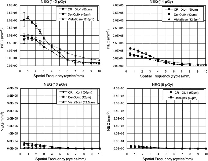
Figure 2 shows the NEQ of each system at the 4 X-ray absorbed doses, that is, as the X-ray absorbed dose was reduced from 143 μGy to 6 μGy. The NEQs of the CR XL-1 in the low frequency range, that is, under 3 cycles/mm, were better than those of the other two systems at all X-ray absorbed doses. When the X-ray absorbed dose was relatively high (143 μGy and 44 μGy), the NEQs of the VistaScan over the 5 cycles/mm were higher than those of the CR XL-1 and DenOptix QST. However, the NEQ of the VistaScan was markedly lower at the low X-ray absorbed dose of 13 μGy. Furthermore, when the X-ray absorbed dose was 6 μGy, the VistaScan was not able to even form an X-ray image. The NEQs of the DenOptix QST had almost the same or slightly lower values than the other two systems, when the X-ray absorbed dose was relatively high (143 μGy and 44 μGy). However, the NEQ of the DenOptix QST was comparatively stable when the X-ray absorbed dose was low (13 μGy and 6 μGy).
Figure 2.
Noise equivalent number of quanta (NEQ) of the three kinds of photostimulable phosphor luminescence imaging systems at the four X-ray absorbed doses. Error bars indicate ±1 SD
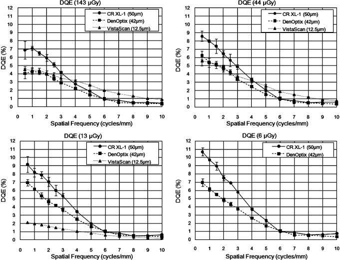
The DQEs at the four X-ray absorbed doses used are shown in Figure 3. The DQE results had similar traits and tendencies to the NEQ results, with the characteristic NEQ result of each PSPL imaging system being emphasised more in the DQE findings. The DQE of the CR XL-1 increased as the X-ray absorbed dose decreased. The DQE of the DenOptix increased when the X-ray absorbed dose decreased from 143 μGy to 13 μGy, and then it remained at an almost constant value. The DQE of the VistaScan also increased a little as the X-ray absorbed dose decreased from 143 μGy to 44 μGy, but then it markedly decreased as the X-ray absorbed dose decreased.
Figure 3.
Detective quantum efficiency (DQE) of the three kinds of photostimulable phosphor luminescence imaging systems at the four X-ray absorbed doses. Error bars indicate ±1 SD
Subjective visual evaluation
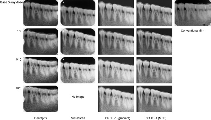
The X-ray images from each system used for visual evaluation are shown in Figure 4. The noise components on the images of all systems were not conspicuous when the X-ray absorbed dose was the same as the base X-ray absorbed dose and 1/3 of the clinical imaging conditions for conventional periapical X-ray film. When the X-ray absorbed dose was 1/10, the images of the DenOptix QST and VistaScan were very coarse images that included a lot of noise. The image from the DenOptix QST at the 1/20 X-ray absorbed dose was also very coarse, and fine anatomical structures such as the trabecular bone and periodontal space were very indistinct. The VistaScan was not able to form an image at the 1/20 X-ray absorbed dose. When the X-ray absorbed dose was 1/10, the image from the CR XL-1 with simple gradient processing for adjusting an X-ray image density was relatively smooth; however, when the X-ray absorbed dose was 1/20, the noise component of this type of image was conspicuous and the image was coarse. The MFP and FNC-processed images of the CR XL-1 were smoother and had less noise than the CR XL-1 images with simple gradient processing and the DenOptix QST images when the X-ray absorbed doses were 1/10 and 1/20.
Figure 4.
Evaluation images from the three kinds of photostimulable phosphor luminescence imaging systems at the four X-ray absorbed doses. VistaScan could not form an X-ray image at the 1/20 base X-ray absorbed dose
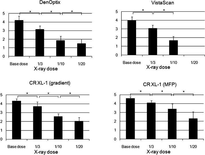
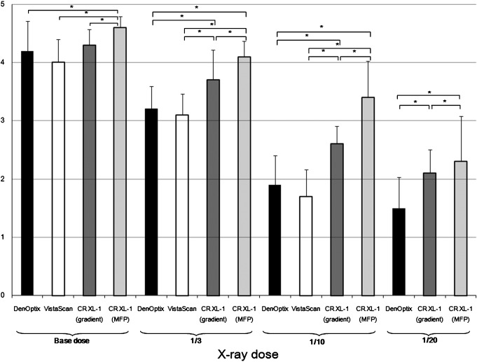
The results of the subjective visual assessment by six oral and maxillofacial radiologists for the images of each system and the different image processing conditions are shown in Figure 5. The diagnostic utility of the DenOptix QST images were slightly better than that of the conventional periapical X-ray film image at the base X-ray absorbed dose, and was almost equivalent to that of the film image when the X-ray absorbed dose was reduced to 1/3 of the base X-ray absorbed dose. The diagnostic utility of the VistaScan images was almost the same as that of the DenOptix QST images. The VistaScan could not form an image when the X-ray absorbed dose was 1/20 of the base X-ray absorbed dose. The diagnostic utility of the CR XL-1 images with simple gradient processing was more than slightly better at the base X-ray absorbed dose, and was close to slightly better at 1/3 of the base X-ray absorbed dose. When the X-ray absorbed dose was 1/10 and 1/20, the assessment values of simple gradient processed CR XL-1 images were less than equivalent to the film image. Only the MFP and FNC-processed CR XL-1 images had assessment values more than slightly better at the base and 1/3 X-ray absorbed doses. Furthermore, only the MFP and FNC-processed CR XL-1 image had an assessment value more than equivalent to film at the 1/10 X-ray absorbed dose. However, at the 1/20 X-ray absorbed dose even the MFP and FNC-processed CR XL-1 image did not receive an assessment value equivalent to film.
Figure 5.
Subjective visual assessment results for the three kinds of photostimulable phosphor luminescence imaging systems and a multi-objective frequency processing (MFP) and flexible noise control-processed image. Error bars indicate ±1 SD, and an asterisk (*) shows a statistically significant difference (P < 0.05) (n = 6)
Figure 6 shows a comparison of assessment results of all conditions at each X-ray absorbed dose. When the X-ray absorbed dose was the same as conventional X-ray film, the images from all conditions were evaluated as slightly better or greater than the film image. The CR XL-1 image with MFP and FNC had a better result than the other three kinds of images at all X-ray absorbed doses. In particular, only the MFP and FNC-processed CR XL-1 image was evaluated as being more than equivalent to the conventional film image, even at the 1/10 X-ray absorbed dose. However, when the X-ray absorbed dose was reduced to 1/20, the values of all images were lower than equivalent to the conventional film image, even the MFP and FNC-processed CR XL-1 image.
Figure 6.
Comparison of subjective visual assessment for the three kinds of photostimulable phosphor luminescence imaging systems and a multobjective frequency processing (MFP) and flexible noise control-processed image. Error bars indicate ±1 SD, and an asterisk (*) shows a statistically significant difference (P < 0.05) (n = 6)
There were no statistically significant differences in intraobserver and interobserver variability.
Discussion
The influence of X-ray dose reduction on the image quality of three PSPL imaging systems was evaluated in the present study. Before the subjective visual evaluation was performed, the fundamental performance of each system was evaluated by a physical experiment. The results of the subjective visual evaluation corresponded with the results of the physical evaluation. Namely, the scanning pixel size of the VistaScan was about four times finer than the other two systems, while the MTF was not as high. The subjective visual evaluation scores of the three systems also showed no significant difference at the base X-ray absorbed dose. These findings suggest that the MTF of a PSPL imaging system is not always dependent on the scanning pixel size, and the image quality of a PSPL imaging system in which the scanning pixel size is small is not necessarily good. When the X-ray absorbed dose was reduced to one-third or less, the visual evaluation scores of the CR XL-1 were significantly higher than those of the DenOptix and VistaScan. This finding cannot be explained from the results of MTF, but may be explained by the results of the NEQ and DQE analyses. The NEQ and DQE of the CR XL-1 were higher than those of the other two systems at 3 or 4 cycles/mm at all X-ray absorbed doses. A previous study found that the normal anatomical structures evaluated in the present study could be satisfactorily reproduced in images with a spatial resolution of less than 3 cycles/mm.14 It was found that diagnostic ability was also improved by enhancing the spatial frequency around 2 cycles/mm.15 Accordingly, the visual evaluation scores of the CR XL-1 were highest at the low X-ray absorbed dose, as this system had the highest NEQ and DQE under 3 cycles/mm at every X-ray absorbed dose. These results do not contradict the authors' past experimental result that the image quality and diagnostic ability were not improved even if the pixel size was improved from 50 μm to 25 μm.16 Thus, not only a reduction in pixel size, but also overall improvement in the system, including improvement of the signal to noise (S/N) ratio, is important for the amelioration of PSPL imaging systems. The DQE parameter, which is one of the physical evaluation methods used for digital X-ray images, may have inherent problems, as reported by Neitzel et al.17 Nonetheless, the DQE is an important physical evaluation method for the comparison of digital X-ray imaging systems, and it agrees with the result of visual evaluation of X-ray images comparatively well.18,19
The MFP and FNC have been reported to improve the diagnostic ability of X-ray images of PSPL imaging systems.8–11 The results of the present study indicate that MFP and FNC also improve the diagnostic utility of intraoral radiography using PSPL imaging systems. The MFP and FNC-processed images of the CR XL-1 had the highest visual evaluation scores at all X-ray absorbed doses. In particular, even at the 1/10 X-ray absorbed dose, the evaluation score of the MFP and FNC-processed image of the CR XL-1 were more than fair compared to conventional film images. Motohashi et al10 evaluated the usefulness of panoramic digital X-ray images obtained with MFP under different conditions to determine the anatomical structure of the dentomaxillofacial region. Although some differences were recognized between different MFP processing conditions, they concluded that MFP was useful to determine anatomical structures such as the apex of the dental root, periodontal membrane space, pulp canal, dentinoenamel junction, etc. on panoramic digital X-ray images. Their study differed from the present study in that they applied MFP to panoramic radiography, whereas in this study MFP was applied to intraoral radiography; however, the respective study results had similar tendencies.
In the present study, the diagnostic utility of X-ray images was evaluated only for normal anatomical structures of a single mandibular bone phantom. Therefore, it is necessary to investigate the diagnostic utility of such X-ray imaging systems for plural maxilla and mandibular bones. It will also be necessary to evaluate the diagnosability for various diseases, as the changes in contrast and changes in the fine structure of bones caused by diseases, such as dental caries, apical periodontitis and osteomyelitis, affect the diagnosis. The results of the present study suggest that PSPL imaging systems can reduce the X-ray dose to 1/3 of that of conventional periapical X-ray film, and that this can be further reduced to 1/10, and the resultant images improved, using MFP and FNC.
References
- 1.Borg E, Gröndahl HG. On the dynamic range of different X-ray photon detectors in intra-oral radiography. A comparison of image quality in film, charge-coupled device and storage phosphor systems. Dentomaxillofac Radiol 1996;25:82–88 [DOI] [PubMed] [Google Scholar]
- 2.Brettle DS, Workman A, Ellwood RP, Launders JH, Horner K, Davies RM. The imaging performance of a storage phosphor system for dental radiology. Br J Radiol 1996;69:256–261 [DOI] [PubMed] [Google Scholar]
- 3.Wenzel A. Digital imaging for dental caries. Dent Clin North Am 2000;44:319–338 [PubMed] [Google Scholar]
- 4.Wakoh M, Kuroyanagi K. Digital imaging modalities for dental practice. Bull Tokyo Dent Coll 2001;42:1–14 [DOI] [PubMed] [Google Scholar]
- 5.Versteeg CH, Sanderink GCH, van derStelt PF. Efficacy of digital intra-oral radiography in clinical dentistry. J Dent 1997;25:215–224 [DOI] [PubMed] [Google Scholar]
- 6.Ogoda M, Hishinuma K, Yamada M, Shimura K. Unsahrp masking technique using multiresolution analysis for computed radiography image enhancement. J Digit Imaging 1997;10:185–189 [DOI] [PMC free article] [PubMed] [Google Scholar]
- 7.Shimura K. Aim at ideal X-ray imaging: multi-objective frequency processing. Nippon Hoshasen Gijutsu Gakkai Zasshi 2001;57:796–802 [Google Scholar]
- 8.Tagashira H, Arakawa K, Yoshimoto M, Mochizuki T, Murase K, Yoshida H. Improvement of lung abnormality detection in computed radiography using multi-objective frequency processing: Evaluation by receiver operating characteristics (ROC) analysis. Eur J Radiol 2008;65:473–477 [DOI] [PubMed] [Google Scholar]
- 9.Yamada S, Murase K. Effectiveness of flexible noise control image processing for digital portal images using computed radiography. Br J Radiol 2005;78:519–527 [DOI] [PubMed] [Google Scholar]
- 10.Motohashi J, Mori S, Lee K, Kato M, Okano Y, Yamashiro M, et al. Assessment of panoramic radiograph using computed radiography with multi-objective frequency processing. Dent Radiol 2003;43:184–191 [Google Scholar]
- 11.Hagiwara S, Takizawa T, Osako M, Kaneda T, Kasai K. Evaluation of cephalogram using multi-objective frequency processing. Nichidai Koko Kagaku 2002;28:227–235 [Google Scholar]
- 12.International Electrotechnical Commission, Intrernational standard IEC62220-1: Medical electrical equipment - Characteristics of digital imaging devices-Part 1: Determination of the detective quantum efficiency. Geneva, Switzerland, 2003. [Google Scholar]
- 13.International Electrotechnical Commission, Intrernational standard IEC 61267 Ed 2. 0 b: Medical diagnostic X-ray equipment - Radiation conditions for use in the determination of characteristics. Geneva, Switzerland, 2005 [Google Scholar]
- 14.Kawahara E, Sakurai T. Spatial frequency components of normal radiographic anatomical features on intraoral computed radiography. Oral Radiol 1995;11:87–96 [Google Scholar]
- 15.Isu T, Sakurai T. Spatial frequency processing conditions on intraoral computed radiography. Oral Radiol 1996;12:27–38 [Google Scholar]
- 16.Sakurai T, Inagaki M, Asai H, Koyama A, Kashima I. Effects of scanning resolution and digital image magnification on photostimulable phosphor imaging system. Oral Radiol 2000;16:73–84 [Google Scholar]
- 17.Neitzel U, Günther-Kohfahl S, Borasi G, Samei E. Determination of the detective quantum efficiency of a digital x-ray detector: comparison of three evaluations using a common image data set. Med Phys 2004;31:2205–2211 [DOI] [PubMed] [Google Scholar]
- 18.Samei E, Flynn MJ. An experimental comparison of detector performance for computed radiography systems. Med Phys 2002;29:447–459 [DOI] [PubMed] [Google Scholar]
- 19.Vandre RH, Pajak JC, Abdel-Nabi H, Farman TT, Farman AG. Comparison of observer performance in determining the position of endodontic files with physical measures in the evaluation of dental X-ray imaging systems. Dentomaxillofac Radiol 2000;29:216–222 [DOI] [PubMed] [Google Scholar]


