Abstract
Objective
The present research was undertaken to study the accuracy and reliability of the foramen magnum (FM) and some cranial measurements in gender classification through the use of reconstructed helical CT images.
Methods
88 patients (43 males and 45 females; age range, 20–49 years) were selected for this study. FM sagittal diameter, transverse diameter, area and circumference were measured and data were subjected to discriminant analysis for gender using multiple regression analysis.
Results
FM circumference and area were the best discriminant parameters that could be used to study sexual dimorphism with an overall accuracy of 67% and 69.3%, respectively. By using multivariate analysis, 90.7% of FM dimensions of males and 73.3% of FM dimensions of females were sexed correctly.
Conclusion
It can be concluded that the reconstructed CT image can provide valuable measurements for the FM and could be used for sexing when other methods are inconclusive.
Keywords: foramen magnum dimensions, sexual dimorphism, helical computed tomography, craniometrics, sexing, forensics
Introduction
Gender determination in unidentified skeletons is not always an easily and correctly performed procedure. In explosions, warfare and other mass disasters, identification may be extremely complicated because of skeletal fragmentation.1 The skull, pelvis and femora are the most useful for radiological determination of gender. Radiography can assist in giving accurate dimensions for which certain formulae can be applied to determine gender.2 The length and the height of the head, the circumference of the head, the circumference of the occipital condyles and the foramen magnum (FM) have been used to determine gender in unidentifiable human remains.3-8 The FM is an important landmark of the skull base and is of particular interest in anthropology, anatomy, forensic medicine and other medical fields. Catalina-Herrera9 indicated that the sagittal and transverse dimensions of the FM were significantly higher in human male than in human female skulls. Zaidi and Dayal10 classified a sample of Indian skulls according to the shape and dimensions of the FM, reporting gender differences which were similar to those reported among Brazilian skulls.11 Günay et al4 examined the usefulness of determining the dimensions of the FM in the diagnosis of sex and noted that the diameters were of some use while the total area was not a good indicator. Yusal et al12 reported sexual dimorphism by analysing the dimensions of the FM in three-dimensional (3D) CT with 81% accuracy in determining the gender. The purpose of this study was to evaluate the accuracy of FM dimensions alone and/or in combination with other craniometric measurements in gender determination using helical CT scanning and to investigate the resultant accuracy among a sample of Iraqi adults.
Materials and methods
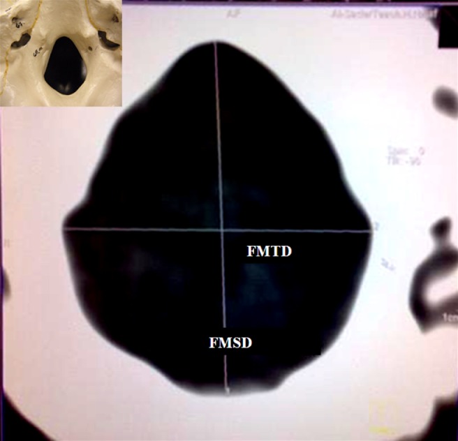
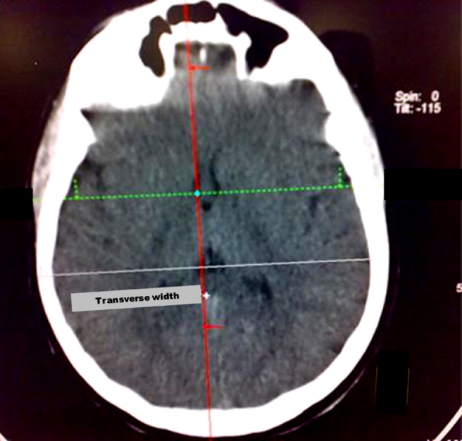
The studied sample consisted of 88 consensual patients (43 males and 45 females; age range, 20–49 years). They were referred to the radiology department in the Al-Najaf Teaching Hospital, Iraq, for the purpose of imaging the brain for several reasons. The study protocol was approved by the Local Ethics Committee of the Al-Najaf Teaching Hospital. Patients with history of trauma, surgery or pathological lesions in the region of the FM were excluded from the study. FM measurements (sagittal, transverse, circumference and area) were obtained from reformatted axial sections using helical CT scan (Somatom Emotion, Siemens, AG, Erlangen, Germany) with 5 mm thickness, 130 kVp, 200–230 mAs, 1800 AU window levels and 35–45 s scan time. All sections selected were parallel to the plane of the FM in order to select the best image of the foramen (Figure 1). The FM sagittal diameter (FMSD) was recorded as the greatest anteroposterior dimension of the FM and the FM transverse diameter (FMTD) was recorded as the greatest width of the FM. The circumference (FMC) and the area (FMA) were automatically given after tracing the bony margin of the FM on the CT image using a 3D program on the CT workstation with a resolution of 1280×1042 full screen format and picture size of 360×288 mm. Head width was also measured from the axial sections as a maximum transverse width at the Euryon point13 (Figure 2). Head circumference was measured clinically by a metric tape at the level of the glabella when the patient was in an upright position. To determine the reliability and the reproducibility of the FM and other craniometric measurements, an intra- and interexaminer calibration was done. This calibration was carried out by 2 radiologists (JF and AU), who compared the greatest measurements of 10 randomly selected radiographs. The radiographic images were examined on the computer monitor in dim light. To predict the gender based on the value of selected skull measurements, discriminant analysis was used. This type of analysis provides the following parameters:
Figure 1.

Sagittal and transverse diameter of the foramen magnum
Figure 2.

Head width measured from the axial section
P (regression model): assesses the statistical significance of each independent variable included in the model.
Wilk's lambda: a value range between 1 and 0. The closer the value is to zero, the more important the model is in discriminating between males and females.
Discriminant score (D): the value calculated from the provided equation using the value of different skull measurements provided. Its value is compared with an equivocal value of D (provided by the model). If it is smaller than the equivocal value, a female gender is expected; if it is greater, a male gender is expected. The more extreme the calculated D value is, the higher the probability that the predicted gender is correct.
Percentage of accurately predicted group membership: used to show the validity of the model in accurately predicting group membership (gender). It is equal to: (number of accurately predicted subjects of one gender type/number of total subjects belonging to that gender)×100%.
Functions at group centroid: the value of calculated D when the mean value of a selected skull measurement for a specific gender is used in the equation. It reflects how far apart the calculated D of each gender is from each other. The more distant the two functions, the more valid the equation.
Comparison of the intra- and interexaminer measurements showed no significant statistical differences (p>0.05) when paired t-test was applied. All data were subjected to descriptive and discriminate analysis using SPSS version 17 (IBM, Armonk, NY).
Results
A total of 88 individuals were studied (45 females and 43 males with an age range of 20–49 years); the results were based on 2 study samples. Six measurements were made by a senior radiologist and the metric parameters are shown in Tables 1 and 2. All measurements were significantly greater in males than in females.
Table 1. Gender difference for foramen magnum and other craniometric measurements.
| Female |
Males |
||||||||
| Variables | Range | Mean | SD | SE | Range | Mean | SD | SE | p Value (t-test) |
| Foramen magnum sagittal diameter (mm) | 26.9–38 | 32.9 | 2 | 0.31 | 29.3–40.8 | 34.9 | 2 | 0.3 | <0.001 |
| Foramen magnum transverse diameter (mm) | 22.3–31.8 | 27.3 | 2.2 | 0.33 | 24–34.8 | 29.5 | 2.5 | 0.39 | <0.001 |
| Foramen magnum circumference (mm) | 75.3–106.8 | 92.6 | 6.5 | 0.97 | 85.2–119.2 | 99.3 | 6.2 | 0.94 | <0.001 |
| Foramen magnum area (mm) | 455–900 | 670.2 | 93.7 | 13.97 | 536–1087 | 765.2 | 98 | 14.95 | <0.001 |
| Head circumference (cm) | 53.3–59.1 | 56.3 | 1.1 | 0.17 | 54.8–60.8 | 57.4 | 1.5 | 0.24 | <0.001 |
| Head width (cm) | 12.6–14.7 | 13.7 | 0.5 | 0.07 | 13.3–15.8 | 14.3 | 0.6 | 0.09 | <0.001 |
SD, standard deviation; SE, standard error.
Table 2. Correlation among tested variables of female group.
| Females | Foramen magnum sagittal diameter (mm) | Foramen magnum transverse diameter (mm) | Foramen magnum circumference (mm) | Foramen magnum area (mm) | Head circumference (cm) | Head Width (cm) |
| Foramen magnum sagittal diameter (mm) | 1 | 0.776** | 0.911** | 0.897** | 0.236 | −0.086 |
| Foramen magnum transverse diameter (mm) | 0.776** | 1 | 0.826** | 0.924** | 0.205 | 0.107 |
| Foramen magnum circumference (mm) | 0.911** | 0.826** | 1 | 0.951** | 0.078 | −0.103 |
| Foramen magnum area (mm) | 0.897** | 0.924** | 0.951** | 1 | 0.15 | −0.053 |
| Head circumference (cm) | 0.236 | 0.205 | 0.078 | 0.15 | 1 | 0.194 |
| Head width (cm) | −0.086 | 0.107 | −0.103 | −0.053 | 0.194 | 1 |
** Correlation is significant at the 0.01 level.
Pearson's correlation equation was applied for all FM measurements. All measurements were significantly correlated with each other (p<0.01). The strongest correlation was between FMC and FMA for males and females (r=0.972 and 0.951, respectively) and between FMSD and FMC (r=0.816 and 0.911 for males and females, respectively). The weakest correlations were between FMTD and FMSD (r=0.449 and 0.776 for males and females, respectively) (Tables 2 and 3). Craniometric measurements did not show any correlation with any FM measurements in both genders; however, significant direct relation was found between head circumference and head width in the male group. Multiple linear regression analysis combining all variables revealed highly significant differences between genders and all tested variables. By using multiple regression formulae and giving the variable (gender) a value (female =0 and male =1), with the application of craniometric and FM measurements as classification variables, the discriminant function scores were obtained from different formulae.
Table 3. Correlation among all tested variables of male group.
| Males | Foramen magnum sagittal diameter (mm) | Foramen magnum transverse diameter (mm) | Foramen magnum circumference (mm) | Foramen magnum area (mm) | Head circumference (cm) | Head Width (cm) |
| Foramen magnum sagittal diameter (mm) | 1 | 0.449** | 0.816** | 0.781** | −0.009 | −0.002 |
| Foramen magnum transverse diameter (mm) | 0.449** | 1 | 0.766** | 0.752** | −0.256 | 0.069 |
| Foramen magnum circumference (mm) | 0.816** | 0.766** | 1 | 0.972** | −0.172 | 0.085 |
| Foramen magnum area (mm) | 0.781** | 0.752** | 0.972** | 1 | −0.178 | 0.078 |
| Head circumference (cm) | −0.099 | −0.256 | −0.172 | −0.178 | 1 | 0.311* |
| Head width (cm) | −0.002 | 0.069 | 0.085 | 0.078 | 0.311* | 1 |
* Correlation is significant at the 0.05 level (two-tailed).
** Correlation is significant at the 0.01 level (two-tailed).
The equation provided by the model to calculate D will aid in the prediction process of gender by substituting the values of the specific measurement(s) in the equation. The resulting value of D is compared with a reference value (also provided by the model). A value of calculated D greater than reference D indicates male gender, while a value less than the reference value indicates female gender. The more extreme the calculated D value from the reference value, the higher the probability that the predicted gender is correct. The model calculated for all the parameters was statistically significant. Among the skull measurements included, FMC was the best discriminator, followed by FMA (Tables 4 and 5). Adding all FM measurements to the regression model gave the highest overall classification accuracy for gender (81.8 %). The equation provided for calculating D was as follows: D=−12.273+(0.136×FMSD)+(0.078×FMTD)+(0.165×FMC)+(−0.008×FMA); this is useful in classifying an unknown skull (after obtaining the selected measurements) into either male (if the discriminant score is>0.018) or female (if the discriminant score is<0.018). The confidence in male diagnosis is higher when the value of D is much higher than the decision value of 0.018, and the confidence in female diagnosis is higher when the value of calculated D is much lower (in the negative direction) than the decision value of 0.018 (Table 6).
Table 4. Discriminant analysis using FM and other craniometric measurements to discriminate between genders.
| FMSD | |||
| D=−16.826+0.496 × FMSD | |||
| Wilks' Lambda=0.802, p<0.001 | |||
| Percentage of accurately predicted group membership | Female | Male | Overall |
| 64.4% | 74.4% | 69.3% | |
| Functions at group centroids | Female | Male | Classified as male if D> |
| −0.48 | 0.503 | 0.012 | |
| FMTD | |||
| D=−11.912+0.42 × FMTD | |||
| Wilks' Lambda=0.83, p<0.001 | |||
| Percentage of accurately predicted group membership | Female | Male | Overall |
| 66.7% | 69.8% | 68.2% | |
| Functions at group centroids | Female | Male | Classified as male if D> |
| −0.437 | 0.457 | 0.01 | |
| FMC | |||
| D=−15.109+0.158 × FMC | |||
| Wilks' Lambda=0.781, p<0.001 | |||
| Percentage of accurately predicted group membership | Female | Male | Overall |
| 62.2% | 72.1% | 67% | |
| Functions at group centroids | Female | Male | Classified as male if D> |
| −0.512 | 0.536 | 0.012 | |
| FMA | |||
| D=−7.478+0.01 × FMA | |||
| Wilks' Lambda=0.799, p<0.001 | |||
| Percentage of accurately predicted group membership | Female | Male | Overall |
| 62.2% | 76.7% | 69.3% | |
| Functions at group centroids | Female | Male | Classified as male if D> |
| −0.484 | 0.507 | 0.012 |
D, discriminant score; FM, foramen magnum; FMA, FM area; FMC, FM circumference; FMSD, FM sagittal diameter; FMTD, FM transverse diameter.
Table 5. Discriminant analysis using craniometric measurements to discriminate between genders.
| Head circumference (head c) | |||
| D=−41.952+0.738 × head c | |||
| Wilks' Lambda=0.855, p<0.001 | |||
| Percentage of accurately predicted group membership | Female | Male | Overall |
| 62.2% | 62.8% | 62.5% | |
| Functions at group centroids | Female | Male | Classified as male if D> |
| −0.398 | 0.416 | 0.009 | |
| Head width (head w) | |||
| D=−27.079+1.94 × head w | |||
| Wilks' Lambda=0.729, p<0.001 | |||
| Percentage of accurately predicted group membership | Female | Male | Overall |
| 80% | 72.1% | 76.1% | |
| Functions at group centroids | Female | Male | Classified as male if D> |
| −0.589 | 0.616 | 0.014 |
D, discriminant score.
Table 6. Discriminant analysis using foramen magnum measurements to discriminate between males and females.
| Standardized coefficient | |||
| Foramen magnum sagittal diameter (FMSD) | 0.273 | ||
| Foramen magnum transverse diameter (FMTD) | 0.186 | ||
| Foramen magnum circumference (FMC) | 1.049 | ||
| Foramen magnum area (FMA) | −0.776 | ||
| Wilk's Lambda=0.601, p<0.001 | |||
| Percentage of accurately predicted group membership | Female | Male | Overall |
| 73.3 | 90.7 | 81.8 | |
| D=−12.273+(0.136 × FMSD)+(0.078 × FMTD)+(0.165 × FMC)+(−0.008 × FMA) | |||
| Functions at group centroids | Female | Male | Classified as male if D > |
| −0.787 | 0.823 | 0.018 | |
Discussion
Identification of skeletal and decomposing human remains is one of the most difficult skills in forensic medicine. Sex determination is also an important problem in the identification. If almost all the bones composing the skeleton are present, sex estimation is not difficult. When the skeleton exists completely, sex can be determined with 100% accuracy. This estimation rate is 98% in the existence of the pelvis and cranium, 95% with only the pelvis or the pelvis and long bones and 80–90% with only the long bones. However, in explosions, warfare and other mass disasters like aircraft crashes, identification and sex determination are not very easy.14 The study of anthropometric characteristics is of fundamental importance when solving problems related to identification. Craniometric features are included among these characteristics, which are closely connected to forensic medicine since they can be used to aid in identifying an individual from a skull found detached from its skeleton.15 Next to the pelvis, the skull is the most easily sexed portion of the skeleton, but the determination of the sex from the skull is not reliable until well after puberty. The craniofacial structures have the advantage of being composed largely of hard tissue, which is relatively indestructible.13 Sex estimation can be accomplished using either morphological or metric methodologies. Statistical methods using metric traits are becoming more popular, with most of the bones having been subjected to linear discriminant classification.16 Murshed et al17 studied FM dimensions using spiral CT and recorded the mean value of the FMSD (37.2 mm ± 3.43 mm in males and 34.6 mm ± 3.16 mm in females) and of the FMTD (31.6 mm ± 2.99 mm in males and 29.3 mm ± 2.19 mm in females). These results were higher than those recorded in the present study where FMSD was 34.9 mm ± 2 mm in males and 32.9 mm ± 2 mm in females and FMTD was 29.5 mm ± 2.5 mm in males and 27.3 mm ± 2.2 in females. This variation might be due to the different measurement techniques followed in their study (they used a millimetric sliding calliper). It was obvious that the mean value of FMSD and FMTD in males was significantly higher than in females among all studies of the FM.
Regarding FMC, the mean values were greater in males than in females (99.3 mm ± 6.2 mm vs 92.6 mm ± 6.5 mm). To our knowledge, this study was the first that used this measurement variable and no literature has previously discussed it. Catalina-Herrera9 reported that the mean values of the FMA found in male and female skulls were 888.4 mm2 and 801 mm2, respectively. These results were slightly higher than those of the present study. Günay et al4 measured the FMA directly on Turkish skulls, estimating it by considering it as a “circle” whose “radius” was obtained as the mean value between the half measurements of the length and the breadth; the results showed a mean value of 909.91 mm2 ± 126.02 mm2 for males and 819.01 mm2 ± 117.24 mm2 for females. These values were higher than those reported in the present study; such variation may be due to differences between the anatomic and radiographic methods. FM measurements on the dry skull done by Wanebo and Chicoine18 were very close to our results. Regarding craniometric measurements, there was a highly significant statistical difference in head width measurements between genders. Deshmukh and Devershi19 measured head width using sliding vernier callipers directly on the crania, which resulted in mean values of 13.1 cm ± 0.49 cm for males and 12.7 cm ± 0.49 cm for females. These values were lower than those recorded in the present study. Deshmukh and Devershi19 measured head circumference using standard flexible steel tape directly on crania which resulted in mean values of 49.6 cm ± 1.33 cm in males and 47.9 cm ± 1.55 cm in females. These values were much higher than those of the current study, which might be because of the difference between the anatomical and radiographic methods. For both genders, all FM measurements (sagittal diameter, transverse diameter, circumference and area) were positively correlated with each other (p<0.01). Murshed et al17 stated that the “area of the FM showed highly significant correlations with both sagittal diameter (r=0.847; p<0.01) and transverse diameter (r=0.834; p<0.01)”; these results agree with those of the present study. Among FM measurements, FMC was the best discriminator (Wilk's lambda =0.781 and overall accuracy =67%) followed by FMA (Wilk's lambda=0.799 and overall accuracy =69.3%). The accuracy rate of gender determination from FM measurements was 62.2% in females and 76.7% in males, with an overall accuracy rate of 69.3%. Gunay et al5 assessed the usefulness of FM size for gender determination and the accuracy rate was found to be 64.0% in females and 64.5% in males. Compared with the present study, the accuracy rate in females was higher by 1.8%, while the accuracy rate in males was lower than the present study by 12.2%. This may be owing to a result of the variation in the studied samples. Uysal et al12 studied the value and accuracy of the measurements of the FM by using 3D CT, taking seven measurements of the FM on 3D images. Using Fisher's linear discriminant functions test, the mean values of FM diameters were found to be statistically different in each sex (p<0.001), with a sex determination accuracy rate of 81%. These results were much better than those of the current study (69.3%) and this can be attributed to the fact that Uysal et al12 took 7 measurements of the FM using 3D CT, which yields better results.
The discriminant analysis of all the variables used in this study provided the highest accuracy of correct sex classification. By substituting the values of the measured variables, the accuracy rate would be 73.3% in females and 90.7% in males, with an overall accuracy rate of 81%, as seen in the following equation:
 |
Fernandes20 performed a gender-discriminant analysis using maxillary sinus measurements in addition to nasal cavity width, total distance across the sinuses, head circumference, head width, bizygomatic width at the zygion, a glabellar/nasion/nasal bone angle and a left and right lateral canthal angle, and he found that 79.2% of the skulls were correctly classified. Deshmukh and Devershi19 studied sexual dimorphism in adult human cranium by using 16 parameters measured directly on 74 crania of known sex including FM diameters, head width and head circumference. By using multivariate discriminant analysis, 90% of male crania and 85.29% of female crania were sexed correctly. Dayal et al16 used traditional anthropometric measurements (14 cranial and 6 mandibular measurements) for the assessment of sex using 120 skulls of black South Africans, and the application of discriminant function analyses resulted in average accuracies between 80% and 85%. These results were similar to those of the present study. In the field of forensic identification, these measurements can be taken without much difficulty. FM measurements can be taken with great speed and accuracy on a CT machine and standard instruments of measure. The results of the present study provide average accuracies that are comparable with those obtained using more complex techniques.
Conclusion
FM measurements are valuable in studying sexual dimorphism in forensic investigations. FM dimensions tend to stabilize after the second decade of life and the reconstructed CT images can provide reliable measurement of these dimensions.
References
- 1.Holland TD. Use of the cranial base in the identification of fire victims. J Forensic Sci 1984;29:1087–1093 [PubMed] [Google Scholar]
- 2.Di Vella G, Campobasso CP, Dragone M, Introna F., Jr Skeletal sex determination by scapular measurements. Boll Soc Ital Biol Sper 1994;70:299–305 [PubMed] [Google Scholar]
- 3.Quatrehomme G, Fronty P, Sapanet M, Grevin G, Bailet P, Olier A. Identification by frontal sinus pattern in forensic anthropology. Forensic Sci Int 1996;83:147–153 [DOI] [PubMed] [Google Scholar]
- 4.Günay Y, Altinkök M, Çagdir S, Kirangil B. Gender determination with skull measurements (in Turkish). J Forensic Med 1997;13:13–19 [Google Scholar]
- 5.Günay Y, Altinkök M, Çagdir S, Sari H. Is foremen magnum size useful for gender determination (in Turkish). Bull Legal Med 1998;3:41–45 [Google Scholar]
- 6.Cameriere R, Ferrante L, Mirtella D, Rollo UF, Cingolani M. Frontal sinuses for identification: quality of classifications, possible error and potential corrections. J Forensic Sci 2005;50:770–773 [PubMed] [Google Scholar]
- 7.Rogers LT. Determining the sex of human remains through cranial morphology. J Forensic Sci 2005;50:493–500 [PubMed] [Google Scholar]
- 8.Gruber P, Henneberg M, Böni T, Rühli F. Varaibility of human foramen magnum size. The Anat Rec 2009;292:1713–1719 [DOI] [PubMed] [Google Scholar]
- 9.Catalina-Herrera CJ. Study of the anatomic metric values of the foramen magnum and its relation to sex. Acta Anat 1987;130:344–347 [DOI] [PubMed] [Google Scholar]
- 10.Zaidi SH, Dayal SS. Variations in the shape of foramen magnum in Indian skulls. Anat Anz Jena 1988;167:338–340 [PubMed] [Google Scholar]
- 11.Manoel C, Prado FB, Caria PHF, Groppo FC. Morphometric analysis of the foramen magnum in human skulls of Brazilian individuals: its relation to gender. Braz J Morphol Sci 2009;26:104–108 [Google Scholar]
- 12.Uysal S, Gokharman D, Kacar M, Tuncbilek I, Kosa U. Estimation of sex by 3D CT measurements of the foramen magnum. J Forensic Sci 2005;50:1310–1314 [PubMed] [Google Scholar]
- 13.Patil KR, Mody RN. Determination of sex by discriminant function analysis and stature by regression analysis: a lateral cephalometric study. Am J Orthod Dentofacial Orthop 2005;128:157–160 [DOI] [PubMed] [Google Scholar]
- 14.Krogman WM, Iscan MY. The human skeleton in forensic medicine. 2nd edn Springfield, Illinois: Charles C Thomas Publishing; 1986 [Google Scholar]
- 15.Ono I, Ohura T, Narumi E, Kawashima K, Matsuno I, Nakamura S, et al. Analysis of craniofacial bones using three dimensional computed tomography. J Cranio Maxillofacial Surg 1992;20:49–60 [DOI] [PubMed] [Google Scholar]
- 16.Dayal MR, Bidmos MA. Discriminating sex in South African blacks using patella dimensions, J Forensic Sci 2005;50:209–221 [PubMed] [Google Scholar]
- 17.Murshed KA, Çıçekcıibaşi AE, Tuncer I. Morphometric evaluation of the foramen magnum and variations in its shape: a study on computerized tomographic images of normal adults. Turk J Med Sci 2003;33:301–306 [Google Scholar]
- 18.Wanebo JE, Chicoine MR. Quantitative analysis of the transcondylar approach to the foramen magnum. Neurosurgery 2001;49:934–941 [DOI] [PubMed] [Google Scholar]
- 19.Deshmukh AG, Devershi DB. Comparison of cranial sex determination by univariate and multivariate analysis. J Anat Soc India 2006;55:1–5 [Google Scholar]
- 20.Fernandes CL. Forensic ethnic identification of crania. The role of the maxillary sinus—a new approach. Am J Forensic Med Pathol 2004;25:302–313 [DOI] [PubMed] [Google Scholar]


