Abstract
Objective
The purpose of this study was to review the clinical and radiographic features of solitary and COD-associated SBCs.
Methods
Archived imaging reports from the Special Procedures Clinic in Oral and Maxillofacial Radiology at the Faculty of Dentistry at the University of Toronto between 1 January 1989 and 31 December 2009 revealed 23 COD-associated SBCs and 68 solitary SBCs.
Results
Almost all solitary and COD-associated SBCs were found in the mandible. Furthermore, 87.0% of COD-associated SBCs were found in females in their fifth decade of life (P < 0.001) while solitary SBCs were found in equal numbers in both sexes in their second decade of life (P < 0.005). COD-associated SBCs were also more likely to cause thinning of the endosteal cortex, bone expansion and scalloping of the superior border between teeth (all P < 0.001) than solitary SBCs that are classically described as having these characteristics. Finally, COD-associated SBC demonstrated a loss of lamina dura more often (P < 0.05) than solitary SBCs.
Conclusions
Knowledge of the sporadic association between COD and SBC and their potential radiographic appearances should prevent inappropriate treatment and management of these patients.
Keywords: jaw disease, jaw cysts, non-odontogenic cysts
Introduction
The simple bone cyst (SBC) is a pseudocyst that may be found in both the jaws and the long bones.1-4 SBCs present radiographically as well-defined radiolucent entities with delicate cortical borders. In the jaws, SBCs are described as having a characteristic scalloping pattern around the roots of nearby teeth, leaving the lamina durae, periodontal ligament spaces or follicular spaces undisturbed.2 Some or all of these features are considered radiographically pathognomonic of the entity. In the long bones, simple bone cysts may present with pain should the lesion cause pathological bone fracture.5 In the jaws, SBCs are often identified incidentally in young patients in whom imaging is obtained as part of orthodontic diagnosis or treatment planning for third molar extraction.
The aetiology of the SBC is not known and as such it has been referred to by many different synonyms, including haemmorraghic bone cyst, traumatic bone cyst, solitary bone cyst/cavity, idiopathic bone cyst/cavity and unicameral bone cyst/cavity. Currently, there are three hypotheses related to the development of SBCs.6-8 First, that a local disturbance in osteoblast differentiation during bone growth and development, possibly related to mechanical factors of the bone, gives rise to the bone cavity. Second, that a developing tumour undergoes liquifactive degeneration, leaving behind an empty cavity. And finally, a traumatic or microtraumatic event fails to induce a bone fracture. This event is believed to cause a localized thrombosis, giving rise to a focal ischaemic event and aseptic necrosis of the bone. In the jaws, it has been suggested that the mandible is more commonly affected than the maxillae because it is subjected to more microtraumatic events, particularly in the premolar areas.8 Presumably, this correlates with the increased incidence of SBCs here. The end result is that the region of developing necrosis results in the formation of a cavity that may be lined by fibrous connective tissue and contain blood and/or serous fluid. In the jaws, SBCs may contain nothing at all.6
From a treatment standpoint, SBCs may be monitored without intervention or they can be entered surgically, curetted and closed. Fenestration of the bone surface and packing of the cavity with materials such as Gelfoam® gelatin sponge (Pfizer, New York, NY) or gauze has also been performed.12 Suei et al9 recently reviewed 108 solitary SBC cases from the literature and 31 of their own cases in an attempt to correlate radiographic characteristics with rates of SBC recurrence. These workers concluded that SBCs could be classified into two groups based on rates of recurrence. First, in cases with intact lamina dura and smooth margins with smooth or no bone expansion, such lesions healed after surgery and did not recur. In contrast, lesions where the lamina dura was absent, or where there was root resorption, scalloped borders, “nodular” expansion or the presence of a radiopaque mass or multiple lesions, recurrence was shown to occur with greater frequency.
SBC has been reported to occur with fibrous dysplasia and cemento-osseous dysplasias (COD), although reports of these associations are sporadic given the lack of large subject sample sizes.9,10 In the long bones, several authors have suggested that the nature of SBCs occurring in the epiphyses and metaphyses of long bones, as well as those occurring within the jaws, may represent discrete and separate entities given their clinical presentations and rates of recurrence.7-9
The purpose of this study is twofold. First, we aim to describe the clinical and radiographic features of a series of solitary and COD-associated SBCs. And second, based on the clinical and radiographic data obtained for both subject groups, we propose a hypothesis for the development of solitary and COD-associated SBCs.
Materials and methods
In total, 68 subjects with solitary SBCs and 23 subjects with COD-associated SBCs were identified from the Special Procedures Clinic database (Filemaker Pro, Database Software Solutions, Santa Clara, CA) in the Discipline of Oral and Maxillofacial Radiology, Faculty of Dentistry, the University of Toronto between 1 January 1989 and 31 December 2009. Clinical/epidemiological data were obtained from written clinical records, archived imaging reports and available radiographic images. The ethnic backgrounds of our subjects were not collected as these data are generally not collected at our institution for our patients.
Of the 68 cases of solitary SBC, 39 image sets were available for review. All 23 image sets were available for subjects with COD-associated SBCs. Imaging studies consisted of at least a panoramic radiograph; however, periapical, occlusal and skull views as well as CT were reviewed when available. The images were reviewed by two oral and maxillofacial radiologists. The study was approved by the Institutional Health Sciences Review Board of the University of Toronto.
Histopathological reports were only available for a few cases of solitary SBC and for none of the COD-associated SBC cases. Given the nature of SBCs, the majority of solitary lesions were monitored and no surgical intervention occurred. With respect to the COD-associated SBCs, no histopathological information was available since the diagnosis of COD is made radiographically rather than histopathologically.11
Subject sex and age data were collected from clinical records, and the following radiographic features were assessed:
The geometric lesional centre was scored on a graphic representation of the jaws (Figure 1).
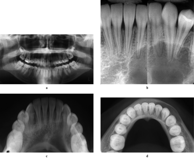
The presence or absence of cortical thinning. Thinning was defined as a loss of the normal thickness of a cortical bone border such that the endosteal surface of this border has undergone a shallow, broad-based thinning (Figure 2).
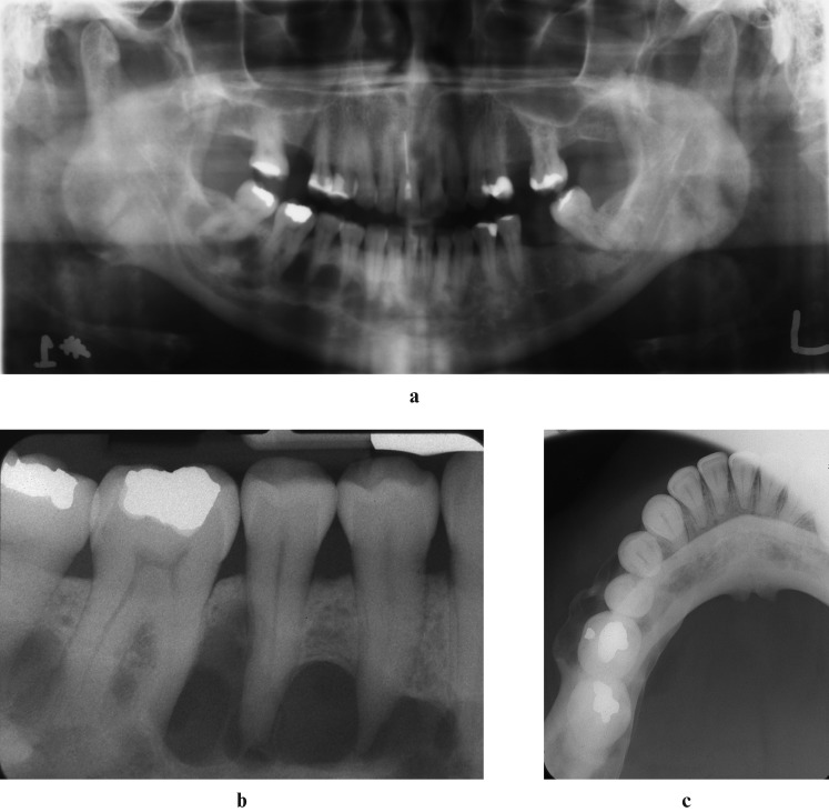
The presence or absence of bone expansion. Expansion was defined as a localized or generalized increase in the area of a bone (Figure 3).
The presence or absence of tooth root scalloping. Scalloping was defined as the appearance of an undulating or sinusoidal-like contour to the superior border of the SBC between or around the roots of adjacent teeth (Figure 3).
The presence or absence of visible lamina dura (Figure 3).
The presence or absence of visible periodontal ligament spaces (Figure 3).
Figure 1.
Distributions of (a) solitary and (b) cemento-osseous dysplasia (COD)-associated simple bone cysts (SBCs). The lesional centre of each SBC is indicated with an “X”
Figure 2.
(a) Panoramic; (b) periapical; (c) anterior occlusal; and (d) true occlusal radiographs of a solitary simple bone cyst (SBC). The SBC has a well-defined corticated border and has caused endosteal scalloping of the buccal and inferior cortices of bone as well as subtle expansion of the mandible. There have been no significant effects on the periodontal ligament spaces and lamina dura of adjacent teeth
Figure 3.
(a) Panoramic; (b) periapical; and (c) true occlusal radiographs of a simple bone cyst (SBC) associated with cemento-osseous dysplasia (COD). Note the well-defined corticated appearance of the SBC in the right posterior mandible. There has been scalloping of the superior border of the SBC around the roots of the premolar teeth and the non-uniform nodular expansion of the buccal cortex of the mandible
Statistical testing of the data was performed using SPSS (SPSS Inc., Chicago, IL). Independent t-tests were used to evaluate differences in sex and mean subject age data. χ2 testing with one degree of freedom was used to evaluate differences between the two subject groups (solitary SBC and COD-associated SBC) with respect to patient sex and the incidences of the radiographic features outlined above. For all tests, statistical relationships were determined to be significant when P < 0.05.
Results
Solitary SBC was identified in equal proportions in males and females, with mean ages of 18.2 years and 18.8 years, respectively (range 11–44 years for males and 11–58 years for females). These differences were not statistically significantly different. COD-associated SBCs were found in more than six times as many females as males (87.0% vs 13.0%). These differences were significant to P < 0.001. Furthermore, COD-associated SBCs occurred more frequently in older subjects than solitary SBCs (P < 0.005). These findings are summarised in Table 1. Furthermore, in our subject populations there were no clinical self-reports of prior tumours or mandibular trauma in areas where SBCs developed.
Table 1. Subject population number and age distribution of solitary and cemento-osseous dysplasia (COD)-associated simple bone cysts (SBCs).
| Sex | Number | Age (range) (years) | |
| Male* | Solitary SBC | 34 | 18.2 (11–44) |
| SBC/COD | 3 | 47.0 (42–52) | |
| Female* | Solitary SBC | 34 | 18.8 (11–58) |
| SBC/COD | 20 | 41.5 (30–52) |
*P < 0.001
Figure 1 shows the distributions of the lesional centres of the SBCs. Of the total 68 solitary SBCs and 23 COD-associated SBCs, only 1 SBC from each group was identified in the maxilla. For solitary SBCs, 98.5% were found in the mandible and for COD-associated SBCs, 95.7% were found in the mandible. Interestingly, we found that solitary SBCs arose in all areas of the mandible whereas COD-associated SBCs were not found in the incisor area of the mandible.
COD-associated SBCs demonstrated thinning of an adjacent bone cortex (Figure 2d), bone expansion (Figure 3c) and tooth root scalloping (Figure 3a,b) more frequently than solitary SBCs (all P < 0.001). Also, COD-associated SBCs resulted in a loss of lamina dura (Figure 3b) of adjacent teeth more frequently than solitary SBCs (P < 0.05). These results are summarised in Table 2.
Table 2. Incidence of radiographic features of solitary and cemento-osseous dysplasia (COD)-associated simple bone cysts (SBCs).
| Feature | Solitary SBC % | SBC/COD % |
| Cortical thinning* | 48.5 | 78.2 |
| Bone expansion* | 17.6 | 60.8 |
| Tooth root scalloping* | 54.4 | 82.6 |
| Loss of periodontal ligament space | 2.9 | 7.9 |
| Loss of lamina dura** | 11.8 | 22.4 |
*P < 0.001; **P < 0.05
Discussion
Several authors have already examined the epidemiological, clinical and histopathological presentations of the solitary SBC in both the jaws and the long bones1,3-5 and their rates of recurrence.9,12,13 These data have enabled some workers to speculate about the nature of gnathic and extra-gnathic SBCs and whether or not they are the same or different entities.5-7 The clinical data from our subject group with solitary SBCs is in general agreement with other previously published case series; that is, SBCs occur in a generally younger age group of patients (mean: 18.2 years in males and 18.8 years in females) and that the male-to-female distribution is 1:1. Previous studies have also mapped the distributions of SBCs to the mandible primarily with few occurrences in the maxilla or away from the tooth-bearing areas of the jaws.1,2 Our data confirm these earlier findings. In only 1 of our solitary SBC cases did we identify an occurrence in the maxilla; the other 67 occurred in and around the tooth-bearing areas of the mandible.
The classic radiographic presentation of the SBC is frequently described as having a well-defined, delicate corticated border. Furthermore, the superior border of the entity is described as having a characteristic scalloping contour between the roots of nearby teeth, leaving periodontal ligament spaces, lamina dura of nearby teeth and the follicles of adjacent unerupted teeth relatively undisturbed.2 Our case series demonstrates that only 48.5% of solitary SBCs had an identifiable corticated border and that 54.4% demonstrated the tooth root scalloping that has been described as being pathognomonic of SBCs. The disagreement of our data with the classical descriptions of SBC may be related to the nature of our specialty referral clinic and the patients we see. Classically appearing examples of SBC may not be referred to our clinic if general dentists or other specialists are comfortable interpreting such entities and managing these patients. More often, patients with SBCs that may not conform to classic appearances may be referred to our clinic for a specialist opinion. A small minority (17.6%) of these subjects' SBCs demonstrated bone expansion, with 2.9% demonstrating a loss of the periodontal ligament space and 11.8% demonstrating loss of lamina dura. The low incidence of bone expansion, lamina dura and periodontal ligament space effects are also well-described features of the solitary SBC.2
Where our study differs from others cited here are the comparisons we are able to make between a sample of solitary SBCs and those arising in association with COD. While this association has been referenced in previously published work,9,10 there are no case series describing the clinical and radiographic features of COD-associated SBCs. In our subject population, 87.0% of COD-associated SBCs occurred in females. This finding is not particularly surprising since COD has a strong predilection for females. Furthermore, the mean ages of subjects presenting with COD-associated SBC were 41.5 years and 47 years for men and women, respectively. The occurrence of COD-associated SBCs in men and women in their fifth decades of life is significantly different than the age demographics of solitary SBCs in our other subject population (P < 0.001). Moreover, the differences in the male-to-female ratios of solitary and COD-associated SBC were also statistically significantly different (P < 0.005).
Solitary SBCs were found incidentally on radiographs made for other purposes and this was also often the case for COD-associated SBCs. In some subjects, COD-associated SBCs were referred to our facility for investigation of a painless mandibular swelling. With respect to the anatomical distributions of COD-associated SBCs, the overwhelming majority (95.7%) occurred in the mandible, with only one SBC occurring in the maxilla. Interestingly, COD-associated SBCs were not found in the incisor areas of the mandible in our population. This suggests that SBCs may not be related to the occurrence of lesions of periapical COD which typically affect the mandibular incisors only. Rather, the association with the premolar and molar teeth primarily suggests a relationship with the florid subtype of COD. In light of the anatomical distribution of solitary SBCs, the dearth of COD-associated SBCs arising in the mandibular incisor area is interesting.
When associated with COD, 78.2% of SBCs demonstrated corticated borders and 82.6% scalloped around the roots of nearby teeth. The occurrence of these imaging features compared with those found for solitary SBCs is statistically significantly different (P < 0.001). Our data suggest that COD-associated SBCs are more often associated with these classic descriptors of SBCs than solitary SBCs. We suggest that these features may occur more frequently in COD-associated SBCs because these SBCs may be larger than solitary SBCs when first identified. Solitary SBCs may be smaller because they are found earlier, perhaps because younger patients may have radiographs made more frequently during this period of their lives for orthodontic treatment planning or third molar extraction. This hypothesis is further supported by our other data that shows 60.8% of COD-associated SBCs were found to produce buccal/lingual expansion of the jaws compared with 17.6% of solitary SBCs. These differences are significant to P < 0.001. Finally, COD-associated SBCs appeared to affect the integrity of the lamina dura twice as often as solitary SBCs (P < 0.05), although in general the incidence of these findings are small.
In summary, our data suggest that COD-associated SBCs have a higher incidence in females in their fifth decade of life than solitary SBCs, which are found equally in males and females in the second decade of life. Our data also show that unlike solitary SBCs that arise in all regions of the mandible, COD-associated SBCs do not occur in the mandibular incisor area. And finally, COD-associated SBCs present more frequently with the radiographic appearances that are classically associated with solitary SBCs; that is, their borders are corticated and they scallop around the roots of nearby teeth. Given these differences in the clinical and radiographic presentations of solitary and COD-associated SBCs, are these two populations of SBCs the same entities, or are they separate lesions as some have hypothesized for metaphyseal and epiphyseal SBCs of the long bones and in extragnathic sites?5,7,8
We believe that our data favour the hypothesis that relates the formation of SBCs (whether solitary or COD-associated) to a disturbance of cellular (i.e. osteoblast and osteoclast) differentiation during growth and ossification of the mandible.8 In our subject populations, there were no clinical self-reports of prior tumours or mandibular trauma in areas where SBCs developed. With regard to the hypothesis that SBC formation is related to microtraumatic events occurring in the jaws, Harnet et al8 have suggested that most SBCs arise in the premolar areas because this part of the jaw in particular is subject to higher numbers of microtraumatic events as a result of higher bite forces. We believe this to be an inaccurate supposition in light of bite force data that suggest that the largest bite forces are generated in the first molar and not in the premolar areas. Furthermore, our data found that solitary SBCs also arose in the mandibular incisor areas where bite forces are the lowest anywhere in the jaws.14
We further hypothesize that solitary and COD-associated SBCs may be the same lesion but arise owing to different biological circumstances. In adolescents, we hypothesize that a disturbance or decoupling of normal osteoblast and osteoclast activities may be related to new and constantly changing biomechanical properties of the mandible during growth and development, and the inability of bone cells, in particular osteoblasts, to keep up with these demands. In skeletally mature subjects, and in particular females in whom COD-associated SBCs are significantly more common, the development of SBC may again reflect a decoupling of normal cellular activity in the bone but for a different reason. Here, the decoupling of bone cells may be owing to a dearth of osteoprogenitor cells in the jaws of these subjects at this time of their lives. That is, low or inadequate osteoblast numbers may be more commonplace in the bones of middle aged, potentially osteoporotic women. Consequently, in both adolescents and middle-aged women, the formation of an empty cavity in bone may be related to the inability of osteoblasts to keep up with the demand for bone mineral deposition during normal remodeling processes taking place in the jaws of these two subject populations.
Conclusion
We have presented the clinical and radiographic findings of two case series of subjects with solitary SBC and COD-associated SBCs. Solitary SBCs occurred more commonly in males and females in their second decade of life than COD-associated SBCs, which were more commonly seen in females in the fifth decade of life. Furthermore, COD-associated SBCs were more commonly found in females than males by a factor of almost 7:1. COD-associated SBCs showed the classical radiographic features of SBC more commonly than solitary SBCs, possibly because they were identified later. Given our data, we hypothesize that both solitary and COD-associated SBCs arise as a result of a decoupling of normal osteoblast and osteoclast function.
Acknowledgments
JW Chadwick acknowledges partial support from the Bertha Rosenstadt Fund, Faculty of Dentistry, the University of Toronto.
References
- 1.MacDonald-Jankowski DS. Traumatic bone cysts in the jaws of a Hong Kong Chinese population. Clin Radiol 1995;50:787–791 [DOI] [PubMed] [Google Scholar]
- 2.Pharoah MJ. Cysts and cyst-like lesions of the jaws. In: White SC, Pharoah MJ. Oral radiology: principles and interpretation. Philadelphia, PA: Mosby, 2009, pp 361–364 [Google Scholar]
- 3.Forssell K, Forssell H, Happonen RP, Neva M. Simple bone cyst. Review of the literature and analysis of 23 cases. Int J Oral Maxillofac Surg 1988;17:21–24 [DOI] [PubMed] [Google Scholar]
- 4.Suei Y, Taguchi A, Tanimoto K. A comparative study of simple bone cysts of the jaw and extracranial bones. Dentomaxillofac Radiol 2007;36:125–129 [DOI] [PubMed] [Google Scholar]
- 5.Lokiec F, Wientroub S. Simple bone cyst: etiology, classification, pathology, and treatment modalities. J Pediatr Orthop B 1998;7:262–273 [PubMed] [Google Scholar]
- 6.Shimoyama T, Horie N, Nasu D, Kaneko T, Kato T, Tojo T, et al. So-called simple bone cyst of the jaw: a family of pseudocysts of diverse nature and etiology. J Oral Sci 1999;41:93–98 [DOI] [PubMed] [Google Scholar]
- 7.Abdel-Wanis ME, Tsuchiya H. Simple bone cyst is not a single entity: point of view based on a literature review. Med Hypoth 2002;58:87–91 [DOI] [PubMed] [Google Scholar]
- 8.Harnet J-C, Lombardi T, Klewansky P, Rieger J, Tempe M-H, Clavert J-M. Solitary bone cyst of the jaws: a review of the etiopathogenic hypotheses. J Oral Maxillofac Surg 2008;66:2345–2348 [DOI] [PubMed] [Google Scholar]
- 9.Suei Y, Taguchi A, Nagasaki T, Tanimoto K. Radiographic findings and prognosis of simple bone cysts of the jaws. Dentomaxillofac Radiol 2010;35:55–61 [DOI] [PMC free article] [PubMed] [Google Scholar]
- 10.Melrose RJ, Abrams AM, Mills BG. Florid osseous dysplasia. A clinical-pathologic study of thirty-four cases. Oral Surg Oral Med Oral Pathol 1976;41:62–82 [DOI] [PubMed] [Google Scholar]
- 11.Waldron CA. Fibro-osseous lesions of the jaws. J Oral Maxillofac Surg 1993;51:828–835 [DOI] [PubMed] [Google Scholar]
- 12.Suei Y, Taguchi A, Tanimoto K. Simple bone cyst of the jaws: evaluation of treatment outcomes by review of 132 cases. J Oral Maxillofac Surg 2007;65:918–923 [DOI] [PubMed] [Google Scholar]
- 13.Horner K, Forman GH, Smith NJD. Atypical simple bone cysts of the jaws: recurrent lesions. Clin Radiol 1988;39:53–57 [DOI] [PubMed] [Google Scholar]
- 14.Blamphin CN, Brafield TR, Jobbins B, Fisher J, Watson CJ, Redfern EJ. A simple instrument for the measurement of maximum occlusal force in human dentition. Proc Inst Mech Eng H 1990;204:129–131 [DOI] [PubMed] [Google Scholar]


