Abstract
Objective
The aim of this study was to evaluate the influence of thyroid collars on radiation dose during cone beam CT (CBCT) scanning.
Methods
Average tissue-absorbed dose for a NewTom 9000 CBCT scanner (Quantitative Radiology, Verona, Italy) was measured using thermoluminescent dosemeter chips in a phantom. The scans were carried out with and without thyroid collars. Effective organ dose and total effective dose were derived using International Commission on Radiological Protection 2007 recommendations.
Results
The effective organ doses for the thyroid gland and oesophagus were 31.0 µSv and 2.4 µSv, respectively, during CBCT scanning without a collar around the neck. When the thyroid collars were used loosely around the neck, no effective organ dose reduction was observed. When one thyroid collar was used tightly on the front of the neck, the effective organ dose for the thyroid gland and oesophagus were reduced to 15.9 µSv (48.7% reduction) and 1.4 µSv (41.7% reduction), respectively. Similar organ dose reduction (46.5% and 41.7%) was achieved when CBCT scanning was performed with two collars tightly on the front and back of the neck. However, the differences to the total effective dose were not significant among the scans with and without collars around the neck (p = 0.775).
Conclusions
Thyroid collars can effectively reduce the radiation dose to the thyroid and oesophagus if used appropriately.
Keywords: cone beam computed tomography, radiation, radiation dosimetry, thyroid gland
Introduction
Cone beam CT (CBCT) can provide three-dimensional (3D) information about the facial skeleton and teeth. This technology has been introduced as an alternative imaging technique for diagnostic tasks including oral surgery, oral medicine, endodontics, periodontology, orthodontics and implantology.1-6
The increasing use of the CBCT technique in dentistry carries the risk of over radiation dose to the patient, which should be one of the dentist's great concerns. Radiation dose should be reduced to a minimum without loss of diagnostic information during imaging. Owing to the fact that dose minimization is more important for children and young adults who are more sensitive to radiation, concerns about the effective doses from different CBCT scanners have arisen.7,8 To reduce the radiation dose, some techniques were employed by the manufacturers, such as decreasing tube voltage and current, altering collimation and filtration judiciously and using pulsating technology to make the exposure time shorter.9 In addition, the use of a lead shield device was found to be significantly effective in reducing the absorbed doses to the thyroid and cervical spine during CBCT scanning.7
Thyroid shielding is recommended by the National Council for Radiation Protection and Measurements when it does not interfere with exposure.10 A thyroid collar was found to reduce radiation doses significantly during CT scanning of the head.11–14 However, until now there have been no studies published to evaluate the radioprotective effects of thyroid collars during CBCT scanning. The aim of this study was to evaluate the radiation dose of CBCT when applying thyroid collars.
Materials and methods
CBCT device
The CBCT device employed in the study was the NewTom 9000 (Quantitative Radiology, Verona, Italy).15 This device uses a cone-shaped X-ray beam centred on an area detector. The detector is an image intensifier (9 inch) coupled with a solid-state (CCD) television camera. The tube-detector system performs one 360° rotation scan to acquire images that are then used for the reconstruction of the examined volume. The reconstruction volume is a 15 cm-high cylinder with a diameter of 15 cm. The rotation time is about 36 s. However, the real exposure time is determined by the bone volume and density with a pulsating technique. By using this technique, the tube voltage and tube current are fixed at 110 kV (peak) and 3.5 mA, respectively, by the manufacturer.
Phantom
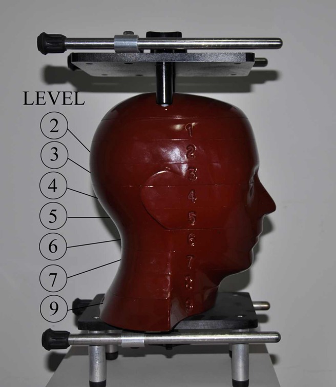
An anthropomorphic adult human male phantom (ART-210; Radiology Support Devices, Inc., Long Beach, CA) (Figure 1) was used in the study. The phantom had tissue equivalent X-ray attenuating characteristics and closely conformed to the International Commission on Radiation Units and Measurements specifications.16
Figure 1.

Anthropomorphic adult human male phantom. Levels correspond to thermoluminescent dosemeter sites identified in Table 1
Thyroid collar shielding technique
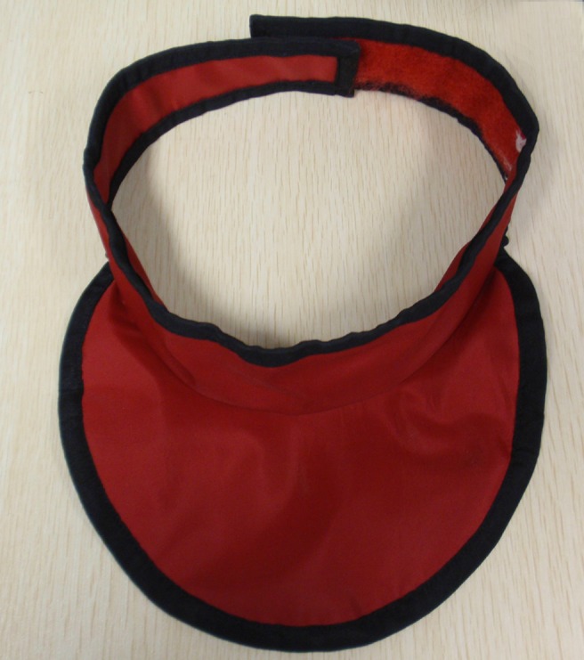
The dentomaxillofacial area (scanning height from nasal root to inferior border of the mandible) of the phantom was scanned with the NewTom 9000 CBCT imaging system with and without applying 0.35 mmPb thyroid collars (model HRNG-I; Beijing Huaren Health Science & Technology Developing Co., Ltd., Beijing, China) (Figure 2). To obtain maximum protection, the collars were placed as close as possible to the neck surface of the phantom. Scans with the collars loosely placed on the neck surface were also carried out. Five scans were completed:
Figure 2.

The thyroid collar used (Model HRNG-I; Beijing Huaren Health Science & Technology Developing Co., Ltd., Beijing, China)
1. without a collar around the neck
2. with one collar loosely on the front of the neck
3. with two collars loosely on the front and back of the neck
4. with one collar tightly on the front of the neck
5. with two collars tightly on the front and back of the neck.
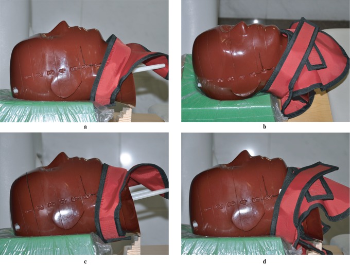
During each scan, a step wedge was placed under the phantom to make the inferior border of the mandible perpendicular to the horizontal plane. The placement of the thyroid collars and the geometric relationship of the phantom, thyroid collars and step wedge are shown in Figure 3.
Figure 3.
The placement of thyroid collars and geometric relation of the phantom, thyroid collars and step wedge. (a) With one collar loosely on the front of the neck; (b) with two collars loosely on the front and back of the neck; (c) with one collar tightly on the front of the neck; and (d) with two collars tightly on the front and back of the neck
Absorbed dose measurement
The absorbed doses were measured using thermoluminescent dosemeter (TLD) chips (LiF:Mg,Cu,P). Before the study, all dosemeters were calibrated using cobalt-60 source. 3 chips were positioned at 21 locations within the head and neck region of the phantom. The method presented by Ludlow8 was used to position the TLD chips (Table 1). Prior to loading, the TLDs were annealed at 240 °C for 10 min and then cooled immediately to an ambient temperature. All TLDs were read within 90 min after each exposure using a BR2000D reader (Beijing Bochuangte Science & Technology Development Co., Ltd., Beijing, China). The consistency of dose measurement by the TLD system has been evaluated in a previous study.17
Table 1. Locations of thermoluminescent dosemeter (TLD) chips as utilized by Ludlow8 to determine the effective dose.
| TLD ID | Phantom location | Rando level |
| 1 | Calvarium anterior | 2 |
| 2 | Calvarium right | 2 |
| 3 | Calvarium posterior | 2 |
| 4 | Mid brain | 2 |
| 5 | Pituitary | 3 |
| 6 | Right orbit | 4 |
| 7 | Left orbit | 4 |
| 8 | Right lens of eye | 3 |
| 9 | Left lens of eye | 3 |
| 10 | Left cheek | 5 |
| 11 | Right parotid | 6 |
| 12 | Left parotid | 6 |
| 13 | Right ramus | 6 |
| 14 | Centre cervical spine | 6 |
| 15 | Left back of neck | 7 |
| 16 | Right mandible body | 7 |
| 17 | Left mandible body | 7 |
| 18 | Right submandibular gland | 7 |
| 19 | Left submandibular gland | 7 |
| 20 | Thyroid | 9 |
| 21 | Oesophagus | 9 |
ID, identification.
During each scan, six non-irradiated TLDs were kept outside the scanning room to measure the background radiation dose which was subtracted from the measured dose values. To ensure that even small radiation doses could be measured, the phantom was exposed five times during each examination protocol without changing the phantom position. It was assumed that the radiation dose delivered on each exposure was the same when the CBCT machine was well-maintained. Measured values from TLDs at different positions within a tissue or organ were divided by five to express the average tissue-absorbed dose per examination in micro-gray (µGy).
Effective dose calculation
As suggested by Roberts,18 the average absorbed dose and the percentage of a tissue or organ irradiated in an examination (Table 2) were used to calculate the radiation weighted dose (HT) in micro-sievert (µSv). For bone surface, a correction factor based on experimentally determined mass energy attenuation coefficients for bone and muscle irradiated with monoenergetic photons was applied following Ludlow's procedure.19 The effective beam energy for the NewTom 9000 was estimated to be two-thirds of the peak energy of 110 kV. With this, a multiplication factor of 2.38 was calculated.
Table 2. Estimated percentage of tissue irradiated and thermoluminescent dosemeters (TLDs) used to calculate mean absorbed dose to a tissue or organ.
| Fraction irradiated (%) | TLD ID | |
| Bone marrow | 16.5 | |
| Mandible | 1.3 | 13, 16, 17 |
| Calvaria | 11.8 | 1, 2, 3 |
| Cervical spine | 3.4 | 14 |
| Thyroid | 100 | 20 |
| Oesophagus | 10 | 21 |
| Skin | 5 | 8, 9, 10, 15 |
| Bone surface | 16.5 | |
| Mandible | 1.3 | 13, 16, 17 |
| Calvaria | 11.8 | 1, 2, 3 |
| Cervical spine | 3.4 | 14 |
| Salivary glands | 100 | |
| Parotid | 100 | 11, 12 |
| Submandibular | 100 | 18, 19 |
| Brain | 100 | 4, 5 |
| Remainder | ||
| Lymphatic nodes | 5 | 11–14, 16–19, 21 |
| Muscle | 5 | 11–14, 16–19, 21 |
| Extrathoracic airway | 100 | 6, 7, 11–14, 16–19, 21 |
| Oral mucosa | 100 | 11–13, 16–19 |
ID, identification.
Using the 2007 International Commission on Radiological Protection (ICRP)20 recommended tissue weights (bone marrow: 0.12; thyroid: 0.04; oesophagus: 0.04; skin: 0.01; bone surface: 0.01; salivary glands: 0.01; brain: 0.01; remainder tissues/organs: 0.12), the effective organ dose (µSv) was calculated as the product of the equivalent dose and the relevant ICRP tissue weighting factor (wT). The total effective dose (E) was calculated for all the effective organ doses (i.e. E = ∑wT×HT).
Statistical analysis
Effective organ doses and the total effective doses resulting from each protocol were assessed statistically using one-way ANOVA (analysis of variance). A significant difference was considered when p < 0.05.
Results
As displayed in Table 3, the effective organ dose for thyroid gland and oesophagus were 31.0 µSv and 2.4 µSv, respectively, during CBCT scanning without a collar around the neck (Number 1). The shielding methods (Numbers 4 and 5) with thyroid collars tightly around the neck can lead to a significant reduction on the effective organ dose for the thyroid gland (Number 4: 48.7% reduction and Number 5: 46.5% reduction) and oesophagus (Number 4: 41.7% reduction and Number 5: 41.7% reduction), respectively (p = 0.000). When the thyroid collars were positioned loosely on the neck (Numbers 2 and 3), the effective organ doses for thyroid gland (33.9 µSv and 29.3 µSv) and oesophagus (2.8 µSv and 2.4 µSv) were close to those scans without thyroid collars (p = 0.683 for thyroid and p = 0.534 for oesophagus).
Table 3. Effective organ dose and total effective dose (µSv) for the different scanning protocols of the NewTom 9000 (Quantitative Radiology, Verona, Italy).
| Scan number | Bone marrow | Thyroid | Oesophagus | Skin | Bone surface | Salivary glands | Brain | Remainder tissues/organs |
Total | |||
| Lymphatic nodes | Extrathoracic region | Muscles | Oral mucosa | |||||||||
| 1 | 9.4 | 31.0 | 2.4 | 0.6 | 1.9 | 15.5 | 1.8 | 0.8 | 13.6 | 0.8 | 17.5 | 95.3 |
| 2 | 7.9 | 33.9 | 2.8 | 0.5 | 1.6 | 14.0 | 1.6 | 0.7 | 12.3 | 0.7 | 15.8 | 91.8 |
| 3 | 7.8 | 29.3 | 2.4 | 0.4 | 1.5 | 13.3 | 1.2 | 0.7 | 11.8 | 0.7 | 15.4 | 84.5 |
| 4 | 10.9 | 15.9* | 1.4* | 0.6 | 2.2 | 15.9 | 2.2 | 0.8 | 13.7 | 0.8 | 17.6 | 82.0 |
| 5 | 10.0 | 16.6* | 1.4* | 0.5 | 2.0 | 14.8 | 1.6 | 0.8 | 13.2 | 0.8 | 17.4 | 79.1 |
Shielding methods: 1, without collar around the neck; 2, with one collar loosely on the front of the neck; 3, with two collars loosely on the front and back of the neck; 4, with one collar tightly on the front of the neck; 5, with two collars tightly on the front and back of the neck.
* = p < 0.05.
The total effective doses were 95.3 µSv, 91.8 µSv, 85.4 µSv, 82.0 µSv and 79.1 µSv, respectively, for the five scans. No significant differences were found among the five scanning protocols (p = 0.755).
Discussion
This study has examined radiation doses (via a phantom) using the NewTom 9000 CBCT scanner with and without applying thyroid collars. Although the thyroid collars cannot result in a significant reduction on total effective dose, their protection on the thyroid gland and oesophagus should be taken into consideration.
The 2007 ICRP recommendations reduce the weight for the thyroid gland and oesophagus to 0.04. However, the thyroid gland is one of the more radiosensitive organs in the head and neck region and is frequently exposed to scattered radiation and occasionally to primary beam during dental radiography. Therefore, the radiation dose absorbed by the thyroid gland during CBCT scanning for the oral and maxillofacial region still makes a large contribution to the effective dose calculation.
Thyroid shielding was found to reduce radiation doses by 45% and is strongly recommended during CT scanning of the head, especially in younger age groups.11 Although the effective dose from CBCT scanning remained far below that of CT protocols,21 lead collars were also recommended when performing CBCT scanning.22 This is confirmed by the present study that thyroid collar can significantly reduce the dose to the thyroid. However, the practitioners sometimes neglect the use of thyroid collars or put the collars loosely around patients' necks. If the collars are not properly used, their protection of the thyroid glands becomes small (Table 3).
A significant dose reduction of the oesophagus was also observed in the present study when applying thyroid collars tightly around the neck. However, the total amount of dose reduction was very small, only about 1.0 µSv. This limits its contribution to the total effective dose calculation and results in a minimal clinical significance.
Since there is no significant difference on radiation dose reduction between the scan with one collar tightly on the front of the neck (Number 4) and that with two collars tightly on both the front and back of the neck (Number 5; Table 3), the use of one thyroid collar tightly on the front of the neck is recommended during CBCT scanning of the head.
In conclusion, thyroid collars can effectively reduce the radiation dose to the thyroid and oesophagus if used appropriately.
References
- 1.Hamada Y, Kondoh T, Noguchi K, Iino M, Isono H, Ishii H, et al. Application of limited cone beam computed tomography to clinical assessment of alveolar bone grafting: a preliminary report. Cleft Palate Craniofac J 2005;42:128–137 [DOI] [PubMed] [Google Scholar]
- 2.Ogawa T, Enciso R, Memon A, Mah JK, Clark GT. Evaluation of 3D airway imaging of obstructive sleep apnea with cone-beam computed tomography. Stud Health Technol Inform 2005;111:365–368 [PubMed] [Google Scholar]
- 3.Hassan B, Metska ME, Ozok AR, van derStelt P, Wesselink PR. Detection of vertical root fractures in endodontically treated teeth by a cone beam computed tomography scan. J Endod 2009;35:719–722 [DOI] [PubMed] [Google Scholar]
- 4.Grimard BA, Hoidal MJ, Mills MP, Mellonig JT, Nummikoski PV, Mealey BL. Comparison of clinical, periapical radiograph, and cone-beam volume tomography measurement techniques for assessing bone level changes following regenerative periodontal therapy. J Periodontol 2009;80:48–55 [DOI] [PubMed] [Google Scholar]
- 5.Merrett SJ, Drage NA, Durning P. Cone beam computed tomography: a useful tool in orthodontic diagnosis and treatment planning. J Orthod 2009;36:202–210 [DOI] [PubMed] [Google Scholar]
- 6.Razavi T, Palmer RM, Davies J, Wilson R, Palmer PJ. Accuracy of measuring the cortical bone thickness adjacent to dental implants using cone beam computed tomography. Clin Oral Implants Res 2010;21:718–725 [DOI] [PubMed] [Google Scholar]
- 7.Tsiklakis K, Donta C, Gavala S, Karayianni K, Kamenopoulou V, Hourdakis CJ. Dose reduction in maxillofacial imaging using low dose cone beam CT. Eur J Radiol 2005;56:413–417 [DOI] [PubMed] [Google Scholar]
- 8.Ludlow JB, Davies-Ludlow LE, Brooks SL, Howerton WB. Dosimetry of 3 CBCT devices for oral and maxillofacial radiology: CB Mercuray, NewTom 3G and i-CAT. Dentomaxillofac Radiol 2006;35:219–226 [DOI] [PubMed] [Google Scholar]
- 9.Palomo JM, Rao PS, Hans MG. Influence of CBCT exposure conditions on radiation dose. Oral Surg Oral Med Oral Pathol Oral Radiol Endod 2008;105:773–782 [DOI] [PubMed] [Google Scholar]
- 10.National Council for Radiation Protection and Measurements Radiation protection in dentistry. Bethesda, MD: NCRP, 2003. (revised 2004), pp 14–27 [Google Scholar]
- 11.Beaconsfield T, Nicholson R, Thornton A, Al-Kutoubi A. Would thyroid and breast shielding be beneficial in CT of the head? Eur Radiol 1998;8:664–667 [DOI] [PubMed] [Google Scholar]
- 12.Catuzzo P, Aimonetto S, Fanelli G, Marchisio P, Meloni T, Mistretta L, et al. Dose reduction in multislice CT by means of bismuth shields: results of in vivo measurements and computed evaluation. Radiol Med 2010;115:152–169 [DOI] [PubMed] [Google Scholar]
- 13.Leswick DA, Hunt MM, Webster ST, Fladeland DA. Thyroid shields versus z-axis automatic tube current modulation for dose reduction at neck CT. Radiology 2008;249:572–580 [DOI] [PubMed] [Google Scholar]
- 14.Chang KH, Lee W, Choo DM, Lee CS, Kim Y. Dose reduction in CT using bismuth shielding: measurements and Monte Carlo simulations. Radiat Prot Dosimetry 2010;138:382–388 [DOI] [PMC free article] [PubMed] [Google Scholar]
- 15.Mozzo P, Procacci C, Tacconi A, Martini PT, Andreis IA. A new volumetric CT machine for dental imaging based on the cone-beam technique: preliminary results. Eur Radiol 1998;8:1558–1564 [DOI] [PubMed] [Google Scholar]
- 16.International Commission on Radiation Units and Measurements International Commission on Radiation Units and Measurements tissue substitutes in radiation dosimetry and measurement (Report 44). Bethesda, MD: ICRU; 1989 [Google Scholar]
- 17.Qu X, Li G, Ludlow JB, Zhang Z, Ma X. Effective radiation dose of ProMax 3D cone beam computed tomography scanner with different dental protocols. Oral Surg Oral Med Oral Pathol Oral Radiol Endod 2010;110:770–776 [DOI] [PubMed] [Google Scholar]
- 18.Roberts JA, Drage NA, Davies J, Thomas DW. Effective dose from cone beam CT examinations in dentistry. Br J Radiol 2009;82:35–40 [DOI] [PubMed] [Google Scholar]
- 19.Ludlow JB, Ivanovic M. Comparative dosimetry of dental CBCT devices and 64-slice CT for oral and maxillofacial radiology. Oral Surg Oral Med Oral Pathol Oral Radiol Endod 2008;106:106–114 [DOI] [PubMed] [Google Scholar]
- 20.Valentin J. The 2007 Recommendations of the International Commission on Radiological Protection. Publication 103. Ann ICRP 2007;37:1–332 [DOI] [PubMed] [Google Scholar]
- 21.Loubele M, Bogaerts R, Van Dijck E, Pauwels R, Vanheusden S, Suetens P, et al. Comparison between effective radiation dose of CBCT and MSCT scanners for dentomaxillofacial applications. Eur J Radiol 2009;71:461–468 [DOI] [PubMed] [Google Scholar]
- 22.Carter L, Farman AG, Geist J, Scarfe WC, Angelopoulos C, Nair MK, et al. American Academy of Oral and Maxillofacial Radiology executive opinion statement on performing and interpreting diagnostic cone beam computed tomography. Oral Surg Oral Med Oral Pathol Oral Radiol Endod 2008;106:561–562 [DOI] [PubMed] [Google Scholar]


