I would like to draw your attention to a very interesting case of a nasal turbinate lipoma that I encountered recently. This entity has never before been described in the literature.
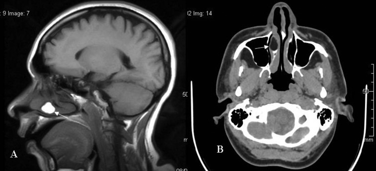
A 40-year-old woman with a known psychiatric disorder came for a routine MRI of the brain. Although there were no abnormal findings in the brain, I noticed a lobulated right nasal turbinate mass of uniform fat intensity (Figure 1a). CT showed a uniform fat-density lesion within the right inferior nasal turbinate, with no calcification or osseous components (Figure 1b). The features were suggestive of a nasal turbinate lipoma. Although histopathological correlation was desired, because of the asymptomatic nature of the lesion and the associated comorbid illness of the patient, no intervention was performed. However, the imaging features were themselves characteristic of a lipoma.
Figure 1.
(A) Sagittal T1 weighted image showing a lobulated hyperintense mass (white arrow) arising from the nasal turbinate (B) Axial CT showing a uniform fat-density lesion within the right inferior nasal turbinate (white arrow).
Lipomas are one of the most common tumours in the head and neck region, however, they are very rarely encountered in the sinonasal tract, and most lipomas are large with osseous metaplasia (termed osteolipoma).1 If the fat density is not conspicuous on imaging, they can resemble an inverted papilloma or a fibrous tumour.1
As lipomas are benign and do not recur, they may undergo ischaemia and develop calcification, in which case they have to be distinguished from an osteolipoma, which is caused by osseous metaplasia rather than dystrophic changes.1 Our case revealed a uniform fat-density lesion arising from the right nasal turbinate, with no osseous components or calcification. This has not been reported in the literature to date.
References
- 1.Abdalla WM, da Motta AC, Lin SY, McCarthy EF, Zinreich SJ. Intraosseous lipoma of the left frontoethmoidal sinuses and nasal cavity. AJNR Am J Neuroradiol 2007;28:615–7 [PMC free article] [PubMed] [Google Scholar]


