Summary
Basi-parallel anatomical scanning (BPAS)-MRI is a simple MRI technique that we designed to reveal the surface appearance of the vertebrobasilar artery within the cistern. Because it requires only 2 cm-thick heavily T2-weighted coronal imaging with gray-scale reversal, we can obtain BPAS-MRI with any MR machine of any company. BPAS-MRI can easily show the outer contour of the vertebrobasilar artery even if occluded. Therefore, BPAS-MRI can also reveal the occluded basilar trunk and the shape of basilar top branching that we cannot see with any other imaging modality before the recanalizing interventional procedure.
To avoid a dangerous blind manipulation of guidewires or micro-catheters, BPAS-MRI should be obtained prior to the interventional procedure in cases of acute basilar artery occlusion.
Key words: MRI, vertebrobasilar system, arterial occlusion, interventional neuroradiology
Introduction
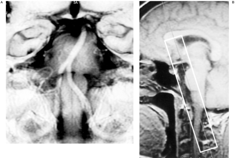
We modified surface anatomical scanning (SAS)-MRI technique1 to reveal the surface appearance of the intracranial vertebrobasilar artery. We named our unique MRI technique basi-parallel anatomical scanning (BPAS)-MRI2. It requires only a 2 cm-thick heavily T2-weighted coronal imaging, parallel to the clivus, with gray-scale reversal in post-processing (figure 1). This technique is so simple that we can obtain BPAS-MRI with any MR machine of any company. BPAS-MRI can show the outer contour of an occluded vertebrobasilar artery within the cistern. We introduce a case of acute basilar artery occlusion in which pre-procedural BPAS-MRI was very useful for the safer interventional recanalizing manipulation.
Figure 1.
A sample image of BPAS-MRI (A), and its scout view (B) planned on a mid-sagittal section.
Methods
We performed BPAS-MRI in a 20-mm thick coronal section parallel to the clivus using fast spin-echo sequence (TR/TE/excitations: 6000/ 250/2, FOV: 19 × 19 cm, matrix: 384 × 224) on a 1.0 Tesla scanner (GE, SIGNA Horizon LX). Acquisition time is one minute 36 seconds by our pulse sequence. We obtain BPAS-MRI routinely because it is a very simple and easy scan, and we always evaluate the vertebrobasilar disease by comparing MR angiogram (MRA) using 3-D time-of-flight with BPAS-MRI. BPAS-MRI is useful especially in visualizing the occluded arteries because it shows an outer contour of the vessels.
Representative Case
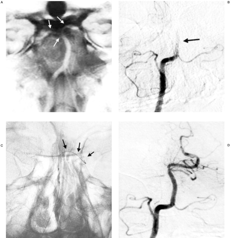
We present a 49-year-old male patient with acute basilar artery occlusion. Although MRA could not be obtained because of the patient's motion, BPAS-MRI showed the entire vertebrobasilar artery in the cistern on initial MR examination (figure 2A). Suspecting acute basilar artery occlusion from his symptoms and the absence of flow void of the basilar artery on axial MR images, we performed cerebral angiography subsequently.
Figure 2.
A) BPAS-MRI on initial MR examination reveals the entire vertebrobasilar artery in the cistern. Bifurcation at the basilar top is clearly shown (arrows). B) Right vertebral angiogram indicates occluded basilar artery at the proximal portion (arrow). C) Using the BPAS-MRI as a referential image, the tip of the micro-guidewire safely reached the left posterior cerebral artery (arrows) crossing through the occluded lesion. D) After the interventional procedure, the basilar trunk and the left PCA are recanalized.
On the right vertebral angiograms, the basilar trunk was occluded and the bilateral posterior cerebral arteries were not shown (figure 2B). Immediately we tried endovascular treatment to recanalize the occluded basilar artery.
At the endovascular session, road-mapping function of the digital subtraction angiography system was not available because of non-opacification of the occluded artery on angiograms. Therefore, we used BPAS-MRI as a referential image for the interventional procedure. It was very useful in crossing the micro-guidewire (Transend, Boston Scientific) through the occluded segment into the PCA. We could pass the occluded segment safely without perforation (figure 2C). In this patient, recanalization of the basilar trunk and the left PCA was successfully achieved by the interventional procedure in acute phase (figure 2D).
Discussion
In the interventional procedure for the recanalization of acute occluded vessels, such as local intra-arterial fibrinolysis or balloon angioplasty, blind manipulation of the microguidewire to cross the lesion is the most dangerous and difficult part of the process. BPAS-MRI, a simple MRI technique that we designed for the precise diagnostic evaluation of the vertebrobasilar system, was very useful as a road map for the recanalizing procedure in a case of acute basilar artery occlusion. Although its application is limited to the posterior circulation, BPAS-MRI can easily be obtained and provides valuable additional information, as in this case. We should apply the BPAS-MRI for not only diagnosis but also intervention in cases of acute basilar artery occlusion.
Conclusions
BPAS-MRI is useful as a road map for the safer interventional procedure in cases of acute basilar artery occlusion.
References
- 1.Katada K. MR imaging of brain surface structures: surface anatomy scanning (SAS) Neuroradiology. 1990;32:439–448. doi: 10.1007/BF00588477. [DOI] [PubMed] [Google Scholar]
- 2.Nagahata M, Hosoya T, et al. Basi-parallel Anatomical Scanning (BPAS) MRI: a simple MRI technique for demonstrating the surface appearance of the intracranial vertebrobasilar artery. Nippon Acta Radiologica. 2003;63:582–584. (in Jpse.) [PubMed] [Google Scholar]


